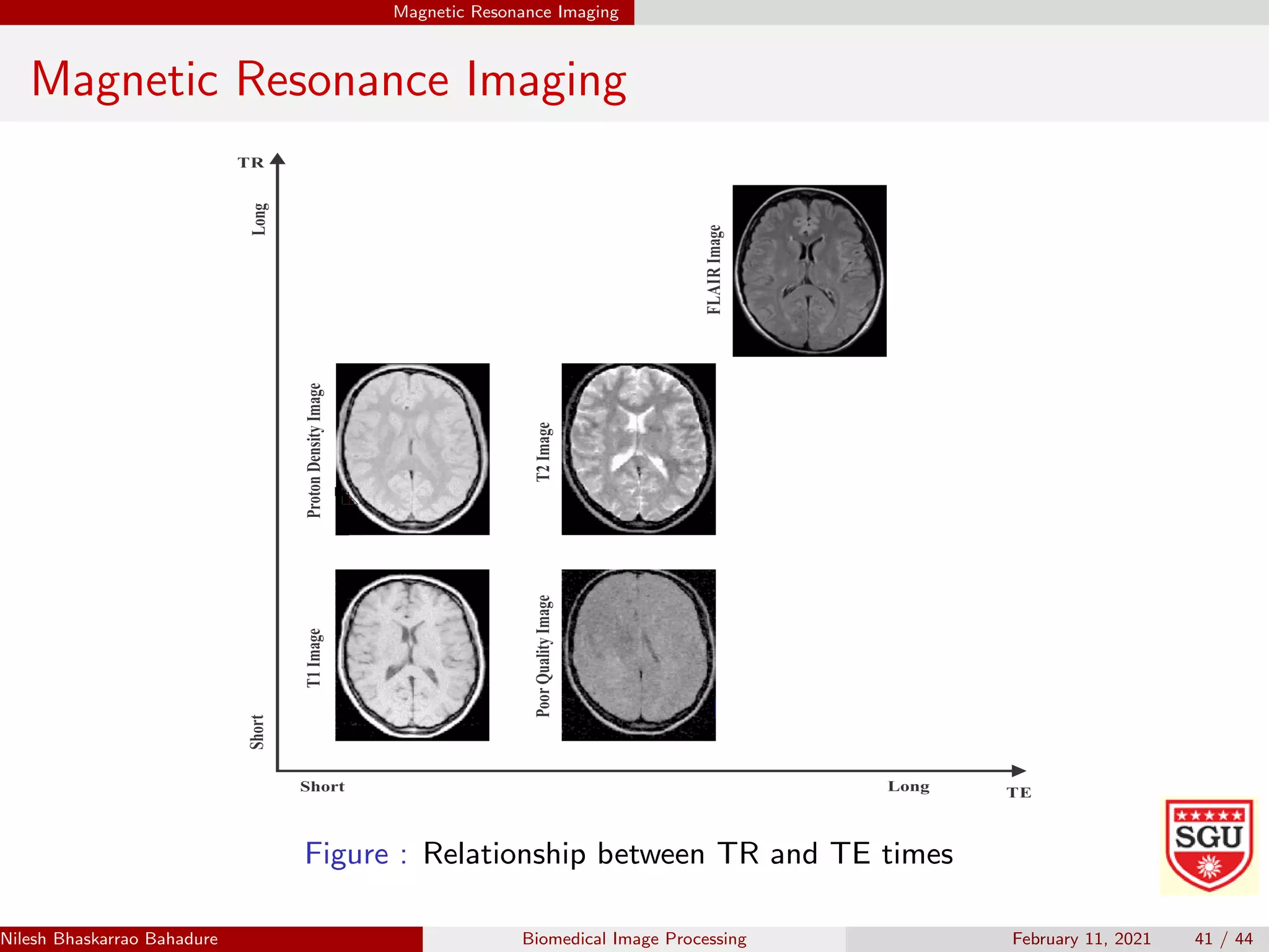
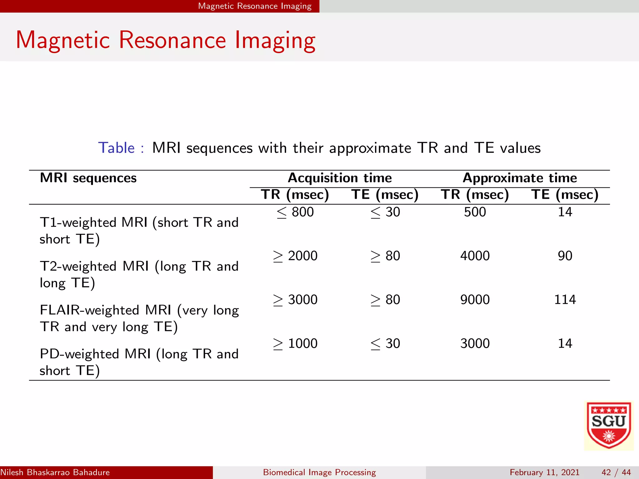
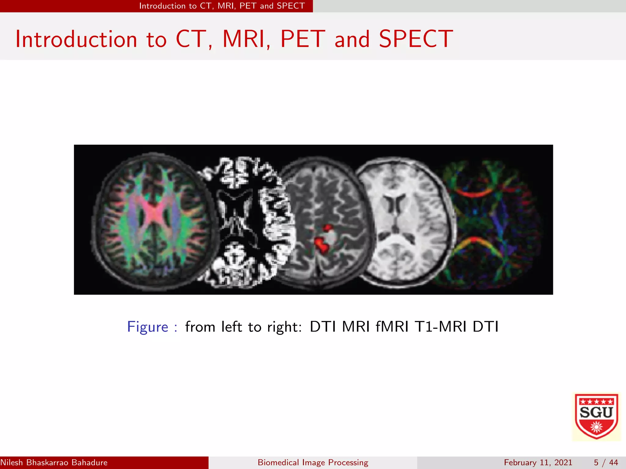
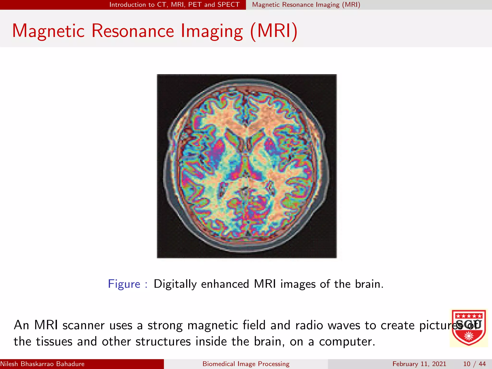
The document provides an overview of various medical imaging techniques used to image the brain including CT, MRI, fMRI, PET, and SPECT. It describes each technique, how they work, what types of images they produce, and what they can be used to detect in the brain. CT uses X-rays to produce 2D images while MRI uses magnetic fields and radio waves to produce detailed 3D images without radiation. fMRI can show which parts of the brain are active during tasks by tracking blood flow and oxygen usage. PET and SPECT involve radioactive tracers to detect biochemical processes.
PET CT combines functional imaging using positron emission tomography (PET) with anatomical imaging using computed tomography (CT). PET detects gamma rays emitted by radiotracers administered to the patient to construct 3D images showing metabolic or biochemical processes. CT provides detailed anatomical images for context. The document discusses the principles and components of PET CT scanning, including radiotracer production and synthesis, scanner design using detector rings, coincidence detection, data acquisition and reconstruction to produce diagnostic images.
Neuroimaging or brain imaging is the use of various techniques to either directly or indirectly image the structure, function, or pharmacology of the nervous system. It is a relatively new discipline within medicine, neuroscience, and psychology
This document summarizes several neuroimaging methods used to study brain function, including EEG, MEG, TMS, PET, MRI, and fMRI. It discusses how each method works, its strengths and limitations, and provides examples of how neuroimaging has been used to study decision making preferences related to uncertainty, probability, and choice.
This document summarizes several brain imaging techniques:
Computerized Tomography uses X-rays to detect lesions, abnormalities, and structural differences. Magnetic Resonance Imaging produces detailed brain images using magnetic fields and radio waves to separate out features not visible on CT scans. Positron Emission Tomography injects radioactive isotopes to trace brain activity and localization of functions like blood flow, glucose uptake, and neurotransmitter binding in response to tasks. New techniques like Magnetic Resonance Spectroscopy and Diffusion Tensor Imaging provide chemical information and map white matter fibers. Electroencephalography measures electrical brain activity patterns using scalp electrodes, including during sleep. Brain Electroactivity Mapping extends EEG by generating activity maps. Event-related potentials measure tiny
Image Processing Technique for Brain Abnormality DetectionCSCJournals
Medical imaging is expensive and very much sophisticated because of proprietary software and expert personalities. This paper introduces an inexpensive, user friendly general-purpose image processing tool and visualization program specifically designed in MATLAB to detect much of the brain disorders as early as possible. The application provides clinical and quantitative analysis of medical images. Minute structural difference of brain gradually results in major disorders such as schizophrenia, Epilepsy, inherited speech and language disorder, Alzheimer's dementia etc. Here the main focusing is given to diagnose the disease related to the brain and its psychic nature (Alzheimer’s disease).
Lithium Filtration for Improved Dose Penetration in BNCTkent.riley
This document summarizes research into adding an optional 6Li filter to an existing epithermal neutron beam used for boron neutron capture therapy (BNCT) to treat brain tumors. Monte Carlo simulations and measurements were used to design and test a removable 8mm thick 6Li filter. The filter improved penetration of thermal neutrons to depths of 9.9cm while maintaining tumor selectivity. Recalculating past treatment plans showed the filter could increase minimum deliverable tumor doses by up to 9% without increasing normal tissue doses. The filter provides an incremental enhancement to the clinical beam that may help establish a therapeutic window for treating deeper tumors.
This document provides information about magnetic resonance imaging (MRI). It begins with background on MRI, noting its wide range of medical applications and over 25,000 scanners in use worldwide. The document then covers MRI goals, definitions, functions, descriptions of how MRI works and the scanning process, precautions, preparations, benefits, disadvantages, risks, and parental concerns. Key points include that MRI uses magnetic fields and radio waves to generate detailed soft tissue images without radiation. Precautions are taken for metal objects and certain implants. MRI is generally safe but may cause claustrophobia or reactions to contrast agents in rare cases.
This document discusses the use of functional MRI (fMRI) in preoperative planning for brain tumor patients. fMRI can help assess the relationship between functionally eloquent brain regions and tumor location to minimize postoperative deficits. It describes different tasks used in fMRI like motor and language tasks. Validation studies show high sensitivity and specificity for motor mapping but more variable results for language mapping. The document also discusses limitations of fMRI like spatial uncertainty and tumor effects on BOLD signal. Overall, fMRI provides useful information to guide surgery when used appropriately with an understanding of its limitations.
This document provides an introduction to medical image processing. It discusses various medical imaging modalities like X-ray, CT, MRI, ultrasound, PET, and angiography. It then describes the basic steps in a medical image processing system: acquisition, preprocessing, segmentation, detection, analysis, and diagnosis. Preprocessing techniques like filtering and denoising are discussed. The document concludes by mentioning some applications of medical image processing like compression, retrieval, and tumor detection.
The document provides an overview of neuroimaging techniques used in psychiatry such as MRI, CT, PET, SPECT, fMRI, DTI, and MRS. It discusses the basic principles, milestones in development, and applications of these techniques. Specifically, it summarizes research using these neuroimaging methods that have found abnormalities in brain structure and function in patients with obsessive-compulsive disorder (OCD), such as reduced serotonin transporter binding in fronto-striatal circuits and differences in brain activity in regions like the thalamus and orbitofrontal cortex.
Imaging Informatics refers to improving the efficiency, accuracy, and reliability of medical imaging services. It involves studying how medical image information is retrieved, analyzed, enhanced, and exchanged within radiology and other areas of medicine. Key areas include PACS, RIS, image processing, 3D visualization, and standards like DICOM that allow integration of imaging technologies. Open source software tools like ImageJ, ITK, and GemIdent provide platforms for medical image analysis.
An evaluation of automated tumor detection techniques of brain magnetic reson...Salam Shah
Image processing is a technique developed by computer and Information technology scientist and being used in all field of research including medical sciences. The focus of this paper is the use of image processing in tumor detection from the brain Magnetic Resonance Imaging (MRI). For the brain tumor detection, Computed Tomography (CT) and Magnetic Resonance Imaging (MRI) are the prominent imaging techniques, but most of the experts prefer MRI over CT. The traditional method of tumor detection in MRI images is a manual inspection which provides variations in the results when analyzed by different experts, therefore, in view of the limitations of the manual analysis of MRI, there is a need for an automated system that can produce globally acceptable and accurate results. There is enough amount of published literature available to replace the manual inspection process of MRI images with the digital computer system using image processing techniques. In this paper, we have provided a review of digital image processing techniques in the context of brain MRI processing and critically analyzed them for the identification of the gaps and limitations of the techniques so that the gaps can be filled and limitations of various techniques can be improved for precise and better results.
PET-scan uses radioactive tracers to visualize and measure metabolic processes in the body. It is often used to diagnose cancer, prepare for epilepsy surgery, and evaluate neurological conditions like Alzheimer's, brain tumors, Parkinson's disease, Huntington's disease, and addiction disorders. PET scans provide information about the brain's chemical functioning that can help identify these conditions and distinguish between similar disorders.
PET RADIOTRACERS
PET images are based on the detection of a tracer
that is typically injected into the body. By comparing
the distribution of the tracer in a patient to
normal templates, a physician is able to evaluate
how well different organs and systems in the body
are functioning. The tracer consists of two components:
a pharmaceutical and a radioactive label.
This slide includes various neuroimaging methods. Firstly, brief backgrounds of positron emission tomography (PET), diffusion tensor MRI, voxel-based morphometry will be introduced. Secondly, a theoretical explanation of BOLD fMRI and preprocessing will be introduced.
http://skyeong.net
The document discusses opportunities for using medical imaging technologies in business applications. It describes an event hosted by the Space IDEAS Hub at the University of Leicester that focuses on exploring how expertise from space missions can benefit UK industry. The event includes sessions on using space missions for medical imaging, image analysis in pathology, tools for predictive drug screening, and stratified medicine in the UK. The Space IDEAS Hub seeks to transfer technologies and experience from space missions to UK companies for commercial applications.
The document discusses various neuroimaging techniques used to study the brain, including their basic principles and psychiatric applications. It describes CT, MRI, MRS, fMRI, and SPECT, explaining what each measures, how they work, and what tissues appear as on the images. It provides examples of structural images and contrasts the advantages and disadvantages of the different modalities. It also outlines specific indications for neuroimaging in clinical practice and research into psychiatric disorders.
What is PET-CT Scan?? Get a detailed overview & Checkout the real cost of PET...BMS_health
A PET (Positron Emission Tomography) scan is an imaging test that allows your doctor
to check for diseases in your body. The scan uses special dye that has radioactive tracers.
A PET-CT scan combines a CT scan and a PET scan. Doctors combine these tests because a CT scan and PET scan show different things. A CT scan shows detailed pictures of tissues and organs inside the body.A PET scan shows abnormal activity. So, the 2 scans together provide more information about the cancer. You usually have a PET-CT scan in the radiology department as an outpatient. Visit BookMyScans and book your whole body PET-CT Scan in India at 60% discount on market price.
https://www.facebook.com/BookMyScansDotCom
https://www.linkedin.com/in/bookmyscans/
Image Segmentation and Identification of Brain Tumor using FFT Techniques of ...IDES Editor
The image processing tools are extensively used on
the development of new algorithms and mathematical tools
for the advanced processing of medical and biological images.
Given an MRI scan, first segment the tumor region in the
MRI brain image and study the pixel intensity values. A
detailed procedure using Matlab script is written to extract
tumor region in CT scan Brain Image and MRI Scan Brain
Image. MRI Scan has higher resolution and easier
identification compare to CT scan Brain image. Fast Fourier
Transform is used here to study the tumor region of MRI
Brain Image in terms of its pixel intensity. Types of FFT like
Zero padded FFT, Windowed FFT are used to study the signal
converted from the MRI Brain Image. It is found that lesser
spectral leakage for Zero Padded Windowed FFT than other
Types of FFT and hence the tumor cell identification is easier
than other methods. Finally higher pixel intensity values of
the cells gives identification of presence and activeness of
tumor cells.
This document presents a proposed method for automatic brain tumor tissue detection in T1-weighted MR images. The method uses a four-step process: segmentation, morphological operations, feature extraction, and classification. In the training section, MRI images are preprocessed and features are extracted using gray-level co-occurrence matrix (GLCM). The features are then used to train a classifier to detect and classify tumors as normal, abnormal, benign, or malignant. In the testing section, input MRI images also undergo preprocessing, feature extraction with GLCM, and then the trained classifier detects, segments, and classifies any tumor tissues found in the images. The goal is to automatically localize and diagnose brain tumor masses in MRI scans.
IRJET- MRI Brain Image Segmentation using Machine Learning TechniquesIRJET Journal
This document discusses machine learning techniques for segmenting brain MRI images. It presents five machine learning methods - K-means clustering, Fuzzy C-means clustering, Watershed segmentation, Support Vector Machine (SVM) classification, and Convolutional Neural Networks (CNN). The methods are applied to segment brain MRI images into gray matter, white matter and cerebrospinal fluid. Segmented images are compared to a ground truth image to analyze segmentation accuracy of the different methods. Accurate segmentation of brain MRI images is important for medical diagnosis and analysis.
Today, computer aided system is widely used in various fields. Among them, the brain tumor detection is an important task in medical image processing. Early diagnosis of brain tumors plays an important role in improving treatment possibilities and increases the survival rate of the patients. Manual segmentation of brain tumors for cancer diagnosis, from large amount of Magnetic Resonance Imaging MRI images generated in clinical routine, is a difficult and time consuming task or even generates errors. So, the automatic brain tumor segmentation is needed to segment tumor. The purpose of the thesis is to detect the brain tumor quickly and accurately from the MRI brain image. In the system, the average filter is used to remove noise and make smooth an input MRI image and threshold segmentation is applied to segment tumor region from MRI brain images. Region properties method is used to detect the tumor region exactly. And then, the equation of the tumor region in the system is effectively applied in any shape of the tumor region. Moe Moe Aye | Kyaw Kyaw Lin "Brain Tumor Detection System for MRI Image" Published in International Journal of Trend in Scientific Research and Development (ijtsrd), ISSN: 2456-6470, Volume-3 | Issue-5 , August 2019, URL: https://www.ijtsrd.com/papers/ijtsrd27864.pdfPaper URL: https://www.ijtsrd.com/engineering/computer-engineering/27864/brain-tumor-detection-system-for-mri-image/moe-moe-aye
This document summarizes current medical image processing research being conducted at various universities. It describes projects involving ECG compression, MRI using blood oxygen level detection, spinal image fusion to improve diagnosis, segmenting anatomical structures from MRI, and using elasticity imaging to detect kidney transplant rejection. It also lists programs for processing MRI data and retinex image processing, as well as websites with medical image test data and news about diagnostic imaging.
CT and MRI are imaging modalities used to visualize structures in the body. CT uses X-rays while MRI uses strong magnetic fields and radio waves. CT provides spatial detail of bones and some soft tissues. MRI has better contrast resolution and does not use ionizing radiation, allowing it to distinguish between soft tissues and detect abnormalities. Different MRI sequences such as T1-weighted and T2-weighted images provide contrast between tissues like fat, water, and pathology. Functional MRI techniques examine brain activity through blood oxygenation levels.
IRJET- Brain Tumor Detection and Classification with Feed Forward Back Propag...IRJET Journal
This document presents a method for detecting and classifying brain tumors in MRI images using a feed forward back propagation neural network. It first preprocesses MRI images by dividing them into blocks and applying Haar transforms for noise removal and edge preservation. Statistical, GLCM, morphological and edge features are then extracted from each block. These features are used to identify abnormal areas. The blocks are then classified as normal or tumor using a feed forward back propagation neural network, which can model nonlinear relationships and is trained to reduce error rates. The method achieves 98% classification accuracy on a benchmark MRI dataset. It results in high accuracy tumor detection with less iterations, reducing computation time compared to previous methods.
Nilesh Bhaskarrao Bahadure presents information on biomedical image processing and signal analysis. The document discusses biomedical signals, their origin and dynamics, and processing techniques. It explains that physiological processes produce signals that can provide information about health and disease states. Advanced signal processing is needed to extract clinically relevant data from complex biomedical signals. The document also describes computer-aided diagnosis systems, which apply computer technology to medical imaging to assist physicians' clinical decision making and improve diagnostic accuracy.
Significance of Brain imaging in Psychiatry. Most of the major Psychiatric disorders are associated with statistically significant differences on various Neuroimaging measures, when comparing groups of patients and controls.
This document is a project report on diagnostic imaging that was completed by nursing students. It discusses four main types of diagnostic imaging - MRI, CT scan, x-ray, and ultrasound. For each type, it provides the definition, purpose, procedures involved, and risks. MRI uses magnetic fields to produce detailed anatomical images. CT scans use x-rays to create cross-sectional images of the body. X-rays produce images using radiation to detect abnormalities. Ultrasound uses sound waves to examine soft tissues and organs without radiation. While very useful diagnostically, some techniques like CT scans and x-rays involve small radiation exposure risks.
Project report on Diagnostic imaging.docxTINKUGARAI1
This document is a project report on diagnostic imaging that was completed by nursing students. It discusses four main types of diagnostic imaging - MRI, CT scan, x-ray, and ultrasound. For each type, it provides definitions, purposes, procedures, and risks. MRI uses magnetic fields to produce detailed anatomical images. CT scans use x-rays to create cross-sectional images. X-rays produce images using radiation absorption. Ultrasound uses sound waves to assess soft tissues and organs. While very useful diagnostically, some techniques involve small radiation risks. The report aims to inform readers about these important medical imaging tools.
This document discusses various research methods for visualizing and studying the living human brain, including both non-invasive and invasive techniques. Non-invasive methods include CT scans, MRI, PET scans, and EEGs which can image brain structure and activity. Invasive animal research techniques are also outlined, such as stereotaxic surgery to precisely position devices in the brain for lesioning, electrical stimulation, or recording brain activity. The goals of cognitive neuroscience to link brain regions to cognitive functions are also mentioned.
Radiation diagnostics diseases of the brain and spinal cord ShieKh Aabid
Radiology plays an important role in diagnosing brain and spinal cord pathologies in children through various imaging techniques. Early diagnosis is key to treating issues in children's brains. There are three main neuroimaging methods - neurosonography, computed tomography, and magnetic resonance imaging. Sonography is a non-invasive way to evaluate the brain through fontanelles in infants, but has limitations. CT scans are also non-invasive and useful for detecting aneurysms and calcifications. MRI provides the best anatomical images and differentiation of structures, and can also perform spectroscopy to examine biochemical changes in tumors. Various contrast-based techniques like angiography can also evaluate the brain's vasculature in children.
Dear B.Sc MIT Students,
Attached is an essential document featuring comprehensive Questions & Answers for Nuclear medicine 3 marks questions and answers. We encourage you to utilize this resource to deepen your understanding and excel in your studies. Wishing you all the success in your academic endeavors and future careers.
Best regards,
Magnetic Resonance Imaging (MRI) is a medical imaging technique that uses strong magnetic fields and radio waves to produce detailed images of the inside of the body. There are different types of MRI machines, including closed, open, and upright configurations. MRI is used to diagnose conditions such as brain tumors, hemorrhages, and multiple sclerosis. It works by aligning hydrogen atoms in the body using magnetism and radio waves, and analyzing the signals produced to form images of tissues and structures. Advantages include a lack of radiation and ability to image soft tissues, while disadvantages include cost and potential incompatibility with metallic implants.
Nuclear medicine imaging uses small amounts of radioactive tracers and imaging technologies like gamma cameras and PET/SPECT scanners to visualize how the body functions at the cellular level. It can help detect diseases earlier than other methods and guide treatment. Nuclear medicine is commonly used to diagnose and manage cancers, heart conditions, brain disorders and more. It involves minimal radiation exposure and has been safely used for decades. Future developments may include more hybrid imaging combining modalities to provide both anatomical and functional information.
Positron emission tomography (PET) is a nuclear imaging technique that produces 3D images of functional processes in the body. A radioactive tracer isotope is injected and accumulates in tissues of interest, undergoing positron emission decay which produces gamma photons detected by the PET scanner to create images. PET scans are used to detect cancer, evaluate brain abnormalities, and examine heart function by tracking radioisotope-labeled molecules processed by the body.
Magnetoencephalography an emerging biological marker for neurodegenerative an...Adonis Sfera, MD
Magnetoencephalography (MEG) is a technique that measures magnetic fields produced by electrical currents in the brain to map functional areas with great temporal resolution. MEG detects alterations in brain structure correlated with changes in function as seen with MRI. A new combined MEG-MRI device can simultaneously record ultra-low-field MRI and MEG to unprecedentedly locate brain activity. MEG has accurately diagnosed PTSD at 90% by identifying patterns unique to sufferers, and combining with diffusion tensor imaging may further improve diagnostic accuracy.
Emerging MRI and metabolic neuroimaging techniques in mild traumatic brain in...IntesarAldweri
Traumatic brain injury (TBI) is one of the leading causes of death worldwide, and mild traumatic brain injury (mTBI) is the most common traumatic injury.
A SPECT scan uses radioactive tracers and computed tomography to show blood flow to organs and tissues. A small amount of radioactive tracer is injected and detected as it passes through the body. Images are created as the tracer is detected, allowing visualization of blood flow. SPECT scans are commonly used to examine the brain, bones, and to detect tumors.
PET scans use radioactive tracers and detectors to generate 3D images of metabolic processes in the body. They have various applications in neurology for diagnosing and monitoring conditions like dementia, epilepsy, movement disorders, and brain tumors. For example, PET can help differentiate Alzheimer's from other dementias based on patterns of hypometabolism in temporal and parietal lobes. It is also useful for localizing epileptic foci before epilepsy surgery. The document discusses the history, mechanisms, common tracers, and limitations of PET scanning as well as its role in evaluating specific neurological conditions and potential future applications.
A PET scan uses radioactive tracers injected into the body to produce 3D images showing functional processes. A short-lived radioactive tracer, FDG, is injected and detected as it breaks down, showing glucose metabolism levels in tissues. Different metabolism levels appear as different colors, allowing the computer to generate images of functional abnormalities like cancers or brain disorders. PET scans can detect diseases earlier than other scans and help avoid unnecessary surgery by precisely identifying areas needing treatment.
Brain imaging techniques allow researchers and doctors to view the brain without invasive surgery. There are several accepted imaging techniques used in research and hospitals worldwide, including brain lesioning, brain staining, and various brain imaging methods. Brain imaging techniques like MRI, PET, CAT, EEG, DOI, and fMRI non-invasively measure brain structure and function by detecting changes in blood flow, oxygen use, electric fields or other signals during mental activities.
Neurobiology of Everyday Life Final Projectmeric89
1) The document discusses the use of diffusion tensor imaging (DTI) to study the effects of stroke on motor outcomes. DTI uses MRI to measure the diffusion of water molecules in brain tissue to analyze the integrity of axons and neurons.
2) Specifically, the author used DTI to study the cortical spinal tract in stroke patients and how damage to axonal membranes and myelin sheaths affects water diffusion. The aim was to predict future upper and lower limb motor recovery based on early DTI scans.
3) Taking the neurobiology course helped the author better understand the biological basis of the DTI signals and relate imaging findings to motor function by learning about neuronal damage mechanisms and cellular structures involved in motor output
Functional magnetic resonance imaging (fMRI) uses blood flow to map brain activity. It detects increased oxygen use in more active brain regions to identify areas involved in sensory, motor, and cognitive tasks. fMRI is a noninvasive way to study brain functions like perception, emotion, and decision-making. It is currently used to plan surgeries, diagnose disorders like Alzheimer's and epilepsy, and investigate conditions including stroke recovery and brain tumors. Further research aims to expand clinical applications of fMRI and improve our understanding of brain activity.
IJRET : International Journal of Research in Engineering and Technology is an international peer reviewed, online journal published by eSAT Publishing House for the enhancement of research in various disciplines of Engineering and Technology. The aim and scope of the journal is to provide an academic medium and an important reference for the advancement and dissemination of research results that support high-level learning, teaching and research in the fields of Engineering and Technology. We bring together Scientists, Academician, Field Engineers, Scholars and Students of related fields of Engineering and Technology
Non invasive modalities of neurocognitive science used for brain mappingeSAT Journals
This document discusses various non-invasive modalities used for brain mapping in neurocognitive science. It describes electromagnetic techniques like EEG and MEG which measure brain's electromagnetic activity, and hemodynamic techniques like MRI, fMRI, PET and SPECT which record hemodynamic activity by detecting changes in blood flow and oxygen levels. These techniques are classified based on their measurement principles. The document provides details on the working mechanisms of EEG, MRI, fMRI and PET modalities. It explains how each modality is used to capture brain activity and images with varying spatial and temporal resolutions.
Similar to Introduction to Medical Image Processing (20)
The document discusses timers in 8051 microcontrollers. It describes the different modes timers can operate in, including 13-bit, 16-bit, and 8-bit auto-reload modes. It explains the timer-related special function registers TMOD, TCON, THx and TLx. It provides steps for initializing timers, programming timers in mode 1, and calculating time delays. The document is intended to provide an understanding of how to generate time delays, measure time, and count pulses using the timers in 8051 microcontrollers.
Total slides: 73
Universal Asynchronous Receiver Transmitter (UART)
Introduction to Serial Communication
Types of Transmission
Simplex Communication
Duplex Communication
Half Duplex Communication
Full Duplex Communication
Methods of Serial data Transmission
Synchronous serial data transfer
Asynchronous serial data transfer
Differences Synchronous Asynchronous
Data Transfer Rate
Calculation of Baud Rate
SCON Register
SBUF Register
Writing to the Serial port
Reading the Serial port
PCON Register
Programming of transmission byte serially
Programming of reception of byte serially
Examples
Programmable Peripheral Interface (PPI) 8255
Features of 8255
Block Diagram of 8255 PPI
3 Modes of operation of 8255 PPI
BSR Mode of 8255 PPI
Parallel IO of 8255 PPI
IC 8155/8156
Features of 8155/8156
Block Diagram of 8155/8156
Chip Enable Logic & Port Addresses (Peripheral I/O Addressing
Scheme
Control Word Register of 8155
Timers of 8155/8156
Modes of Timers of 8155
IC 8355/8755
Block Diagram of 8155/8156
The document discusses the Microprocessor 8085. It describes the architecture of the 8085, which is divided into registers, an arithmetic logic unit, an instruction decoder, address buffers, interrupt control, and timing/control circuitry. It details the registers of the 8085 including general purpose, temporary, special purpose, and 16-bit registers like the program counter and stack pointer. The document also examines the ALU, instruction decoder, addressing mechanisms, interrupt handling, serial I/O, and timing control circuitry of the 8085 microprocessor.
Addressing Modes of 8051
Symbol or nomenclature used for data or memory
Instruction sets of 8051
Assembler and Assembler Directives
Delay Calculation
Examples on Delay Calculation
The Microcontroller 8051 Family
Features of 8051 Microcontroller
Pin Configuration of 8051 Microcontroller
Ports of 8051 Microcontroller
Architecture of 8051 Microcontroller
Registers of 8051
Special Function Registers (SFR's)
Bit addressable RAM
Register Bank and Stack of 8051
Semiconductor Memory Fundamentals
Memory Types
Memory Structure and its requirements
Memory Decoding
Examples
Input - Output Interfacing
Types of Parallel Data Transfer or I/O Techniques
Introduction to Interrupts
What happens when the interrupt is occurs
Interrupt Vs Polling
Classfication of Interrupts
Hardware Interrupts of 8085
Software Interrupts of 8085
Maskable Interrupts of Microprocessor 8085
Non - Maskable Interrupts of Microprocessor 8085
Vectored Interrupts of Microprocessor 8085
Non - Vectored Interrupts of Microprocessor 8085
8085 Microprocessor Interrupt Structure
Interrupt Structure of Microprocessor 8085
SIM Instruction
Non - Vectored Interrupt
Pending Interrupts
What is Interrupt
Introduction to 8051 Microcontroller Interrupts
Interrupts of 8051 Microcontroller
Interrupt Vs Polling
IE register
IP register
What happens when an interrupt occurs?
What happens when an interrupt Ends?
Programming Timer Interrupt
Serial Interrupt
External Hardware Interrupt
Examples
The document discusses the instruction sets and programming of the 8085 microprocessor. It covers the various addressing modes of the 8085 including immediate, register, direct, indirect, and implicit addressing modes. It also describes the instruction format, opcode format, and the different instruction groups of the 8085 such as data transfer, arithmetic, logical, branching, and machine control instructions. For each instruction group, it provides the list of instructions, number of bytes, and number of clock cycles.
Introduction
Embedded Operating Systems
Applications of Embedded Systems
Characteristics of Embedded Systems
Architecture of Real Embedded Systems
Embedded Operating System
Real Time Operating Systems (RTOS)
Total slides: 102
Depletion Layer in PN Junction
Barrier Potential in a PN junction
Energy Diagram of PN Junction
Biasing The PN Junction
V-I Characteristics of P-N junction Diode
Applications of Diode - Rectiers
Photodiode
Light Emitting Diodes - LED
Zener Diode
Total slides: 75
What is Transducers
Selection Criteria of the Transducers
Basic Requirements of a Transducers
Strain Gauge
Inductive Transducer - LVDT
Load Cell
Temperature Transducers
Photoelectric Transducer
LDR
Photovoltaic Solar Cells
The document provides an overview of the bipolar junction transistor (BJT) including:
1. A BJT has three terminals connected to three doped semiconductor regions, either NPN or PNP.
2. BJTs act as current-controlled switches, regulating the current flowing from emitter to collector in proportion to the base voltage.
3. There are three common configurations - common base has voltage gain but no current gain, common emitter has both gains, and common collector has current gain but no voltage gain. Each configuration has different characteristics.
Total slides: 109
Light Emitting Diodes
Seven Segment LED
LCD Interfacing
Stepper Motor Interfacing
Digital to Analog Converter
ADC Interfacing
Keyboard Interfacing
This document contains 99 questions related to programmable logic controllers (PLCs). The questions cover topics such as PLC components, ladder logic programming, registers, instructions, numbering systems, and applications. They range from basic questions testing understanding of PLC concepts to more complex questions involving designing PLC programs to solve application problems. The questions are divided into three units, with unit one focusing on basic PLC operation, unit two on registers and instructions, and unit three on numbering systems, subroutines, and advanced instructions.
This document contains a question bank for the subject "Microprocessor & Interfaces" provided by Dr. Nilesh Bhaskarrao Bahadure of the Department of Electronics Engineering. It includes short answer, mid-range, and long answer type questions related to the architecture, operation, registers, pins, signals, addressing modes and instructions of the 8085 microprocessor. The questions cover topics such as the components of a computer system, microprocessor architecture, assembly language, interrupts, memory interfacing, I/O techniques and programming of the 8085 microprocessor.
This document contains 76 questions related to linear integrated circuits and applications. The questions cover topics such as calculating output voltages for inverting and non-inverting amplifiers, determining parameters like gain and input/output resistances for operational amplifier circuits, and designing circuits like summers, subtractors, and instrumentation amplifiers using operational amplifiers. The questions range from short calculations to longer problems involving circuit design and analysis.
Comparative analysis between traditional aquaponics and reconstructed aquapon...bijceesjournal
The aquaponic system of planting is a method that does not require soil usage. It is a method that only needs water, fish, lava rocks (a substitute for soil), and plants. Aquaponic systems are sustainable and environmentally friendly. Its use not only helps to plant in small spaces but also helps reduce artificial chemical use and minimizes excess water use, as aquaponics consumes 90% less water than soil-based gardening. The study applied a descriptive and experimental design to assess and compare conventional and reconstructed aquaponic methods for reproducing tomatoes. The researchers created an observation checklist to determine the significant factors of the study. The study aims to determine the significant difference between traditional aquaponics and reconstructed aquaponics systems propagating tomatoes in terms of height, weight, girth, and number of fruits. The reconstructed aquaponics system’s higher growth yield results in a much more nourished crop than the traditional aquaponics system. It is superior in its number of fruits, height, weight, and girth measurement. Moreover, the reconstructed aquaponics system is proven to eliminate all the hindrances present in the traditional aquaponics system, which are overcrowding of fish, algae growth, pest problems, contaminated water, and dead fish.
DEEP LEARNING FOR SMART GRID INTRUSION DETECTION: A HYBRID CNN-LSTM-BASED MODELgerogepatton
As digital technology becomes more deeply embedded in power systems, protecting the communication
networks of Smart Grids (SG) has emerged as a critical concern. Distributed Network Protocol 3 (DNP3)
represents a multi-tiered application layer protocol extensively utilized in Supervisory Control and Data
Acquisition (SCADA)-based smart grids to facilitate real-time data gathering and control functionalities.
Robust Intrusion Detection Systems (IDS) are necessary for early threat detection and mitigation because
of the interconnection of these networks, which makes them vulnerable to a variety of cyberattacks. To
solve this issue, this paper develops a hybrid Deep Learning (DL) model specifically designed for intrusion
detection in smart grids. The proposed approach is a combination of the Convolutional Neural Network
(CNN) and the Long-Short-Term Memory algorithms (LSTM). We employed a recent intrusion detection
dataset (DNP3), which focuses on unauthorized commands and Denial of Service (DoS) cyberattacks, to
train and test our model. The results of our experiments show that our CNN-LSTM method is much better
at finding smart grid intrusions than other deep learning algorithms used for classification. In addition,
our proposed approach improves accuracy, precision, recall, and F1 score, achieving a high detection
accuracy rate of 99.50%.
Electric vehicle and photovoltaic advanced roles in enhancing the financial p...IJECEIAES
Climate change's impact on the planet forced the United Nations and governments to promote green energies and electric transportation. The deployments of photovoltaic (PV) and electric vehicle (EV) systems gained stronger momentum due to their numerous advantages over fossil fuel types. The advantages go beyond sustainability to reach financial support and stability. The work in this paper introduces the hybrid system between PV and EV to support industrial and commercial plants. This paper covers the theoretical framework of the proposed hybrid system including the required equation to complete the cost analysis when PV and EV are present. In addition, the proposed design diagram which sets the priorities and requirements of the system is presented. The proposed approach allows setup to advance their power stability, especially during power outages. The presented information supports researchers and plant owners to complete the necessary analysis while promoting the deployment of clean energy. The result of a case study that represents a dairy milk farmer supports the theoretical works and highlights its advanced benefits to existing plants. The short return on investment of the proposed approach supports the paper's novelty approach for the sustainable electrical system. In addition, the proposed system allows for an isolated power setup without the need for a transmission line which enhances the safety of the electrical network
Harnessing WebAssembly for Real-time Stateless Streaming PipelinesChristina Lin
Traditionally, dealing with real-time data pipelines has involved significant overhead, even for straightforward tasks like data transformation or masking. However, in this talk, we’ll venture into the dynamic realm of WebAssembly (WASM) and discover how it can revolutionize the creation of stateless streaming pipelines within a Kafka (Redpanda) broker. These pipelines are adept at managing low-latency, high-data-volume scenarios.
Embedded machine learning-based road conditions and driving behavior monitoringIJECEIAES
Car accident rates have increased in recent years, resulting in losses in human lives, properties, and other financial costs. An embedded machine learning-based system is developed to address this critical issue. The system can monitor road conditions, detect driving patterns, and identify aggressive driving behaviors. The system is based on neural networks trained on a comprehensive dataset of driving events, driving styles, and road conditions. The system effectively detects potential risks and helps mitigate the frequency and impact of accidents. The primary goal is to ensure the safety of drivers and vehicles. Collecting data involved gathering information on three key road events: normal street and normal drive, speed bumps, circular yellow speed bumps, and three aggressive driving actions: sudden start, sudden stop, and sudden entry. The gathered data is processed and analyzed using a machine learning system designed for limited power and memory devices. The developed system resulted in 91.9% accuracy, 93.6% precision, and 92% recall. The achieved inference time on an Arduino Nano 33 BLE Sense with a 32-bit CPU running at 64 MHz is 34 ms and requires 2.6 kB peak RAM and 139.9 kB program flash memory, making it suitable for resource-constrained embedded systems.
A review on techniques and modelling methodologies used for checking electrom...nooriasukmaningtyas
The proper function of the integrated circuit (IC) in an inhibiting electromagnetic environment has always been a serious concern throughout the decades of revolution in the world of electronics, from disjunct devices to today’s integrated circuit technology, where billions of transistors are combined on a single chip. The automotive industry and smart vehicles in particular, are confronting design issues such as being prone to electromagnetic interference (EMI). Electronic control devices calculate incorrect outputs because of EMI and sensors give misleading values which can prove fatal in case of automotives. In this paper, the authors have non exhaustively tried to review research work concerned with the investigation of EMI in ICs and prediction of this EMI using various modelling methodologies and measurement setups.
Introduction- e - waste – definition - sources of e-waste– hazardous substances in e-waste - effects of e-waste on environment and human health- need for e-waste management– e-waste handling rules - waste minimization techniques for managing e-waste – recycling of e-waste - disposal treatment methods of e- waste – mechanism of extraction of precious metal from leaching solution-global Scenario of E-waste – E-waste in India- case studies.
Using recycled concrete aggregates (RCA) for pavements is crucial to achieving sustainability. Implementing RCA for new pavement can minimize carbon footprint, conserve natural resources, reduce harmful emissions, and lower life cycle costs. Compared to natural aggregate (NA), RCA pavement has fewer comprehensive studies and sustainability assessments.
International Conference on NLP, Artificial Intelligence, Machine Learning an...gerogepatton
International Conference on NLP, Artificial Intelligence, Machine Learning and Applications (NLAIM 2024) offers a premier global platform for exchanging insights and findings in the theory, methodology, and applications of NLP, Artificial Intelligence, Machine Learning, and their applications. The conference seeks substantial contributions across all key domains of NLP, Artificial Intelligence, Machine Learning, and their practical applications, aiming to foster both theoretical advancements and real-world implementations. With a focus on facilitating collaboration between researchers and practitioners from academia and industry, the conference serves as a nexus for sharing the latest developments in the field.
Literature Review Basics and Understanding Reference Management.pptxDr Ramhari Poudyal
Three-day training on academic research focuses on analytical tools at United Technical College, supported by the University Grant Commission, Nepal. 24-26 May 2024
Batteries -Introduction – Types of Batteries – discharging and charging of battery - characteristics of battery –battery rating- various tests on battery- – Primary battery: silver button cell- Secondary battery :Ni-Cd battery-modern battery: lithium ion battery-maintenance of batteries-choices of batteries for electric vehicle applications.
Fuel Cells: Introduction- importance and classification of fuel cells - description, principle, components, applications of fuel cells: H2-O2 fuel cell, alkaline fuel cell, molten carbonate fuel cell and direct methanol fuel cells.
1. Biomedical Image Processing
Introduction to Medical Image Processing
Nilesh Bhaskarrao Bahadure
Ph.D., M.E., B.E.
Sanjay Ghodawat University
Atigre, Kolhapur, Maharashtra
nbahadure@gmail.com
https://www.sites.google.com/site/nileshbbahadure/home
February 11, 2021
Nilesh Bhaskarrao Bahadure Ph.D., M.E., B.E. (Sanjay Ghodawat University)
Biomedical Image Processing February 11, 2021 1 / 44
2. Overview
1 Syllabus - Unit 6
2 Introduction to CT, MRI, PET and SPECT
Computerised Tomography (CT)
Magnetic Resonance Imaging (MRI)
Functional Magnetic Resonance Imaging (fMRI)
Positron Emission Tomography (PET)
Single Photon Emission Computed Tomography (SPECT)
3 Tumor types and their therapy
4 Treatment
Surgery
Radiotherapy
Chemotherapy
Immunotherapy
5 Magnetic Resonance Imaging
6 Questions
7 Thank You
Nilesh Bhaskarrao Bahadure Ph.D., M.E., B.E. (Sanjay Ghodawat University)
Biomedical Image Processing February 11, 2021 2 / 44
3. Syllabus - Unit 6
Syllabus Highlights
1 Introduction to CT, MRI, PET and SPECT,
2 tumor types and their therapy,
3 magnetic resonance imaging.
4 Advancement in healthcare technologies.
5 Case studies of biomedical signal and image processing
Nilesh Bhaskarrao Bahadure Ph.D., M.E., B.E. (Sanjay Ghodawat University)
Biomedical Image Processing February 11, 2021 3 / 44
4. Introduction to CT, MRI, PET and SPECT
Introduction to CT, MRI, PET and SPECT
Brain imaging has greatly advanced in the last 20 years, due to better
understanding of the electromagnetic spectrum and radiofrequency waves, in
relation to protons in individual molecules within the cells of the brain. New
technologies allow non-invasive spatial mapping, (morphology), and observations
of processes within the brain during set tasks. By sequencing scanned sections of
the brain, activity between neurons in different parts of the brain can be observed
and monitored. There are a range of scanning techniques such as CT, MRI, fMRI,
PET, SPECT, DTI, and DOT etc, their purpose and limitations are described
below:
Nilesh Bhaskarrao Bahadure Ph.D., M.E., B.E. (Sanjay Ghodawat University)
Biomedical Image Processing February 11, 2021 4 / 44
5. Introduction to CT, MRI, PET and SPECT
Introduction to CT, MRI, PET and SPECT
Figure : from left to right: DTI MRI fMRI T1-MRI DTI
Nilesh Bhaskarrao Bahadure Ph.D., M.E., B.E. (Sanjay Ghodawat University)
Biomedical Image Processing February 11, 2021 5 / 44
6. Introduction to CT, MRI, PET and SPECT Computerised Tomography (CT)
Computerised Tomography (CT)
Computerised Tomography scans use X-rays to show the structure of the brain,
with details such as blood perfusion, the resultant images are two dimensional and
of comparatively low resolution, however, the quality has been much improved
since 1998. With improved technology, the single section has now become a
multisection and the speed has increased eight times, giving well-defined 3-D
pictures. A CT scan may reveal underdeveloped parts of the brain or sites of
injury from impact, tumours, lesions or infection.
Nilesh Bhaskarrao Bahadure Ph.D., M.E., B.E. (Sanjay Ghodawat University)
Biomedical Image Processing February 11, 2021 6 / 44
7. Introduction to CT, MRI, PET and SPECT Computerised Tomography (CT)
Computerised Tomography (CT)
Figure : detectors for a single-section scanner and a multisection scanner
Nilesh Bhaskarrao Bahadure Ph.D., M.E., B.E. (Sanjay Ghodawat University)
Biomedical Image Processing February 11, 2021 7 / 44
8. Introduction to CT, MRI, PET and SPECT Computerised Tomography (CT)
Computerised Tomography (CT)
Figure : Sample CT image for brain
Nilesh Bhaskarrao Bahadure Ph.D., M.E., B.E. (Sanjay Ghodawat University)
Biomedical Image Processing February 11, 2021 8 / 44
9. Introduction to CT, MRI, PET and SPECT Computerised Tomography (CT)
Computerised Tomography (CT)
1 The blue colour shows total blood perfusion throughout the brain.
2 The red colour shows blood perfusion to the left side of the brain only.
Before a CT scan, the patient may drink but is asked not to eat for four hours
beforehand, and not to take strenuous exercise. A CT brain scan will take about
30 minutes and the patient must lie still for the duration. An EEG may be
attached to monitor heart rate, and for some investigations, a tracer injection
(iodinated contrast fluid), may be required to highlight blood vessels, sometimes
leaving a ’taste’ at the back of the throat for a short time afterwards. The
radiologist needs to know if the patient is diabetic, pregnant or on medication.
The procedure is painless, but does involve exposure to radiation at a very low
level.
Nilesh Bhaskarrao Bahadure Ph.D., M.E., B.E. (Sanjay Ghodawat University)
Biomedical Image Processing February 11, 2021 9 / 44
10. Introduction to CT, MRI, PET and SPECT Magnetic Resonance Imaging (MRI)
Magnetic Resonance Imaging (MRI)
Figure : Digitally enhanced MRI images of the brain.
An MRI scanner uses a strong magnetic field and radio waves to create pictures of
the tissues and other structures inside the brain, on a computer.
Nilesh Bhaskarrao Bahadure Ph.D., M.E., B.E. (Sanjay Ghodawat University)
Biomedical Image Processing February 11, 2021 10 / 44
11. Introduction to CT, MRI, PET and SPECT Magnetic Resonance Imaging (MRI)
Magnetic Resonance Imaging (MRI)
The magnetic field aligns the protons (positively charged particles) in hydrogen
atoms, like tiny magnets. Short bursts of radio waves are then sent to knock the
protons out of position, and as they realign, (relaxation time), they emit radio
signals which are detected by a receiving device in the scanner. The signals
emitted from different tissues vary, and can, therefore, be distinguished in the
computer picture
Nilesh Bhaskarrao Bahadure Ph.D., M.E., B.E. (Sanjay Ghodawat University)
Biomedical Image Processing February 11, 2021 11 / 44
12. Introduction to CT, MRI, PET and SPECT Magnetic Resonance Imaging (MRI)
Magnetic Resonance Imaging (MRI)
An MRI scanner can create clear detailed pictures of the structure of the brain
and detect any abnormalities or tumours. Sometimes a dye, or tracer, such as
gadolinium may be introduced via a vein in the arm, to improve contrast in the
image. Images can be enhanced by differences in the strength of the nuclear
magnetic resonance signal recovered from different locations in the brain. The
relaxation times, T1, T2, and T2* are measured after the scanner’s pulse
sequence, and can be chosen to look at specific tissue within the brain.For
example, at a T2 setting, water and fluid containing tissue appears bright, whilst
fat containing tissue is dark, and this can be used to distinguish damaged tissue
from normal tissue. A T1 setting gives a clear image for the contrast between
white and grey matter in the brain. T2* imaging uses a marker, eg. gadolinium,
to measure cerebral blood volume and flow. It may be seen that by selecting
different relaxation times and manipulating radio frequencies, specific brain tissue
can be highlighted for examination by the physician
Nilesh Bhaskarrao Bahadure Ph.D., M.E., B.E. (Sanjay Ghodawat University)
Biomedical Image Processing February 11, 2021 12 / 44
13. Introduction to CT, MRI, PET and SPECT Magnetic Resonance Imaging (MRI)
Magnetic Resonance Imaging (MRI)
The process of having a scan is painless and safe, there is no exposure to
radiation, but occasionally, a patient may have a reaction to the tracer dye.
Pregnant mothers are not recommended to undertake the procedure unless there
is no alternative, since it is not known whether the effects of a strong magnetic
field may affect the developing baby
Nilesh Bhaskarrao Bahadure Ph.D., M.E., B.E. (Sanjay Ghodawat University)
Biomedical Image Processing February 11, 2021 13 / 44
14. Introduction to CT, MRI, PET and SPECT Magnetic Resonance Imaging (MRI)
Magnetic Resonance Imaging (MRI)
Figure : Dataset for sample MR images
Nilesh Bhaskarrao Bahadure Ph.D., M.E., B.E. (Sanjay Ghodawat University)
Biomedical Image Processing February 11, 2021 14 / 44
15. Introduction to CT, MRI, PET and SPECT Functional Magnetic Resonance Imaging (fMRI)
Functional Magnetic Resonance Imaging (fMRI)
Functional magnetic resonance imaging can show which part of the brain is active,
or functioning, in response to the patient performing a given task, by recording
the movement of blood flow. All atoms and molecules have magnetic resonance,
emitting tiny radio wave signals with movement, because they contain protons.
Different molecules have different magnetic resonance and two components of
blood are tracked to observe brain activity.
Nilesh Bhaskarrao Bahadure Ph.D., M.E., B.E. (Sanjay Ghodawat University)
Biomedical Image Processing February 11, 2021 15 / 44
16. Introduction to CT, MRI, PET and SPECT Functional Magnetic Resonance Imaging (fMRI)
Functional Magnetic Resonance Imaging (fMRI)
Haemoglobin in the blood carries oxygen; oxyhaemoglobin, around the brain and
when it is used up, it becomes desoxyhaemoglobin. Where the oxygen is being
’used up’ shows the site of activity in the brain. The picture is made by
monitoring the ratio of the tiny wave frequencies between these two states whilst
the patient carries out a task, eg. tapping a finger, which highlights the area of
the brain functioning to carry out this task
Nilesh Bhaskarrao Bahadure Ph.D., M.E., B.E. (Sanjay Ghodawat University)
Biomedical Image Processing February 11, 2021 16 / 44
17. Introduction to CT, MRI, PET and SPECT Functional Magnetic Resonance Imaging (fMRI)
Functional Magnetic Resonance Imaging (fMRI)
Figure : An fMRI scan showing regions of activation, including the primary visual cortex
Nilesh Bhaskarrao Bahadure Ph.D., M.E., B.E. (Sanjay Ghodawat University)
Biomedical Image Processing February 11, 2021 17 / 44
18. Introduction to CT, MRI, PET and SPECT Functional Magnetic Resonance Imaging (fMRI)
Functional Magnetic Resonance Imaging (fMRI)
An fMRI scan is painless and harmless and can, therefore, be carried out at
regular intervals to monitor the progress of a patient under treatment.
Nilesh Bhaskarrao Bahadure Ph.D., M.E., B.E. (Sanjay Ghodawat University)
Biomedical Image Processing February 11, 2021 18 / 44
19. Introduction to CT, MRI, PET and SPECT Positron Emission Tomography (PET)
Positron Emission Tomography (PET)
Positron emission tomography scanning produces a three-dimensional image of
functional processes in the brain, (not just the structure). PET is a nuclear
medicine imaging technique which requires the patient to receive a small injection
of radio-active material (a sugar tracer; fluorodeoxyglucose), into the bloodstream.
The radio-active material causes the production of gamma-rays, these are a form
of electromagnetic radiation like X-rays, but of higher energy. The radio-active
material is transported around the body and into the brain. A ring of detectors
outside the head is used to detect pairs of gamma rays emitted indirectly by the
positron-emitting radionuclide (tracer), in each part of the brain under
examination.
Nilesh Bhaskarrao Bahadure Ph.D., M.E., B.E. (Sanjay Ghodawat University)
Biomedical Image Processing February 11, 2021 19 / 44
20. Introduction to CT, MRI, PET and SPECT Positron Emission Tomography (PET)
Positron Emission Tomography (PET)
The areas of the brain that command the greater volumes of blood produce the
most gamma-rays, and it is these areas that are computed and displayed by the
PET scan. As the tracer decays, there is a point when gamma photons are
emitted almost opposite to each other, (’annihilation’ in the Figure 7), the timing
of this event is detected and will ultimately improve the detail of the image. This
system not only identifies the activated area of the brain, but also measures the
degree of activity.
Nilesh Bhaskarrao Bahadure Ph.D., M.E., B.E. (Sanjay Ghodawat University)
Biomedical Image Processing February 11, 2021 20 / 44
21. Introduction to CT, MRI, PET and SPECT Positron Emission Tomography (PET)
Positron Emission Tomography (PET)
Figure : Schematic view of the PET process.
Nilesh Bhaskarrao Bahadure Ph.D., M.E., B.E. (Sanjay Ghodawat University)
Biomedical Image Processing February 11, 2021 21 / 44
22. Introduction to CT, MRI, PET and SPECT Positron Emission Tomography (PET)
Positron Emission Tomography (PET)
A patient may only have one PET scan, due to radiation dosage regulations. PET
has proved to be particularly useful in monitoring visual problems, tumours and
metabolic processes.
Nilesh Bhaskarrao Bahadure Ph.D., M.E., B.E. (Sanjay Ghodawat University)
Biomedical Image Processing February 11, 2021 22 / 44
23. Introduction to CT, MRI, PET and SPECT Single Photon Emission Computed Tomography (SPECT)
Single Photon Emission Computed Tomography (SPECT)
Figure : SPECT scan showing blood perfusion
Nilesh Bhaskarrao Bahadure Ph.D., M.E., B.E. (Sanjay Ghodawat University)
Biomedical Image Processing February 11, 2021 23 / 44
24. Introduction to CT, MRI, PET and SPECT Single Photon Emission Computed Tomography (SPECT)
Single Photon Emission Computed Tomography (SPECT)
The single photon emission computed tomography records the signals from
gamma rays,using two or more synchronised gamma cameras, and the multiple
2-D images are computed, tomographically reconstructed, to 3-D. A section may
be examined from several angles, but is slightly less clear than a PET image. A
SPECT scanner is less expensive than a PET scanner and uses longer-lived, more
easily obtained radioisotopes. Tracing blood flow within the brain identifies where
metabolic activity is occurring, enabling assessment of brain functions.
Nilesh Bhaskarrao Bahadure Ph.D., M.E., B.E. (Sanjay Ghodawat University)
Biomedical Image Processing February 11, 2021 24 / 44
25. Introduction to CT, MRI, PET and SPECT Single Photon Emission Computed Tomography (SPECT)
Single Photon Emission Computed Tomography (SPECT)
The patient will not have to fast before the procedure, but will have to remain
absolutely still for 15 to 20 minutes in a scanner, similar to the MRI. A
radiopharmaceutical (tracer) will be injected via a catheter in the arm. The
amount of radiation the patient will be exposed to is very small, about 1 to 3
times normal human annual exposure to background radiation. The procedure is
painless and the patient may resume normal activities immediately afterwards.
Nilesh Bhaskarrao Bahadure Ph.D., M.E., B.E. (Sanjay Ghodawat University)
Biomedical Image Processing February 11, 2021 25 / 44
26. Tumor types and their therapy
Tumor types and their therapy
Tumor is basically an uncontrolled growth of cancerous cells in the body, whereas
brain tumor is classified as uncontrolled or abnormal growth of cancerous cells in
the brain. Brain tumor can be a benign or a malignant. Any tumor that arises
from the glial cells of the brain or from the supportive tissue of the brain is called
“Glioma”. One class of the Glioma tumor is the astrocytoma1
. According to
World Health Organization and American Brain Tumor Association, the tumor
may also be classified according to the grading mechanism, from grade I to grade
IV. The tumor class belongs to grade I and II also referred as low-grade brain
tumors, whereas grade III and IV are referred as high-grade brain tumors.
1is a star-shaped brain cells in the cerebrum called astrocytes
Nilesh Bhaskarrao Bahadure Ph.D., M.E., B.E. (Sanjay Ghodawat University)
Biomedical Image Processing February 11, 2021 26 / 44
27. Tumor types and their therapy
Tumor types and their therapy
The low-grade brain tumor observed slow growth of cancerous cells, whereas
high-grade brain tumor observed rapid growth of cancerous cells. On the scale of
grading mechanism grade I glioma is surely be considered as benign type of
tumor, and complete surgical excursion in this case is considered to be curative.
Grade II glioma is observed as a low-grade brain tumor and often considered to be
the class of benign tumor, but the patient infected with grade II Gliomas require
serial monitoring by MRI, CT or other modern imaging modalities scan in every 6
to 12 months. If the low-grade brain tumor is left untreated then it is likely to
develop into high-grade brain tumors and hence early detection and diagnosis of
the brain tumor is primary concern by the radiology department. The summary of
benign and malignant tumors are shown in Table 1.
Nilesh Bhaskarrao Bahadure Ph.D., M.E., B.E. (Sanjay Ghodawat University)
Biomedical Image Processing February 11, 2021 27 / 44
28. Tumor types and their therapy
Tumor types and their therapy
Table : Differences between benign and malignant tumors
Benign tumors
Malignant tumors
Non-cancerous
Cancerous
Abnormal cells incapable of spreading
Abnormal cells capable of spreading
Cells multiply slowly
Cells multiply rapidly
Grades I and II
Grades III and IV
Easier to remove and does not recur after
excision
Difficult to remove and recurs after ex-
cision
Mass is mobile
Mass is fixed
Homogeneous in structure
Heterogeneous in structure
Surgical excursion is considered to be cu-
rative
Surgery, radiotherapy, chemotherapy or
combination thereof is needed
Nilesh Bhaskarrao Bahadure Ph.D., M.E., B.E. (Sanjay Ghodawat University)
Biomedical Image Processing February 11, 2021 28 / 44
29. Tumor types and their therapy
Tumor types and their therapy
Figure : Tumor types (a) Benign tumor (b) Malignant tumor
Nilesh Bhaskarrao Bahadure Ph.D., M.E., B.E. (Sanjay Ghodawat University)
Biomedical Image Processing February 11, 2021 29 / 44
30. Treatment
Treatment
To obtain an accurate diagnosis, the patients are process through various stages of
treatment, because to offer a better treatment and to increase the chances of the
survival of the patient, early detection of the tumor stage, its size and location is
very important and plays a critical role in the diagnosis. If the patients are
detected with the benign tumor then only surgery is enough to cure the patient,
but if the malignant tumor is detected then continuous monitoring in every 6 to 12
month is needed with surgery, radiotherapy, chemotherapy or combination thereof.
Nilesh Bhaskarrao Bahadure Ph.D., M.E., B.E. (Sanjay Ghodawat University)
Biomedical Image Processing February 11, 2021 30 / 44
31. Treatment Surgery
Surgery
The extreme case of tumor stage is Glioblastoma and the first step in the
treatment of Glioblastoma is surgery. Now a days with the advancement of
modern techniques, surgery is safe for most patients. The purpose of surgery are
to obtain tumor tissues for diagnosis and further treatment planning. To obtain
the exact location of the tumor in the brain, a biopsy may be done in place of
surgery. The astrocytoma and Glioblastoma kind of malignant tumors have
tentacle-like cells and grows into the surrounding tissue, and because of
heterogeneous in structure and rapid growth, these tumors cannot be removed
completely. So, for partial removal of the tumor and obtained the confirmation of
the tumor type, surgery is employed, and then radiation, chemotherapy and or
immunotherapy or combination thereof are used to treat the remaining tumor.
Nilesh Bhaskarrao Bahadure Ph.D., M.E., B.E. (Sanjay Ghodawat University)
Biomedical Image Processing February 11, 2021 31 / 44
32. Treatment Radiotherapy
Radiotherapy
If the malignant type of tumors such as Glioblastoma and astrocytoma is
detected, then radiation therapy usually follows a biopsy or surgery. There are
different types of radiation is employed at different intervals and schedules.
Conventional fractionated external beam radiation is a standard form of radiation
given to the patient in five days a week for five to six week. This form of radiation
is simple form of radiation and similar to the radiation effect cause by X-ray. To
operate on the tumor’s area directly, intensity modulated radiation therapy is
applied, this is also referred as conformal photon radiation. Image-guided
radiation therapy (IGRT) is the technique of using imaging technology at the time
of each treatment to verify that patients are in the right position within a
millimeter. To protect healthy tissue and reduces overall toxicity, proton bean
therapy is employed as an alternative to the standard radiation.
Nilesh Bhaskarrao Bahadure Ph.D., M.E., B.E. (Sanjay Ghodawat University)
Biomedical Image Processing February 11, 2021 32 / 44
33. Treatment Chemotherapy
Chemotherapy
Chemotherapy is applied for newly diagnosed Glioblastoma type of tumor and it is
a six-week course of Temozolomide2
given concurrently with radiotherapy.
Temozolomide is an alkylating agent with reasonable blood-brain barrier
penetration. The main purpose of taking Temozolomide is to maximize the effect
of radio-sensitization and it is generally taken one hour prior to radiation therapy.
Similar type of treatment has also been routinely applied to the patient detected
with astrocytoma type of tumor. It is also observed that, during chemotherapy
drug normal cells of the patient is also affected and will result in the side effect
such as low white blood cell count, fatigue, and hair loss.
2is an oral chemotherapy drug
Nilesh Bhaskarrao Bahadure Ph.D., M.E., B.E. (Sanjay Ghodawat University)
Biomedical Image Processing February 11, 2021 33 / 44
34. Treatment Immunotherapy
Immunotherapy
Immunotherapy is a new kind of treatment used to improve the immune system of
the patients with an aim to fight with the tumor cells and to stop or slow down
the tumor growth. These kind of therapy suggested some natural treatment by
means of healthy foods, cancer vaccines, antigens etc. To check and differentiate
between the healthy and bad tissues, checkpoint proteins, such as PD-L1
(Programmed death ligand 1) is also inserted in the body. The body needs PD-L1
to keep the immune system stronger and to protect healthy cells from the attack
by cancerous cells. The therapy system based on Immunotherapy is less hazardous
and may represent the next frontiers of the promising therapies in the coming
decade.
Nilesh Bhaskarrao Bahadure Ph.D., M.E., B.E. (Sanjay Ghodawat University)
Biomedical Image Processing February 11, 2021 34 / 44
35. Magnetic Resonance Imaging
Magnetic Resonance Imaging
Now a days radiology department has many option for diagnosis of the tumor
stages from modern imaging modalities, but image contrast is the goal in all
imaging procedures. There are number of different imaging modalities are
available, notably, some of them are, MRI, CT, PET, SPECT. Table 2 outlines
some of the strength and weaknesses of the different imaging modalities. The
imaging modality used in our research is based on MRI, as it provides excellent
spatial resolution, no bone artifact, better contrast with high sensitivity, and low
risk because of no or low radiation in comparison to CT scan and other modern
imaging modalities. For diagnosis of the patient and to check time to time
evaluation and measure continuous monitoring is needed, magnetic resonance
imaging can be used several times on the patients because the absorbed radiation
is lowest in MRI. The magnetic resonance imaging is also called NMR is highly
versatile, because it can be used to study both structural and functional features
of brain with different configurations.
Nilesh Bhaskarrao Bahadure Ph.D., M.E., B.E. (Sanjay Ghodawat University)
Biomedical Image Processing February 11, 2021 35 / 44
36. Magnetic Resonance Imaging
Magnetic Resonance Imaging
Table : Strengths and weaknesses of imaging modalities
Modalities Strength Weaknesses
MRI
no or low radiation cost is very high
excellent spatial resolution used mainly for soft tissues
better contrast
low risk
no bone artifact
detect flowing blood and cryptic vascular
malformations
CT
widely available because of low cost low contrast so poor visibility
excellent spatial resolution problem of bone artifact
higher radiation
PET
fair spatial resolution high cost
brain activation can be measured higher radiation
not widely available
SPECT
low cost poor spatial resolution
widely available higher radiation
regional brain perfusion can be measure not widely available
Nilesh Bhaskarrao Bahadure Ph.D., M.E., B.E. (Sanjay Ghodawat University)
Biomedical Image Processing February 11, 2021 36 / 44
37. Magnetic Resonance Imaging
Magnetic Resonance Imaging
The magnetic resonance imaging uses protons, especially hydrogen protons in the
body as a source of signal. This hydrogen protons induce a small magnetic field
due to the movement of atom’s. The acquisition of the MRI is based on the
following relaxation properties:
1 T1 relaxation (T1-weighted MRI)
2 T2 relaxation (T2-weighted MRI)
3 Proton density (PD-weighted MRI)
4 Fluid attenuated inverse recovery (FLAIR-weighted MRI)
Nilesh Bhaskarrao Bahadure Ph.D., M.E., B.E. (Sanjay Ghodawat University)
Biomedical Image Processing February 11, 2021 37 / 44
38. Magnetic Resonance Imaging
Magnetic Resonance Imaging
In MRI scanning, when a patient is placed in the magnetic field, then the
hydrogen atoms in water of their body tissues will line up along the magnetic field
and then the Radio Frequency (RF) pulse is sent in, this causes spinning the
protons into another plane and allows them to be aligned in relax mode and stays
relax when the pulse is turned off. This process of applying RF pulse, allows
protons in relax alignment and then turned off RF pulse, known as relaxation.
This relaxation time varies from one type of tissues to another type and is used in
MRI to differentiate between normal tissues and infected tissues. Each tissue is
measure and characterized in MRI by two relaxations times, T1 (longitudinal
relaxation time) and T2 (transverse relaxation time). An external magnetic field
at the constant rate is employed to align the randomly oriented protons.
Nilesh Bhaskarrao Bahadure Ph.D., M.E., B.E. (Sanjay Ghodawat University)
Biomedical Image Processing February 11, 2021 38 / 44
39. Magnetic Resonance Imaging
Magnetic Resonance Imaging
This alignment or magnetization of the proton element is next disrupted by
introduction of an external RF energy. By emphasizing through the various
relaxation processes, the protons are return to their resting alignment and doing
so it will emits RF energy. The emitted signals are measured after a certain period
following the initial sequence of RF. By changing and validating the sequence of
RF pulses applied and collected, different types of images are created, this images
are referred to as T1-weighted MR images, T2-weighted MR images, Proton
density weighted MR images and FLAIR-weighted MR images.The timing
constraint i.e. TR and TE are described below:
Nilesh Bhaskarrao Bahadure Ph.D., M.E., B.E. (Sanjay Ghodawat University)
Biomedical Image Processing February 11, 2021 39 / 44
40. Magnetic Resonance Imaging
Magnetic Resonance Imaging
1 Repetition time (TR):
It is the time between the successive RF pulses applied during the
magnetization. A long repetition time allows the protons in all the tissues
such as WM, GM, CSF and other tumor infected tissues to relax back into
relax alignment, whereas short repetition time allows the protons in all the
tissues into not having fully relaxed alignment.
2 Echo time (TE):
It is the time at which the electrical signal induced by the spinning protons is
measured. A long echo time results in reduced signal in tissues, whereas a
short echo time reduces the amount of dephasing occurs in tissues like white
matter and gray matter.
The relationship between repetition time and echo time for the acquisition of the
different MRI sequences are shown in Figure 9
Nilesh Bhaskarrao Bahadure Ph.D., M.E., B.E. (Sanjay Ghodawat University)
Biomedical Image Processing February 11, 2021 40 / 44
41. Magnetic Resonance Imaging
Magnetic Resonance Imaging
Figure : Relationship between TR and TE times
Nilesh Bhaskarrao Bahadure Ph.D., M.E., B.E. (Sanjay Ghodawat University)
Biomedical Image Processing February 11, 2021 41 / 44
42. Magnetic Resonance Imaging
Magnetic Resonance Imaging
Table : MRI sequences with their approximate TR and TE values
MRI sequences Acquisition time Approximate time
TR (msec) TE (msec) TR (msec) TE (msec)
T1-weighted MRI (short TR and
short TE)
≤ 800 ≤ 30 500 14
T2-weighted MRI (long TR and
long TE)
≥ 2000 ≥ 80 4000 90
FLAIR-weighted MRI (very long
TR and very long TE)
≥ 3000 ≥ 80 9000 114
PD-weighted MRI (long TR and
short TE)
≥ 1000 ≤ 30 3000 14
Nilesh Bhaskarrao Bahadure Ph.D., M.E., B.E. (Sanjay Ghodawat University)
Biomedical Image Processing February 11, 2021 42 / 44
43. Questions
Questions
1 Differentiate between benign and malignant tumors
2 Explain the treatments involved in brain tumor detection.
3 Explain PET and SPECT
4 Explain Magnetic Resonance Imaging and also give Strengths and weaknesses
of imaging modalities (CT, MRI, PET and SPECT)
5 Write short notes on
1 CT
2 MRI
3 PET
4 SPECT
6 Outlines the differences between different imaging modalities
7 Explain the TR and TE in MRI in details
8 Explain radiation effects in CT, MRI, PET and SPECT
9 Show the MRI sequences with their approximate TR and TE values
10 Explain what to avoid during CT, MRI and PET examination
Nilesh Bhaskarrao Bahadure Ph.D., M.E., B.E. (Sanjay Ghodawat University)
Biomedical Image Processing February 11, 2021 43 / 44
44. Thank You
Thank you
Please send your feedback at nilesh.bahadure@sanjayghodawatuniversity.ac.in
This is Nilesh B. Bahadure from SGU Kolhapur
Thanks for joining
Goodbye, Have a nice day.
For download and more information Click Here
Nilesh Bhaskarrao Bahadure Ph.D., M.E., B.E. (Sanjay Ghodawat University)
Biomedical Image Processing February 11, 2021 44 / 44