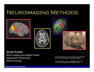
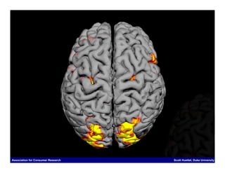
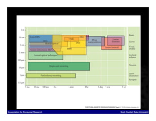
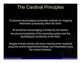
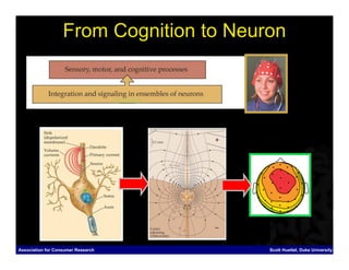
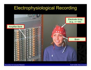
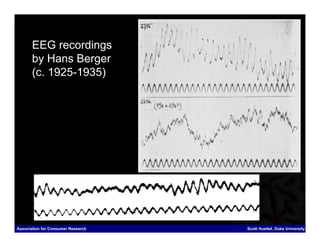
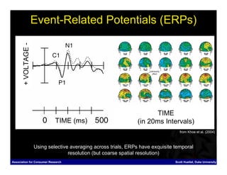
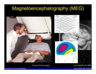
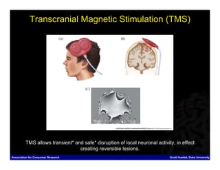
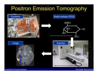
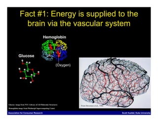
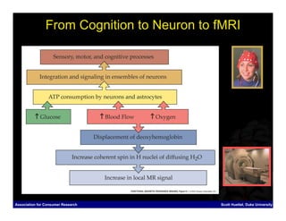
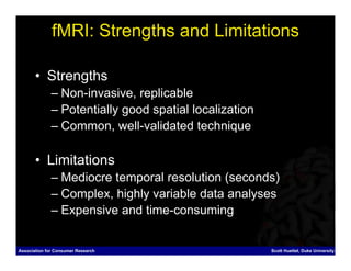
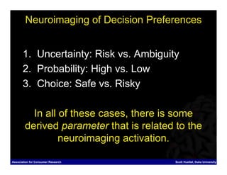
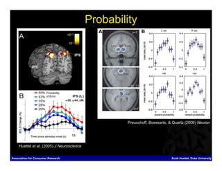
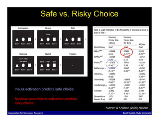
This document summarizes several neuroimaging methods used to study brain function, including EEG, MEG, TMS, PET, MRI, and fMRI. It discusses how each method works, its strengths and limitations, and provides examples of how neuroimaging has been used to study decision making preferences related to uncertainty, probability, and choice.
fNIRS and Brain Computer Interface for CommunicationInsideScientific
LIVE WEBINAR: June 8, 2017
Dr. Ujwal Chaudhary and Dr. Bettina Sorger present groundbreaking research in the field of fNIRS-based BCI for communication with healthy subjects and patients in completely locked-in states.
Neural activity is accompanied by a hemodynamic (vascular) responses that is sensitive to a host of features of coordinated brain function. Relating these measures to the seemingly endless breadth of human behavior is a principal aim of many scientific investigations. Fortunately, learning, language acquisition, sensory and motor functions, emotion, social interactions, and the influence of a host of disease processes can all be explored from measures of the functional near-infrared spectroscopy (fNIRS) signal. Wearable fNIRS technology exists that is portable, safe and easy to use, resistant to motion artifacts and can be employed in a subjects natural environment.
A promising application for fNIRS is the design of brain-computer interfaces (BCIs) for communication with completely locked-in patients. In the so called ‘locked-in’ state, fully conscious and awake patients are unable to communicate naturally due to severe motor paralysis. These patients are, however, able to modulate their brain activity which can be decoded and understood by exploring the fNIRS signal.
In this exclusive webinar sponsored by NIRx Medical Technologies, experts present the basic principles of fNIRS and BCI, technical setup and guidelines for running a successful fNIRS study and a comparison of fNIRS with other functional neuroimaging methods. Presenters highlight groundbreaking research in the field of fNIRS-based BCI for communication with healthy subjects and patients in a completely locked-in state. Specifically, Dr. Ujwal Chaudhary (University of Tübingen) shares results of his research with healthy participants and patients with locked-in syndrome due to amyotrophic lateral sclerosis (ALS). Dr. Bettina Sorger (Maastricht University) presents data from a recent study demonstrating the feasibility of a multiple-choice fNIRS-based communication BCI using differently-timed motor imagery as an information-encoding strategy.
This slide includes various neuroimaging methods. Firstly, brief backgrounds of positron emission tomography (PET), diffusion tensor MRI, voxel-based morphometry will be introduced. Secondly, a theoretical explanation of BOLD fMRI and preprocessing will be introduced.
http://skyeong.net
Optical Coherence Tomography: Technology and applications for neuroimagingManish Kumar
Optical coherence tomography (OCT) is an emerging imaging technology with applications in biology, medicine, and materials investigations. Attractive features include high cellular-level resolution, real-time acquisition rates, and spectroscopic feature extraction in a compact noninvasive instrument. OCT can perform ‘‘optical biopsies’’ of tissue, producing images approaching the resolution of histology without having to resect and histologically process tissue specimens for characterization and diagnosis.
High Precision And Fast Functional Mapping Of Cortical Circuitry Through A No...Taruna Ikrar
Taruna Ikrar, MD., PhD. Author at (High Precision and Fast Functional Mapping of Cortical Circuitry Through a Novel Combination of Voltage Sensitive Dye Imaging and Laser Scanning Photostimulation)
Multimodality Molecular Imaging – An Overview With Special Focus on PET/CTApollo Hospitals
Imaging capabilities have evolved from those that provide anatomical pictures to those that capture functional information and, more recently, molecular information (nuclear medicine, PET, SPECT, PET/CT, SPECT/CT, MRS, contrast-enhanced ultrasound, fluorescence and bioluminescence imaging). Multimodality imaging has emerged as a technology that utilizes the strengths of different modalities and yields a hybrid imaging platform with benefits superior to those of any of its individual components, considered alone. Leading edge hybrid imaging (combining multiple, complementary imaging technologies such as PET and CT) offer unique opportunities to “view” the molecular biology of disease, and the use of this equipment is on the rise.
Dear B.Sc MIT Students,
Attached is an essential document featuring comprehensive Questions & Answers for Nuclear medicine 3 marks questions and answers. We encourage you to utilize this resource to deepen your understanding and excel in your studies. Wishing you all the success in your academic endeavors and future careers.
Best regards,
https://www.snmclub.com/presentation
PET/MRI Current & Future Status
DALE BAILEY PhD , Principal Physicist
Departement of Nuclear Medicine, Royal North Shore Hospital
Professor in Medical Radiation Sciences, University of Sydney
Sydney, Australia
icrm2018
Significance of Brain imaging in Psychiatry. Most of the major Psychiatric disorders are associated with statistically significant differences on various Neuroimaging measures, when comparing groups of patients and controls.
Part 2 of 2 of lecture series introducing undergraduate neuroscience students to the core electrophysiological and imaging techniques used to study neuronal activity.
June 3, 2024 Anti-Semitism Letter Sent to MIT President Kornbluth and MIT Cor...Levi Shapiro
Letter from the Congress of the United States regarding Anti-Semitism sent June 3rd to MIT President Sally Kornbluth, MIT Corp Chair, Mark Gorenberg
Dear Dr. Kornbluth and Mr. Gorenberg,
The US House of Representatives is deeply concerned by ongoing and pervasive acts of antisemitic
harassment and intimidation at the Massachusetts Institute of Technology (MIT). Failing to act decisively to ensure a safe learning environment for all students would be a grave dereliction of your responsibilities as President of MIT and Chair of the MIT Corporation.
This Congress will not stand idly by and allow an environment hostile to Jewish students to persist. The House believes that your institution is in violation of Title VI of the Civil Rights Act, and the inability or
unwillingness to rectify this violation through action requires accountability.
Postsecondary education is a unique opportunity for students to learn and have their ideas and beliefs challenged. However, universities receiving hundreds of millions of federal funds annually have denied
students that opportunity and have been hijacked to become venues for the promotion of terrorism, antisemitic harassment and intimidation, unlawful encampments, and in some cases, assaults and riots.
The House of Representatives will not countenance the use of federal funds to indoctrinate students into hateful, antisemitic, anti-American supporters of terrorism. Investigations into campus antisemitism by the Committee on Education and the Workforce and the Committee on Ways and Means have been expanded into a Congress-wide probe across all relevant jurisdictions to address this national crisis. The undersigned Committees will conduct oversight into the use of federal funds at MIT and its learning environment under authorities granted to each Committee.
• The Committee on Education and the Workforce has been investigating your institution since December 7, 2023. The Committee has broad jurisdiction over postsecondary education, including its compliance with Title VI of the Civil Rights Act, campus safety concerns over disruptions to the learning environment, and the awarding of federal student aid under the Higher Education Act.
• The Committee on Oversight and Accountability is investigating the sources of funding and other support flowing to groups espousing pro-Hamas propaganda and engaged in antisemitic harassment and intimidation of students. The Committee on Oversight and Accountability is the principal oversight committee of the US House of Representatives and has broad authority to investigate “any matter” at “any time” under House Rule X.
• The Committee on Ways and Means has been investigating several universities since November 15, 2023, when the Committee held a hearing entitled From Ivory Towers to Dark Corners: Investigating the Nexus Between Antisemitism, Tax-Exempt Universities, and Terror Financing. The Committee followed the hearing with letters to those institutions on January 10, 202
Honest Reviews of Tim Han LMA Course Program.pptxtimhan337
Personal development courses are widely available today, with each one promising life-changing outcomes. Tim Han’s Life Mastery Achievers (LMA) Course has drawn a lot of interest. In addition to offering my frank assessment of Success Insider’s LMA Course, this piece examines the course’s effects via a variety of Tim Han LMA course reviews and Success Insider comments.
Normal Labour/ Stages of Labour/ Mechanism of LabourWasim Ak
Normal labor is also termed spontaneous labor, defined as the natural physiological process through which the fetus, placenta, and membranes are expelled from the uterus through the birth canal at term (37 to 42 weeks
Macroeconomics- Movie Location
This will be used as part of your Personal Professional Portfolio once graded.
Objective:
Prepare a presentation or a paper using research, basic comparative analysis, data organization and application of economic information. You will make an informed assessment of an economic climate outside of the United States to accomplish an entertainment industry objective.
Thesis Statement for students diagnonsed withADHD.ppt
Neuroimaging methods
1. Neuroimaging Methods
Scott Huettel
Brain Imaging and Analysis Center
All uncredited figures are from Huettel, Song, & McCarthy
Department of Psychiatry (2004). Functional Magnetic Resonance Imaging.
Duke University This presentation, save for credited figures from other
sources, is copyrighted by Scott Huettel (2006).
Association for Consumer Research Scott Huettel, Duke University
3. Methods for Creating Images of
(Human) Brain Function
1. Electroencephalography (EEG)
2. Magnetoencephalography (MEG)
3. Transcranial Magnetic Stimulation (TMS)
4. Positron Emission Tomography (PET)
5. Structural Magnetic Resonance Imaging (MRI)
6. Functional Magnetic Resonance Imaging (fMRI)
7. Examples: Neuroimaging of Choice
Association for Consumer Research Scott Huettel, Duke University
5. The Cardinal Principles
Functional neuroimaging comprises methods for mapping
information processing within the brain.
All functional neuroimaging is limited by two factors:
the physical properties of the recording system and the
physiological constraints of the brain.
Images of brain activity only have meaning when acquired
using the correct experimental design and interpreted using
the correct analyses.
Association for Consumer Research Scott Huettel, Duke University
7. From Cognition to Neuron
Association for Consumer Research Scott Huettel, Duke University
8. Electrophysiological Recording
Electrode Array
(e.g., n = 64)
Amplifier Bank
Brain
Association for Consumer Research Scott Huettel, Duke University
9. EEG recordings
by Hans Berger
(c. 1925-1935)
Association for Consumer Research Scott Huettel, Duke University
10. + VOLTAGE -
Event-Related Potentials (ERPs)
TIME
TIME (ms) (in 20ms Intervals)
from Khoe et al. (2004)
Using selective averaging across trials, ERPs have exquisite temporal
resolution (but coarse spatial resolution)
Association for Consumer Research Scott Huettel, Duke University
12. Magnetoencephalography (MEG)
Courtesy 4D Neuroimaging from Woldorff et al. (1999)
Association for Consumer Research Scott Huettel, Duke University
13. 3. Transcranial Magnetic Stimulation
(TMS)
Association for Consumer Research Scott Huettel, Duke University
14. Transcranial Magnetic Stimulation (TMS)
TMS allows transient* and safe* disruption of local neuronal activity, in effect
creating reversible lesions.
Association for Consumer Research Scott Huettel, Duke University
15. 4. Positron Emission Tomography
(PET)
Association for Consumer Research Scott Huettel, Duke University
17. Positron Emission Tomography
Cyclotron Radio-isotope (FDG)
http://www.med-ed.virginia.edu/courses/rad/PETCT/Emission.html
Image from Utah Center for Advanced Imaging Research
Image Scanner
http://www.idac.tohoku.ac.jp/dep/nmr/pet1.jpg
Association for Consumer Research Scott Huettel, Duke University
18. PET: Strengths and Limitations
• Strengths
– Uses a simple physiological mechanism
– Provides absolute, quantitative data
– Allows imaging of anything that can be tagged
• Limitations
– Poor temporal resolution (many minutes)
– Poor spatial resolution (several centimeters)
– Requires injection of radioactive material
Association for Consumer Research Scott Huettel, Duke University
20. MRI Scanning Hardware
“Imaging”
(Weak Gradient “Magnetic”
Magnetic Fields) (Strong Static Magnetic Field)
“Resonance”
(Radiofrequency Energy)
Association for Consumer Research Scott Huettel, Duke University
22. 6. Functional MRI (fMRI)
Association for Consumer Research Scott Huettel, Duke University
23. Fact #1: Energy is supplied to the
brain via the vascular system
Hemoglobin
Glucose
(Oxygen)
Glucose image from NYU Library of 3-D Molecular Structures
From Duvernoy et al., 1982
Hemoglobin image from Pittsburgh Supercomputing Center
Association for Consumer Research Scott Huettel, Duke University
24. Fact #2: More hemoglobin is supplied than needed,
causing a decrease in deoxygenated hemoglobin.
From Mandeville et al., 1999
Association for Consumer Research Scott Huettel, Duke University
25. Fact #3: Deoxygenated hemoglobin reduces
some forms (T2*) of MR signal.
Baseline
Blood-Oxygenation-Level
Dependent Contrast
(BOLD Contrast)
Task
Association for Consumer Research Scott Huettel, Duke University
26. From Cognition to Neuron to fMRI
Association for Consumer Research Scott Huettel, Duke University
27. fMRI: Strengths and Limitations
• Strengths
– Non-invasive, replicable
– Potentially good spatial localization
– Common, well-validated technique
• Limitations
– Mediocre temporal resolution (seconds)
– Complex, highly variable data analyses
– Expensive and time-consuming
Association for Consumer Research Scott Huettel, Duke University
28. Neuroimaging of Decision Preferences
1. Uncertainty: Risk vs. Ambiguity
2. Probability: High vs. Low
3. Choice: Safe vs. Risky
In all of these cases, there is some
derived parameter that is related to the
neuroimaging activation.
Association for Consumer Research Scott Huettel, Duke University
29. Dissociable Systems for Risk and Ambiguity
Risky - Certain
Parietal Cortex Ambiguity Preference Risk Preference
Risky - Risky
Ambiguity preference (1-α) Risk preference (β)
Ambiguity Preference Risk Preference
Prefrontal cortex
Ambiguity preference (1-α) Risk preference (β)
Huettel et al. (2006) Neuron
Association for Consumer Research Scott Huettel, Duke University
30. Probability
Probability
of Error
Preuschoff, Boessarts, & Quartz (2006) Neuron
Huettel et al. (2005) J Neuroscience
Association for Consumer Research Scott Huettel, Duke University
31. Safe vs. Risky Choice
Insula activation predicts safe choice.
Nucleus accumbens activation predicts
risky choice.
Kuhnen & Knutson (2005) Neuron
Association for Consumer Research Scott Huettel, Duke University
32. Summary
• Neuroimaging techniques create maps of brain
function.
• The most common approaches measure
neuronal activity (EEG, ERP, MEG) or brain
hemodynamics (PET, FMRI).
• The neuroimaging approaches relevant for
consumer research involve relating
neuroimaging data to economic parameters.
Association for Consumer Research Scott Huettel, Duke University
33. Acknowledgments
Recommended Readings:
• Huettel, Song, & McCarthy (2004). Functional Magnetic Resonance Imaging.
• Buxton (2002). Introduction to fMRI.
• Luck (2005). An Introduction to the ERP Technique.
• Purves et al. (2004). Neuroscience, 3rd Edition.
FMRI education colleagues:
• Allen Song (Duke University), Gregory McCarthy (Yale University)
Laboratory members:
• Bethany Weber, Dharol Tankersley, John Clithero, Luke Vicens, Lily Kinross-
Wright, Parker Goyer, Jason Chen
neuroeconomics.duke.edu
Association for Consumer Research Scott Huettel, Duke University