1-definition of SPECT :Single Photon Emission Computed Tomography.
2-differs from BET scan and SPECT.
3-divaice of SPECT.
4-SPECT scan for brain.
5-clinical application
6-patient preparation
7-ADVANTAGE & DISADVANTAGE
1-definition of SPECT :Single Photon Emission Computed Tomography.
2-differs from BET scan and SPECT.
3-divaice of SPECT.
4-SPECT scan for brain.
5-clinical application
6-patient preparation
7-ADVANTAGE & DISADVANTAGE
What is pet scan, it's principle, components of pet, pet working , cases of pet , pet clinical applications PET/CT, Disadvantages and accuracy.#PETSCAN
Nuclear Medicine.................
Radioactivity………………
Gamma camera………………
PET scan and SPECT scan…...........
Nuclear Medicine Studies…………..
Nuclear Medicine Team……………
Safety in Nuclear Medicine…………
This pdf is about the Positron Emission Tomography (PET) technique.
For more details visit on YouTube; @SELF-EXPLANATORY;
PET; https://youtu.be/rlwGbFGS6wg
Thanks...!
Factory Supply Best Quality Pmk Oil CAS 28578–16–7 PMK Powder in Stockrebeccabio
Factory Supply Best Quality Pmk Oil CAS 28578–16–7 PMK Powder in Stock
Telegram: bmksupplier
signal: +85264872720
threema: TUD4A6YC
You can contact me on Telegram or Threema
Communicate promptly and reply
Free of customs clearance, Double Clearance 100% pass delivery to USA, Canada, Spain, Germany, Netherland, Poland, Italy, Sweden, UK, Czech Republic, Australia, Mexico, Russia, Ukraine, Kazakhstan.Door to door service
Hot Selling Organic intermediates
Ozempic: Preoperative Management of Patients on GLP-1 Receptor Agonists Saeid Safari
Preoperative Management of Patients on GLP-1 Receptor Agonists like Ozempic and Semiglutide
ASA GUIDELINE
NYSORA Guideline
2 Case Reports of Gastric Ultrasound
Ethanol (CH3CH2OH), or beverage alcohol, is a two-carbon alcohol
that is rapidly distributed in the body and brain. Ethanol alters many
neurochemical systems and has rewarding and addictive properties. It
is the oldest recreational drug and likely contributes to more morbidity,
mortality, and public health costs than all illicit drugs combined. The
5th edition of the Diagnostic and Statistical Manual of Mental Disorders
(DSM-5) integrates alcohol abuse and alcohol dependence into a single
disorder called alcohol use disorder (AUD), with mild, moderate,
and severe subclassifications (American Psychiatric Association, 2013).
In the DSM-5, all types of substance abuse and dependence have been
combined into a single substance use disorder (SUD) on a continuum
from mild to severe. A diagnosis of AUD requires that at least two of
the 11 DSM-5 behaviors be present within a 12-month period (mild
AUD: 2–3 criteria; moderate AUD: 4–5 criteria; severe AUD: 6–11 criteria).
The four main behavioral effects of AUD are impaired control over
drinking, negative social consequences, risky use, and altered physiological
effects (tolerance, withdrawal). This chapter presents an overview
of the prevalence and harmful consequences of AUD in the U.S.,
the systemic nature of the disease, neurocircuitry and stages of AUD,
comorbidities, fetal alcohol spectrum disorders, genetic risk factors, and
pharmacotherapies for AUD.
These lecture slides, by Dr Sidra Arshad, offer a quick overview of physiological basis of a normal electrocardiogram.
Learning objectives:
1. Define an electrocardiogram (ECG) and electrocardiography
2. Describe how dipoles generated by the heart produce the waveforms of the ECG
3. Describe the components of a normal electrocardiogram of a typical bipolar leads (limb II)
4. Differentiate between intervals and segments
5. Enlist some common indications for obtaining an ECG
Study Resources:
1. Chapter 11, Guyton and Hall Textbook of Medical Physiology, 14th edition
2. Chapter 9, Human Physiology - From Cells to Systems, Lauralee Sherwood, 9th edition
3. Chapter 29, Ganong’s Review of Medical Physiology, 26th edition
4. Electrocardiogram, StatPearls - https://www.ncbi.nlm.nih.gov/books/NBK549803/
5. ECG in Medical Practice by ABM Abdullah, 4th edition
6. ECG Basics, http://www.nataliescasebook.com/tag/e-c-g-basics
HOT NEW PRODUCT! BIG SALES FAST SHIPPING NOW FROM CHINA!! EU KU DB BK substit...GL Anaacs
Contact us if you are interested:
Email / Skype : kefaya1771@gmail.com
Threema: PXHY5PDH
New BATCH Ku !!! MUCH IN DEMAND FAST SALE EVERY BATCH HAPPY GOOD EFFECT BIG BATCH !
Contact me on Threema or skype to start big business!!
Hot-sale products:
NEW HOT EUTYLONE WHITE CRYSTAL!!
5cl-adba precursor (semi finished )
5cl-adba raw materials
ADBB precursor (semi finished )
ADBB raw materials
APVP powder
5fadb/4f-adb
Jwh018 / Jwh210
Eutylone crystal
Protonitazene (hydrochloride) CAS: 119276-01-6
Flubrotizolam CAS: 57801-95-3
Metonitazene CAS: 14680-51-4
Payment terms: Western Union,MoneyGram,Bitcoin or USDT.
Deliver Time: Usually 7-15days
Shipping method: FedEx, TNT, DHL,UPS etc.Our deliveries are 100% safe, fast, reliable and discreet.
Samples will be sent for your evaluation!If you are interested in, please contact me, let's talk details.
We specializes in exporting high quality Research chemical, medical intermediate, Pharmaceutical chemicals and so on. Products are exported to USA, Canada, France, Korea, Japan,Russia, Southeast Asia and other countries.
Pulmonary Thromboembolism - etilogy, types, medical- Surgical and nursing man...VarunMahajani
Disruption of blood supply to lung alveoli due to blockage of one or more pulmonary blood vessels is called as Pulmonary thromboembolism. In this presentation we will discuss its causes, types and its management in depth.
Couples presenting to the infertility clinic- Do they really have infertility...Sujoy Dasgupta
Dr Sujoy Dasgupta presented the study on "Couples presenting to the infertility clinic- Do they really have infertility? – The unexplored stories of non-consummation" in the 13th Congress of the Asia Pacific Initiative on Reproduction (ASPIRE 2024) at Manila on 24 May, 2024.
MANAGEMENT OF ATRIOVENTRICULAR CONDUCTION BLOCK.pdfJim Jacob Roy
Cardiac conduction defects can occur due to various causes.
Atrioventricular conduction blocks ( AV blocks ) are classified into 3 types.
This document describes the acute management of AV block.
MANAGEMENT OF ATRIOVENTRICULAR CONDUCTION BLOCK.pdf
SPECT Scan
1. > 1
Overview
A Single Photon Emission Computed Tomography
(SPECT) scan is a type of nuclear imaging test that
shows how blood flows to tissues and organs.
How does a SPECT scan work?
A SPECT scan integrates two technologies to view
your body: computed tomography (CT) and a
radioactive material (tracer). The tracer is what
allows doctors to see how blood flows to tissues and
organs.
Before the SPECT scan, you are injected with a
chemical that is radiolabled, meaning it emits
gamma rays that can be detected by the scanner.
The computer collects the information emitted by
the gamma rays and translates them into two-
dimensional cross-sections. These cross-sections
can be added back together to form a 3D image of
your brain.
The radioisotopes typically used in SPECT to label
tracers are iodine-123, technetium-99m, xenon-
133, thallium-201, and fluorine-18. These
radioactive forms of natural elements will pass
safely through your body and be detected by the
scanner. Various drugs and other chemicals can be
labeled with these isotopes.
The type of tracer used depends on what your
doctor wants to measure. For example, if your
doctor is looking at a tumor, he or she might use
radiolabled glucose (FDG) and watch how it is
metabolized by the tumor.
The test differs from a PET scan in that the tracer
stays in your blood stream rather than being
absorbed by surrounding tissues, thereby limiting
the images to areas where blood flows. SPECT
scans are cheaper and more readily available than
higher resolution PET scans.
What does a SPECT scan show?
A SPECT scan is primarily used to view how blood
flows through arteries and veins in the brain. Tests
have shown that it might be more sensitive to brain
injury than either MRI or CT scanning because it
can detect reduced blood flow to injured sites.
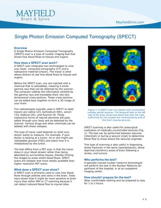
SPECT scanning is also useful for presurgical
evaluation of medically uncontrolled seizures (Fig.
1). The test can be performed between seizures
(interictal) or during a seizure (ictal) to determine
blood flow to areas where the seizures originate.
This type of scanning is also useful in diagnosing
stress fractures in the spine (spondylolysis), blood
deprived (ischemic) areas of brain following a
stroke, and tumors.
Who performs the test?
A specially trained nuclear medicine technologist
will perform the test in the Nuclear Medicine de-
partment of the hospital, or at an outpatient
imaging center.
How should I prepare for the test?
Wear comfortable clothing and be prepared to stay
for 1 to 2 hours.
Single Photon Emission Computed Tomography (SPECT)
Figure 1. A SPECT scan of a patient with uncontrolled
complex partial seizures. The temporal lobe on the left
side of the brain shows less blood flow than the right,
confirming for the surgeon the nonfunctioning area of
the brain causing seizures.