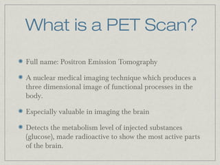
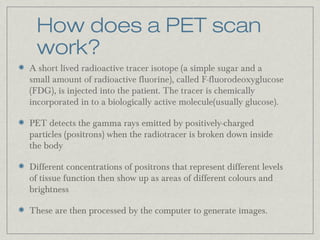
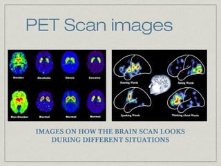
PROJECT

Positron Emission Tomography
PET SCAN

DATE

NOV.1ST 2013

CLIENT

THERESA YIP & MICHELLE MA
What is a PET Scan?
Full name: Positron Emission Tomography
A nuclear medical imaging technique which produces a
three dimensional image of functional processes in the
body.
Especially valuable in imaging the brain
Detects the metabolism level of injected substances
(glucose), made radioactive to show the most active parts
of the brain.
How does a PET scan
work?
A short lived radioactive tracer isotope (a simple sugar and a
small amount of radioactive fluorine), called F-fluorodeoxyglucose
(FDG), is injected into the patient. The tracer is chemically
incorporated in to a biologically active molecule(usually glucose).
PET detects the gamma rays emitted by positively-charged
particles (positrons) when the radiotracer is broken down inside
the body
Different concentrations of positrons that represent different levels
of tissue function then show up as areas of different colours and
brightness
These are then processed by the computer to generate images.
PET Scan images

IMAGES ON HOW THE BRAIN SCAN LOOKS
DURING DIFFERENT SITUATIONS
Strengths
Early PET scan can diagnose any abnormalities in activity level e.g. Can help
detect cancer, brain disorders, heart conditions and other diseases
By identifying changes in the body at the cellular level, PET imaging may detect
the early onset of disease before it is evident on other imaging tests such as CT
or MRI.
PET scan can avoid unnecessary surgery
For many diseases, nuclear medicine scans yield the most useful information
needed to make a diagnosis or to determine appropriate treatment, if any.
Nuclear medicine is less expensive and may yield more precise information than
exploratory surgery.
The information provided by nuclear medicine examinations is unique and
often unattainable using other imaging procedures.
Limitations
Time-consuming
Not as precise as fMRI scan
PET scan might be dangerous depending on each individual’s
heart conditions
PET scan can sometimes show up areas of high activity which my
be mistake for cancers
The radioactive substance has a very short decay and therefore
appointments must run on schedule.
PET scans are a very expensive form of imaging, and are not
readily available all the time.
Bibliography

•

Brain Positron Emission Tomography. (n.d.). In
Wikipedia. Retrieved from
http://en.wikipedia.org/wiki/Brain_positron_emission_tom
ography

•

Raichle. M. (n.d.). Retrieved from
http://www.alz.org/living_with_alzheimers_your_brain.asp

•

Silverman D. (Doctor) (2011, July 18). What is a PET
Sc an? Podcast retrieved from
http://www.youtube.com/watch?v=scoOTHl879A

•

(n.d.) Retrieved from http://www.snm.org/index.cfm?
PageID=7988

PET scan

  • 1.
    PROJECT Positron Emission Tomography PETSCAN DATE NOV.1ST 2013 CLIENT THERESA YIP & MICHELLE MA
  • 2.
    What is aPET Scan? Full name: Positron Emission Tomography A nuclear medical imaging technique which produces a three dimensional image of functional processes in the body. Especially valuable in imaging the brain Detects the metabolism level of injected substances (glucose), made radioactive to show the most active parts of the brain.
  • 3.
    How does aPET scan work? A short lived radioactive tracer isotope (a simple sugar and a small amount of radioactive fluorine), called F-fluorodeoxyglucose (FDG), is injected into the patient. The tracer is chemically incorporated in to a biologically active molecule(usually glucose). PET detects the gamma rays emitted by positively-charged particles (positrons) when the radiotracer is broken down inside the body Different concentrations of positrons that represent different levels of tissue function then show up as areas of different colours and brightness These are then processed by the computer to generate images.
  • 4.
    PET Scan images IMAGESON HOW THE BRAIN SCAN LOOKS DURING DIFFERENT SITUATIONS
  • 5.
    Strengths Early PET scancan diagnose any abnormalities in activity level e.g. Can help detect cancer, brain disorders, heart conditions and other diseases By identifying changes in the body at the cellular level, PET imaging may detect the early onset of disease before it is evident on other imaging tests such as CT or MRI. PET scan can avoid unnecessary surgery For many diseases, nuclear medicine scans yield the most useful information needed to make a diagnosis or to determine appropriate treatment, if any. Nuclear medicine is less expensive and may yield more precise information than exploratory surgery. The information provided by nuclear medicine examinations is unique and often unattainable using other imaging procedures.
  • 6.
    Limitations Time-consuming Not as preciseas fMRI scan PET scan might be dangerous depending on each individual’s heart conditions PET scan can sometimes show up areas of high activity which my be mistake for cancers The radioactive substance has a very short decay and therefore appointments must run on schedule. PET scans are a very expensive form of imaging, and are not readily available all the time.
  • 7.
    Bibliography • Brain Positron EmissionTomography. (n.d.). In Wikipedia. Retrieved from http://en.wikipedia.org/wiki/Brain_positron_emission_tom ography • Raichle. M. (n.d.). Retrieved from http://www.alz.org/living_with_alzheimers_your_brain.asp • Silverman D. (Doctor) (2011, July 18). What is a PET Sc an? Podcast retrieved from http://www.youtube.com/watch?v=scoOTHl879A • (n.d.) Retrieved from http://www.snm.org/index.cfm? PageID=7988

Editor's Notes