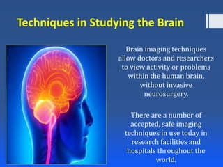
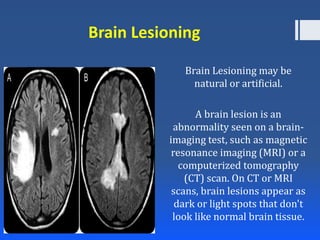
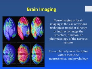
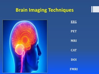
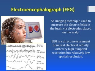
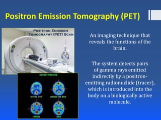
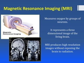
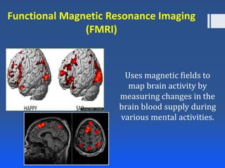
Model Call Girl in Bikash Puri Delhi reach out to us at 🔝9953056974🔝
Techniques in measuring Brain Function
1. Techniques in Studying the Brain
Prepared by:
Orlando A. Pistan, MAEd
Psychology Instructor
2. Techniques in Studying the Brain
Brain imaging techniques
allow doctors and researchers
to view activity or problems
within the human brain,
without invasive
neurosurgery.
There are a number of
accepted, safe imaging
techniques in use today in
research facilities and
hospitals throughout the
world.
4. Brain Lesioning
Brain Lesioning may be
natural or artificial.
A brain lesion is an
abnormality seen on a brain-
imaging test, such as magnetic
resonance imaging (MRI) or a
computerized tomography
(CT) scan. On CT or MRI
scans, brain lesions appear as
dark or light spots that don't
look like normal brain tissue.
5. Brain Staining
Staining the brain is a
technique used by
psychologists to pinpoint which
neurons are connected and
activated by each other.
Because neuronal membranes
contain ion channels that open
and close whenever a neuron is
activated, stains like horse dish
peroxidase enter these
channels and paint the path
neurotransmitters take to
transmit information to
neighboring neurons.
6. Brain Imaging
Neuroimaging or brain
imaging is the use of various
techniques to either directly
or indirectly image the
structure, function, or
pharmacology of the nervous
system.
It is a relatively new discipline
within medicine,
neuroscience, and psychology
8. Electroencephalograph (EEG)
An imaging technique used to
measure the electric fields in
the brain via electrodes placed
on the scalp.
EEG is a direct measurement
of neural electrical activity
with very high temporal
resolution but relatively low
spatial resolution.
9. Positron Emission Tomography (PET)
An imaging technique that
reveals the functions of the
brain.
The system detects pairs
of gamma rays emitted
indirectly by a positron-
emitting radionuclide (tracer),
which is introduced into the
body on a biologically active
molecule.
10. Magnetic Resonance Imaging (MRI)
Measures oxygen by groups of
neurons.
It represents a three
dimensional image of the
living brain.
MRI produces high resolution
images without exposing the
brain to radiation.
11. Computed Axial Tomography (CAT)
Uses a series of X-rays of the
head taken from different
directions.
This is used for quick
assessment of brain injuries.
12. Diffuse Optical Imaging (DOI)
Non-invasive techniques that
utilize light in the near infrared
spectral region to measure the
optical properties of
physiological tissue.
DOI have been applied in various
deep-tissue applications
including breast cancer imaging,
brain functional imaging, stroke
detection, muscle functional
studies, photodynamic therapy,
and radiation therapy
monitoring.
13. Functional Magnetic Resonance Imaging
(FMRI)
Uses magnetic fields to
map brain activity by
measuring changes in the
brain blood supply during
various mental activities.