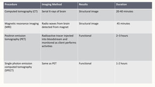
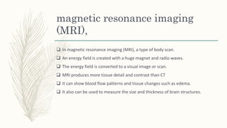
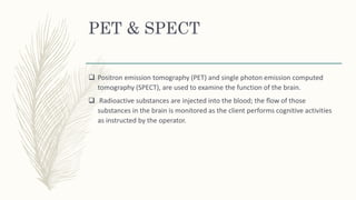
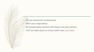
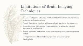
BRAIN
IMAGING
TECHNIQUES
Procedure Imaging Method Results Duration
Computed tomography (CT) Serial X-rays of brain Structural image 20-40 minutes
Magnetic resonance imaging
(MRI)
Radio waves from brain
detected from magnet
Structural image 45 minutes
Positron emission
tomography (PET)
Radioactive tracer injected
into bloodstream and
monitored as client performs
activities
Functional 2–3 hours
Single photon emission
computed tomography
(SPECT)
Same as PET Functional 1-2 hours
Computed tomography
 Computed tomography (CT, also called computed axial tomography or CAT scan)
is a procedure in which a precise x-ray beam takes cross-sectional images
(slices) layer by layer.
 A computer reconstructs the images on a monitor and also stores the images
on magnetic tape or film
 CT can visualize the brain’s soft tissues
 CT is used to diagnose primary tumors, metastases, and effusions and to
determine the size of the ventricles of the brain
 Eg, in schizophrenia have been shown to have enlarged ventricles
procedure
– The person undergoing a CT scan must lie motionless on a stretcher-like table
for about 20 to 40 minutes as the stretcher passes through a “ring” while the
serial x-rays are taken.
magnetic resonance imaging
(MRI),
 In magnetic resonance imaging (MRI), a type of body scan.
 An energy field is created with a huge magnet and radio waves.
 The energy field is converted to a visual image or scan.
 MRI produces more tissue detail and contrast than CT
 It can show blood flow patterns and tissue changes such as edema.
 It also can be used to measure the size and thickness of brain structures.
procedure
– The person undergoing an MRI must lie in a small, closed chamber and remain
motionless during the procedure, which takes about 45 minutes.
– Those who feel claustrophobic or have increased anxiety may require sedation
before the procedure.
– Clients with pacemakers or metal implants, such as heart valves or orthopedic
devices, cannot undergo MRI
PET & SPECT
 Positron emission tomography (PET) and single photon emission computed
tomography (SPECT), are used to examine the function of the brain.
 Radioactive substances are injected into the blood; the flow of those
substances in the brain is monitored as the client performs cognitive activities
as instructed by the operator.
– PET uses two photons simultaneously;
– SPECT uses a single photon.
– PET provides better resolution with sharper and clearer pictures
– A PET scan takes about 2 to 3 hours; SPECT takes 1 to 2 hours
Limitations of Brain Imaging
Techniques
– The use of radioactive substances in PET and SPECT limits the number of times a
person can undergo these tests.
– There is the risk that the client will have an allergic reaction to the substances.
– Some clients may find receiving intravenous doses of radioactive material
frightening or unacceptable.
– Imaging equipment is expensive to purchase and maintain, so availability can be
limited.
– Some persons cannot tolerate these procedures because of fear or
claustrophobia

Brain imaging techniques

  • 1.
  • 2.
    Procedure Imaging MethodResults Duration Computed tomography (CT) Serial X-rays of brain Structural image 20-40 minutes Magnetic resonance imaging (MRI) Radio waves from brain detected from magnet Structural image 45 minutes Positron emission tomography (PET) Radioactive tracer injected into bloodstream and monitored as client performs activities Functional 2–3 hours Single photon emission computed tomography (SPECT) Same as PET Functional 1-2 hours
  • 3.
    Computed tomography  Computedtomography (CT, also called computed axial tomography or CAT scan) is a procedure in which a precise x-ray beam takes cross-sectional images (slices) layer by layer.  A computer reconstructs the images on a monitor and also stores the images on magnetic tape or film
  • 4.
     CT canvisualize the brain’s soft tissues  CT is used to diagnose primary tumors, metastases, and effusions and to determine the size of the ventricles of the brain  Eg, in schizophrenia have been shown to have enlarged ventricles
  • 5.
    procedure – The personundergoing a CT scan must lie motionless on a stretcher-like table for about 20 to 40 minutes as the stretcher passes through a “ring” while the serial x-rays are taken.
  • 6.
    magnetic resonance imaging (MRI), In magnetic resonance imaging (MRI), a type of body scan.  An energy field is created with a huge magnet and radio waves.  The energy field is converted to a visual image or scan.  MRI produces more tissue detail and contrast than CT  It can show blood flow patterns and tissue changes such as edema.  It also can be used to measure the size and thickness of brain structures.
  • 7.
    procedure – The personundergoing an MRI must lie in a small, closed chamber and remain motionless during the procedure, which takes about 45 minutes. – Those who feel claustrophobic or have increased anxiety may require sedation before the procedure. – Clients with pacemakers or metal implants, such as heart valves or orthopedic devices, cannot undergo MRI
  • 8.
    PET & SPECT Positron emission tomography (PET) and single photon emission computed tomography (SPECT), are used to examine the function of the brain.  Radioactive substances are injected into the blood; the flow of those substances in the brain is monitored as the client performs cognitive activities as instructed by the operator.
  • 9.
    – PET usestwo photons simultaneously; – SPECT uses a single photon. – PET provides better resolution with sharper and clearer pictures – A PET scan takes about 2 to 3 hours; SPECT takes 1 to 2 hours
  • 10.
    Limitations of BrainImaging Techniques – The use of radioactive substances in PET and SPECT limits the number of times a person can undergo these tests. – There is the risk that the client will have an allergic reaction to the substances. – Some clients may find receiving intravenous doses of radioactive material frightening or unacceptable. – Imaging equipment is expensive to purchase and maintain, so availability can be limited. – Some persons cannot tolerate these procedures because of fear or claustrophobia