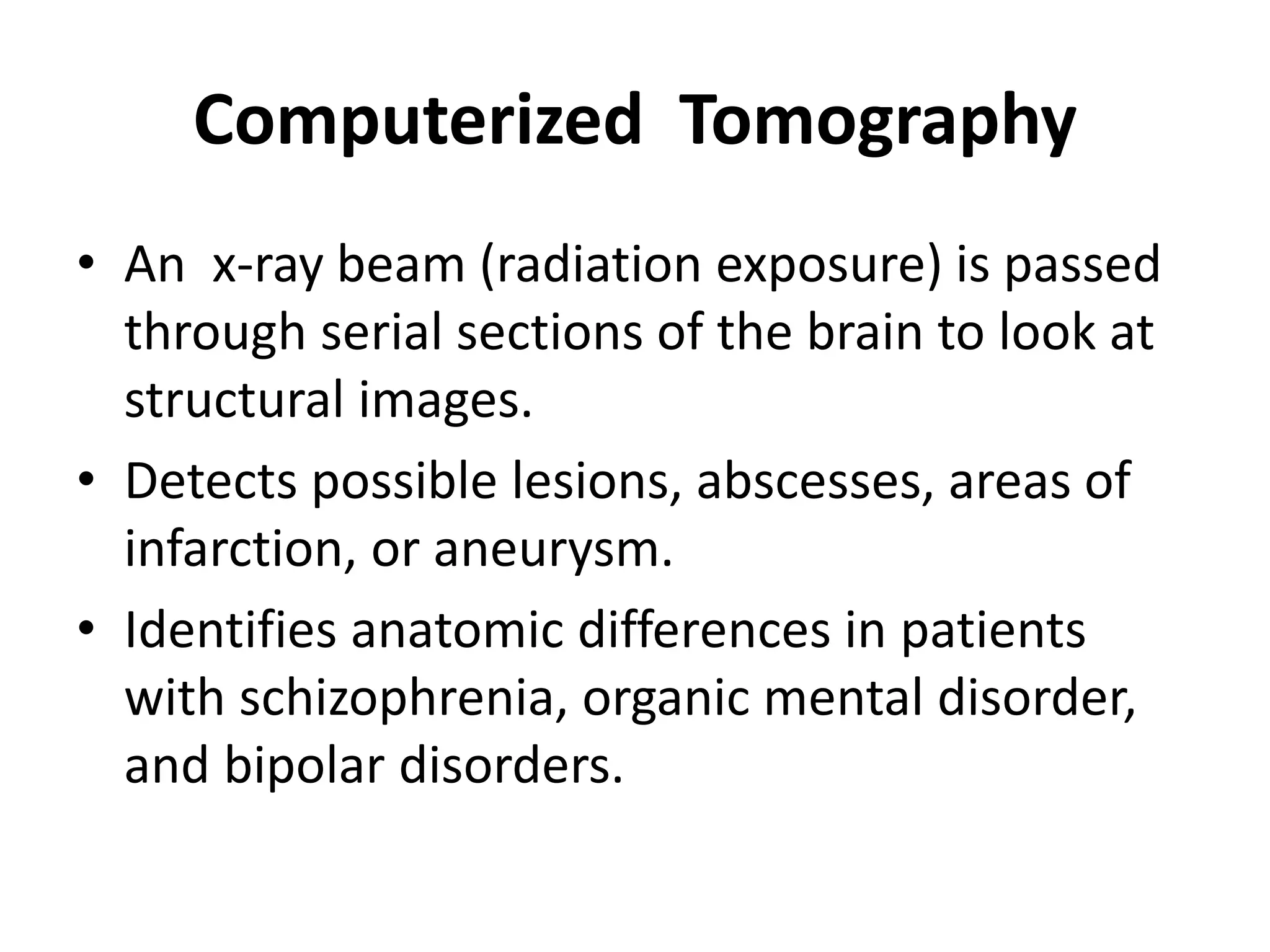
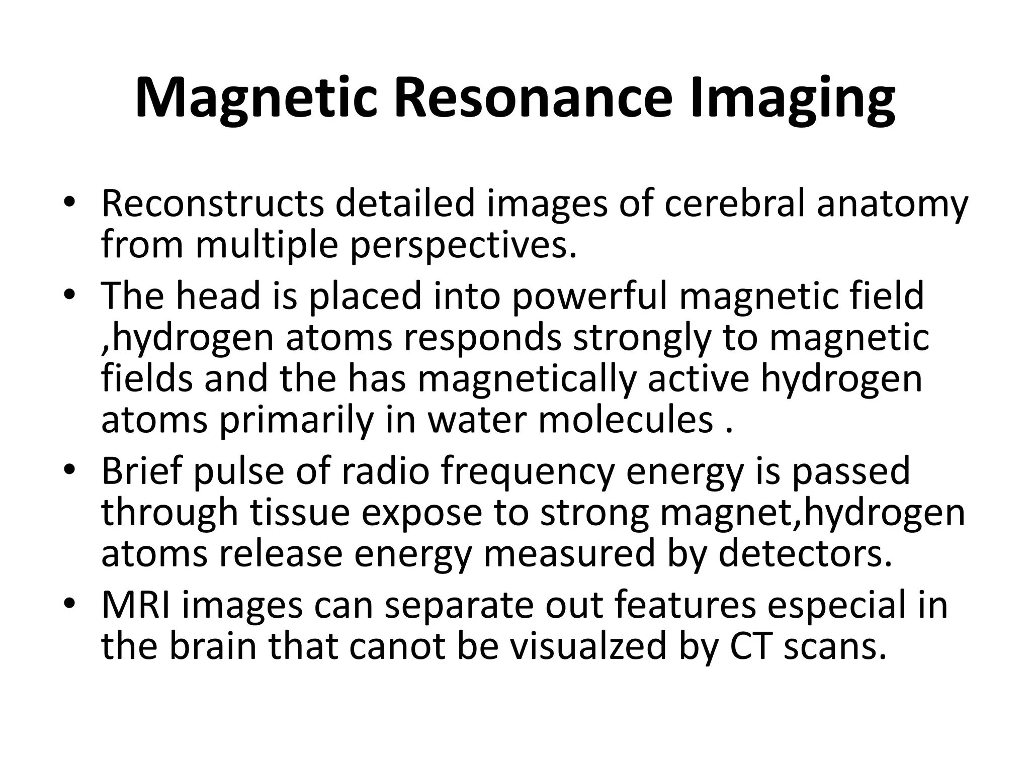
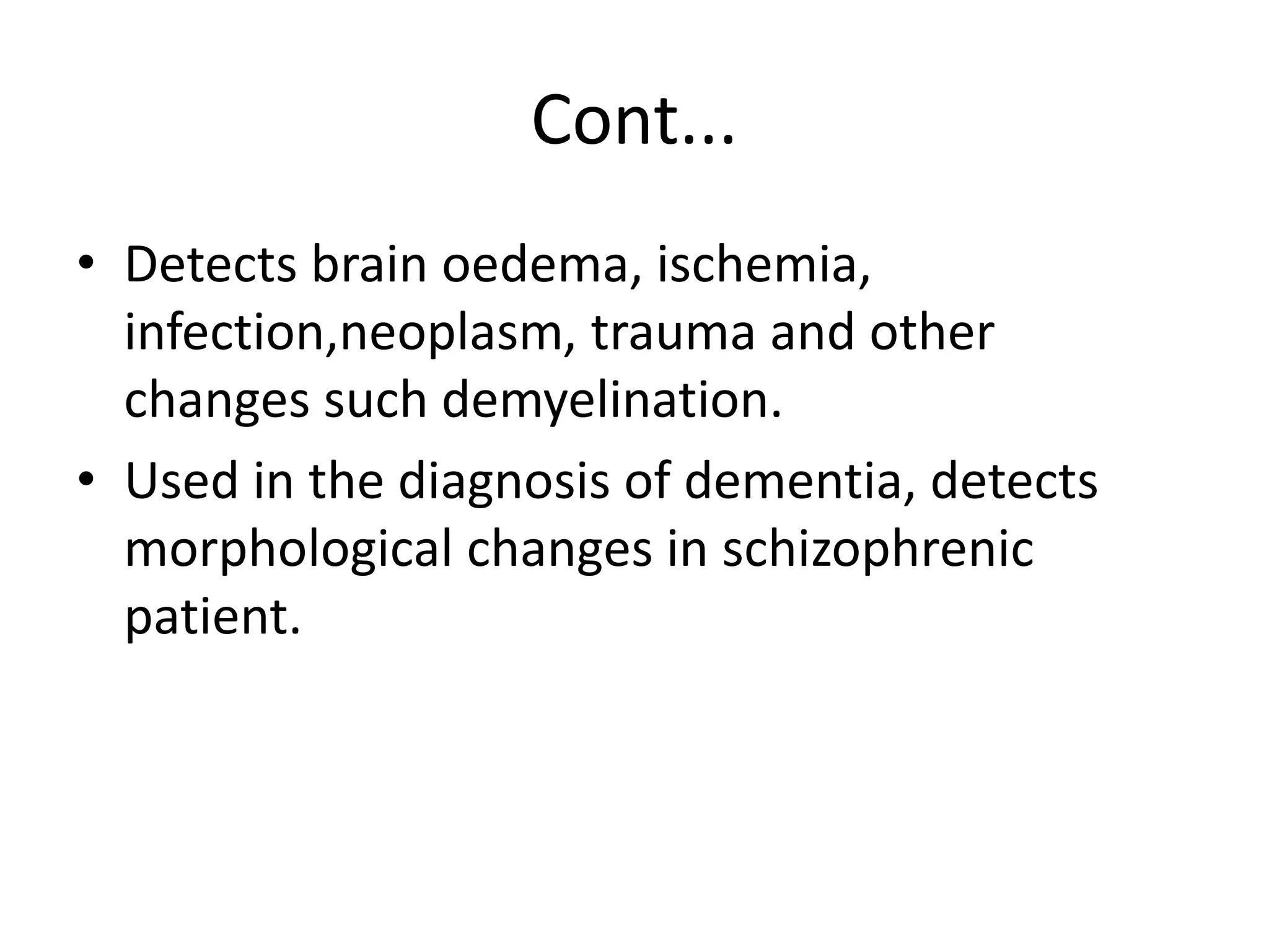
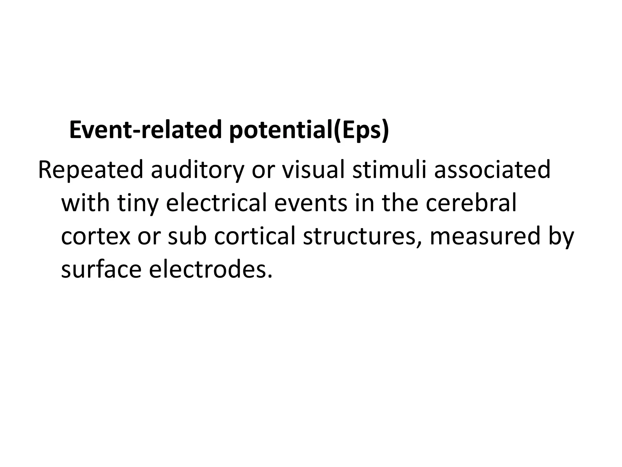
Brain Imaging
• Imaging is a generic word to describe a range
of remarkable techniques to visualize internal
organs.
Computerized Tomography
• An x-ray beam (radiation exposure) is passed
through serial sections of the brain to look at
structural images.
• Detects possible lesions, abscesses, areas of
infarction, or aneurysm.
• Identifies anatomic differences in patients
with schizophrenia, organic mental disorder,
and bipolar disorders.
Magnetic Resonance Imaging
• Reconstructs detailed images of cerebral anatomy
from multiple perspectives.
• The head is placed into powerful magnetic field
,hydrogen atoms responds strongly to magnetic
fields and the has magnetically active hydrogen
atoms primarily in water molecules .
• Brief pulse of radio frequency energy is passed
through tissue expose to strong magnet,hydrogen
atoms release energy measured by detectors.
• MRI images can separate out features especial in
the brain that canot be visualzed by CT scans.
Cont...
• Detects brain oedema, ischemia,
infection,neoplasm, trauma and other
changes such demyelination.
• Used in the diagnosis of dementia, detects
morphological changes in schizophrenic
patient.
Positron Emission Tomography
• Unlike CT scanning PET scan requires the
injection of small amount of radioactive isotope
that is localized in brain tissue.
• PET scan allows the location of that isotope to be
traced while subject performs certain tasks.
• How it works
• Uses positrons as radioactive tracers or more
precisely uses positron generated tracers that are
bound to molecules(including glucose and certain
neurotransmitter analogues)
• Positrons are a form of antimatter electrons with positive
charge when antimatter and matter (in this case an
electron) meet they are both annihilated and produce two
gamma particles traveling in nearly opposite directions .
• This production of two diverging particles allows the
precise location of the original collision to be determined.
• Since human tissue are full of electrons, the collision occurs
very close to the site at which the positron generating
substance was concentrated.
• PET scan allows scientists to map function
such as the flow of blood within brain the
uptake of glucose and the binding of
neurotransmitters to synaptic membranes all
in response to specific real time.
• For example in normal individual, there are
major differences in where brain function
localize depending on whether an individual
does the following activities:
• Look at word :The metabolic activity is limited
to the visual cortex.
• Listen to word: The metabolic activity is
limited to parts of the temporal cortex.
• Speaks a word: The metabolic activity is
limited to the medial frontal cortex.
• Think about word meaning activity is widely
spread over areas of the association cortex
• PET scan provides a remarkable confimation
of the aspects of the biophysiological theory .
• Brain functions do link to mental processes
and are highly localized within the cortex.
Magnetic Resonance Spectroscopy
and Diffusion Tensor Imaging
• Two new MRI derived scanning techniques .
• MRS does not produce visual brain images but
allows scientists to determine which
neurotransmitters and other chemicals are
present at specific brain sites .DTI is an
extension of MRI that allows bran white
matter fibers.
Electroencephalogram(EEG)
• Measures electrical activity patterns of the
brain from leads connected to the surface
electrodes placed on the scalp and
nasopharyngeal area.
Polysomnography (sleep EEG)
• Measures electrial brain activity data during
all-night sleep.
Brain Electroactivity Mapping (BEAM)
• Extends theEEG by generating computerized
maps of brain electrial to produce
images;permits visualuzation of the brain
performing tasks or specific functions. Useful
in children.
Event-related potential(Eps)
Repeated auditory or visual stimuli associated
with tiny electrical events in the cerebral
cortex or sub cortical structures, measured by
surface electrodes.
END
THANK YOU

Brain imaging

  • 1.
    Brain Imaging • Imagingis a generic word to describe a range of remarkable techniques to visualize internal organs.
  • 2.
    Computerized Tomography • Anx-ray beam (radiation exposure) is passed through serial sections of the brain to look at structural images. • Detects possible lesions, abscesses, areas of infarction, or aneurysm. • Identifies anatomic differences in patients with schizophrenia, organic mental disorder, and bipolar disorders.
  • 3.
    Magnetic Resonance Imaging •Reconstructs detailed images of cerebral anatomy from multiple perspectives. • The head is placed into powerful magnetic field ,hydrogen atoms responds strongly to magnetic fields and the has magnetically active hydrogen atoms primarily in water molecules . • Brief pulse of radio frequency energy is passed through tissue expose to strong magnet,hydrogen atoms release energy measured by detectors. • MRI images can separate out features especial in the brain that canot be visualzed by CT scans.
  • 4.
    Cont... • Detects brainoedema, ischemia, infection,neoplasm, trauma and other changes such demyelination. • Used in the diagnosis of dementia, detects morphological changes in schizophrenic patient.
  • 5.
    Positron Emission Tomography •Unlike CT scanning PET scan requires the injection of small amount of radioactive isotope that is localized in brain tissue. • PET scan allows the location of that isotope to be traced while subject performs certain tasks. • How it works • Uses positrons as radioactive tracers or more precisely uses positron generated tracers that are bound to molecules(including glucose and certain neurotransmitter analogues)
  • 6.
    • Positrons area form of antimatter electrons with positive charge when antimatter and matter (in this case an electron) meet they are both annihilated and produce two gamma particles traveling in nearly opposite directions . • This production of two diverging particles allows the precise location of the original collision to be determined. • Since human tissue are full of electrons, the collision occurs very close to the site at which the positron generating substance was concentrated.
  • 7.
    • PET scanallows scientists to map function such as the flow of blood within brain the uptake of glucose and the binding of neurotransmitters to synaptic membranes all in response to specific real time. • For example in normal individual, there are major differences in where brain function localize depending on whether an individual does the following activities:
  • 8.
    • Look atword :The metabolic activity is limited to the visual cortex. • Listen to word: The metabolic activity is limited to parts of the temporal cortex. • Speaks a word: The metabolic activity is limited to the medial frontal cortex. • Think about word meaning activity is widely spread over areas of the association cortex
  • 9.
    • PET scanprovides a remarkable confimation of the aspects of the biophysiological theory . • Brain functions do link to mental processes and are highly localized within the cortex.
  • 10.
    Magnetic Resonance Spectroscopy andDiffusion Tensor Imaging • Two new MRI derived scanning techniques . • MRS does not produce visual brain images but allows scientists to determine which neurotransmitters and other chemicals are present at specific brain sites .DTI is an extension of MRI that allows bran white matter fibers.
  • 11.
    Electroencephalogram(EEG) • Measures electricalactivity patterns of the brain from leads connected to the surface electrodes placed on the scalp and nasopharyngeal area. Polysomnography (sleep EEG) • Measures electrial brain activity data during all-night sleep.
  • 12.
    Brain Electroactivity Mapping(BEAM) • Extends theEEG by generating computerized maps of brain electrial to produce images;permits visualuzation of the brain performing tasks or specific functions. Useful in children.
  • 13.
    Event-related potential(Eps) Repeated auditoryor visual stimuli associated with tiny electrical events in the cerebral cortex or sub cortical structures, measured by surface electrodes.
  • 14.