Imaging in mediastinal masses by Dr. Milan Silwal
•Download as PPT, PDF•
7 likes•1,466 views

Imaging in mediastinal masses

Recommended
More Related Content
What's hot
What's hot (20)
Similar to Imaging in mediastinal masses by Dr. Milan Silwal
Similar to Imaging in mediastinal masses by Dr. Milan Silwal (20)
More from Milan Silwal
More from Milan Silwal (20)
Recently uploaded
Recently uploaded (20)
Imaging in mediastinal masses by Dr. Milan Silwal
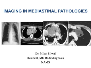
- 1. IMAGING IN MEDIASTINAL PATHOLOGIES Dr. Milan Silwal Resident, MD Radiodiagnosis NAMS
- 2. Presentation Outline • Relevant radiological anatomy • Mediastinum: Division • Imaging methods for mediastinum • Approach to mediastinal pathologies • Pathologies according to the divisions • Summary
- 3. • The mediastinum is a space in the thorax that lies in the midline of the chest between the pleura of each lung and extends from the sternum to the vertebral column. Relations • superiorly - continuous with the loose connective tissue of the neck • anteriorly - chest wall • laterally - lungs and pleura • posteriorly - thoracic spine • inferiorly - diaphragm
- 4. Divisions • Numerous methods; the most common of which include the Shields’ classification scheme used in clinical practice and the traditional, Fraser and Pare, Felson, Heitzman, Zylak, and Whitten models used in radiologic practice Anatomical classification Sutton classification Felson classification ITMIG classification
- 5. A. Anatomical classification • superior mediastinum: above upper level of the pericardium and plane of Ludwig • inferior mediastinum: below the plane of Ludwig – anterior mediastinum: anterior to the pericardium – middle mediastinum: within the pericardium – posterior mediastinum: posterior to the pericardium
- 6. B. Sutton classification • Anterior division: in front of the anterior pericardium and trachea, • Middle division: within the pericardial cavity but including the trachea. • posterior division: lies behind the posterior pericardium and trachea.
- 7. C. Felson classification: • Anterior: from the sternum to the posterior aspect of the heart and great vessels. • Middle: The compartment posterior to the heart and great vessels, to a line drawn 1 cm posterior to the anterior edge of the thoracic vertebrae. • Posterior: The space behind the posterior limit of the middle mediastinum.
- 10. Heitzman classification It describes 6 compartments: the thoracic inlet, the anterior mediastinum, the superior aortic area (above the aortic arch), the infra-aortic area (below the aortic arch), the supra-azygos area, and the infra-azygos area
- 11. Imaging methods • CXR • Barium Swallow • CT • MRI • USG • Radionuclide scan • PET
- 12. • Mediastinal abnormalities often manifest initially at conventional radiography. • CT will be made to further analyze and characterize anterior and middle mediastinal masses. • An MRI is usually made to analyze masses located in the posterior compartment because the majority of these masses turn out to be neurogenic in nature.
- 13. CXR • high kVp (120-150 kVp) technique • A specially designed wedge filter with its thickest portion over the lung and thinnest portion over the mediastinum achieves appropriate exposure of the mediastinum without over exposing the lungs. • May appear normal or present as mediastinal widening
- 14. Barium swallow • oesophageal displacement help localize a mass • hernias and oesophageal tumours can be definitively diagnosed.
- 15. Computed tomography (CT) • Useful for – localizing mass – demonstrating the extent masses – Characterize nature of mass • Fat and some fluid-filled masses are readily differentiated from solid tumours • aneurysms and anomalous vessels can be precisely diagnosed in CECT.
- 16. • CT better demonstrates – Calcification within a mass – invasion of the mediastinal fat, indicating malignant rather than benign disease – resectibility of mediastinal masses • Also helpful – for guided FNAC/biopsy – To guide treatment and F/U for the response to therapy.
- 17. MRI • major uses: – to demonstrate the status of the larger intrathoracic blood vessels, notably in patients with superior vena caval obstruction or aortic aneurysms, – Particularly helpful for posterior mediastinal masses. – also to differentiate between vessels and LN where contrast injection is contraindicated • MRI is useful for demonstrating any intraspinal extension of mediastinal masses
- 18. MRA • to demonstrate intrinsic disorders of the major intrathoracic blood vessels and • to demonstrate stenosis, distortion, and displacement of vessels by mediastinal masses and other mediastinal processes
- 19. USG • particularly helpful in distinguishing cysts from solid masses in contact with the intercostal spaces; • to distinguish cardiac from paracardiac masses • to guide mediastinal biopsy • Approaches: Transthoracic, parasternal or suprasternal
- 20. Endoscopic US • demonstrates lymph nodes and other masses adjacent to the oesophagus • can show the aortic arch and descending aorta, notably for the diagnosis of aortic dissection and aortic aneurysms. • Future applications may include – bronchoscopic US for staging assessment and needle aspiration of mediastinal and hila nodes.
- 21. Radionuclide examinations • rarely been used for mediastinal disorders, • Conventional techniques can be helpful in deciding whether a mediastinal mass is due to ectopic thyroid tissue by using sodium iodide • Neuroendocrine tumours and phaeochromocytomas can be demonstrated by somatostatin receptor scintigraphy
- 22. • radio-iodine MIBG(metaiodobenzylguanidine) imaging is very useful when searching for extra- adrenal intrathoracic phaeochromocytomas. • Gallium scanning may be of use in evaluation of the mediastinum in adults with sarcoidosis or children with lymphoma.
- 23. PET • PET scanning using [fluorine 18]flouro-2-deoxy-D- glucose (FDG) • more accurate than CT or MRI for diagnosing mediastinal lymph node metastases from bronchogenic carcinoma.
- 24. Related pathology • Broadly, pathology that affects the mediastinum can be divided into those that result in a focal mass, or those that result in diffuse disease involving the mediastinum. • Additionally, air may track into the mediastinum in pneumomediastinum.
- 25. • D/D of a focal mediastinal mass is highly dependant on its location within the mediastinum, resulting in specific differential lists for • thoracic inlet masses • anterior mediastinal masses • middle mediastinal masses • posterior mediastinal masses
- 26. Diffuse mediastinal disease can be separated into groups depending on whether the mediastinal disease is smooth or lobulated: o smooth mediastinal lipomatosis mediastinal malignant infiltration mediastinal haemorrhage mediastinitis o lobulated mediastinal lymphadenopathy thymic mass germ cell neoplasm mediastinal vascular abnormality mediastinal neurofibromatosis
- 28. Approach to a mediastinal mass Localize to mediastinum Localize within the mediastinum Characterise with CT or MRI
- 29. A. Localize to the mediastinum • Characteristics indicate that a lesion originates within the mediastinum: – Unlike lung lesions, a mediastinal mass will not contain air bronchograms. – The margins with the lung will be obtuse. (A lung mass abutts the mediastinal surface and creates acute angles with the lung) – Mediastinal lines (azygoesophageal recess, anterior and posterior junction lines) will be disrupted. – There can be associated spinal, costal or sternal abnormalities.
- 30. • CXR showing sign of lung mass and mediastinal mass
- 31. B. Localize within the mediastinum • The mediastinum can be divided into anterior, middle and posterior compartments. • It is important to remember that there is no tissue plane separating these compartments.
- 32. Anterior mediastinum • The anterior mediastinum contains: thymus, lymph nodes, ascending aorta, pulmonary artery, phrenic nerves and thyroid. • The most common lesions seen in the anterior mediastinum will either be of thymic or lymph node origin. Even the germ cell tumors arise from the pluripotent cells of the thymus. Before we want to biopsy an anterior mediastinal mass, do not forget that some of these lesions can be vascular in origin. • The four T's make up the mnemonic for anterior mediastinal masses:: – Thymus – Teratoma (germ cell) – Thyroid – Terrible Lymphoma
- 34. • On conventional radiographs look for the signs listed in the table above • The finding of an obliterated retrosternal clear space is not so helpfull anymore, since nowadays many patients are obese. In these patients the retrosternal space can be filled with fat.
- 35. Hilum overlay sign • When there is a mediastinal mass and we still can see the hilar vessels through the mass, then we know the mass does not arise from the hilum. This is known as the hilum overlay sign. • Because of the geometry of the mediastinum most of these masses will be located in the anterior mediastinum.
- 36. • Hilum overlay sign in a patient with lymphoma Hilum overlay sign in a patient with lymphoma: (a)PA CXR clearly depicts the hila (white arrow), which indicates that the mass is either anterior or posterior to the hila. In addition, the descending aorta is clearly seen (black arrow), indicating that the mass is not within the posterior mediastinum. (b) Chest CT scan demonstrates an anterior mediastinal mass. The anterior junction line is obliterated
- 37. Mediastinal mass (Diagnosed as Hodgkins lymphoma): Wide upper mediastinum (arrowheads) Poorly defined aortic knuckle - indicating adjacent disease Wide right paratracheal stripe (*) Normal lungs
- 38. Axial CT image showing homogenously enhancing soft tissue anterior mediastinal mass (arrow) replacing the thymus in a case of thymic lymphoma.
- 39. Anterior junction line • Anterior mediastinal masses in the prevascular region can obliterate the anterior junction line, although it is usually the preservation of more posterior lines at radiography that helps identify the location of an anterior mediastinal mass.
- 40. • Obliteration of cardiophrenic angle by Epicardial fat pad. • (a) Posteroanterior chest radiograph shows loss of the cardiac silhouette at the border of the right side of the heart and an epicardial fat pad with relatively low density (arrow). • (b) CT scan shows the fat pad (arrow) as an area of homogeneous fat attenuation adjacent to the right border of the heart.
- 41. Middle mediastinum • The middle mediastinum contains: lymph nodes, trachea, esophagus, azygos vein, vena cavae, posterior heart and the aortic arch. • The majority of middle mediastinal masses will consist of foregut duplication cysts (eg oesophageal duplication or bronchogenic cysts) or lymphadenopathy. • Aortic arch anomalies can also present as middle mediastinal masses. • Fluid containing lesions are usually duplication cysts or necrotic lymph nodes. • A pancreatic fluid collection due to pancreatitis may also present as a mediastinal mass. • A fibrovascular esophageal polyp is a mesenchymal lesion which almost always contains fat. • Vascular lesions are arch anomalies, azygous continuation due to interrupted inferior vena cava or hyperenhancing lymph nodes.
- 43. • Displaced azygoesophageal recess will be seen on the right. • On the left we may have a pseudoparavertebral line. This is a new interface that looks like a paravertebral line.
- 44. • Lymphadenopathy. (a) On CXR PA view , the right paratracheal stripe is not seen, having been obliterated by a right paratracheal mass (arrowheads). (b) CT scan demonstrates right paratracheal lymphadenopathy (arrow), which obliterates the air–soft tissue interface between the right lung and the tracheal wall.
- 45. AP window lymphadenopathy. (a) Chest radiograph shows the AP window with an abnormal convex border (arrow). (b) CT scan demonstrates lymphadenopathy (arrow), which accounts for the distortion of the AP window in a.
- 46. •(a) PA CXR demonstrates a subcarinal abnormality with increased opacity (*), splaying of the carina, and abnormal convexity of the upper and middle thirds of the azygoesophageal line (arrowheads). •(b) Corresponding CT scan helps confirm a subcarinal mass (arrow), which proved to be a bronchogenic cyst.
- 47. Pitfalls in assessment of middle mediastinum: • A variety of normal vascular variants may be mistaken for middle mediastinal disease at CXR. • Right-sided aortic arch may mimic paratracheal lymphadenopathy because it obliterates the right paratracheal stripe – the absence of the aortic knuckle on the left should help correctly identify this variant. • Left sided SVC may create an additional mediastinal line lateral to the aortic arch at radiography. • Azygos continuation of the IVC - provides an alternate route for systemic venous return to the heart. – results in an enlarged azygos vein, which may be mistaken for lymphadenopathy.
- 48. • Right-sided aortic arch. (a) Posteroanterior chest radiograph demonstrates an abnormality in the right paratracheal region (arrow) with loss of the paratracheal stripe. Note, however, the absence of the aortic knuckle on the left. (b) CT scan shows a right-sided aortic arch (arrow), which explains the findings in a.
- 49. Left-sided SVC. (a) Collimated PA CXR shows an additional line (arrow) lateral to the aortic arch. (b) Venogram demonstrates a left-sided SVC, which explains the finding in a. (c, d) CT scans obtained at the levels of the aortic arch (c) and pulmonary trunk (d) show the left-sided SVC (arrow), which drains into the coronary sinus.
- 50. Azygos continuation of the IVC. (a) Collimated posteroanterior chest radiograph shows enlargement of the azygos vein at the inferior margin of the right paratracheal stripe (arrowheads), a finding that mimics lymphadenopathy. (b) CT scan also shows enlargement of the azygos vein (arrow). This finding is the result of azygos continuation of the IVC.
- 51. Posterior mediastinum • The posterior mediastinum contains: sympathetic ganglia, nerve roots, lymph nodes, parasympathetic chain, thoracic duct, descending thoracic aorta, small vessels and the vertebrae. • Most masses in the posterior mediastinum are neurogenic in nature. These can arise from the sympathetic ganglia (eg neuroblastoma) or from the nerve roots (eg schwannoma or neurofibroma). • Others masses are: lymphadenopathy, the vertebrae , paravertebral lesions, and the descending thoracic aorta as potential causes for posterior mediastinal masses. • Cystic lesions will be either neuroenteric cysts, schwannomas or meningoceles. • Fat containing lesions will be extramedullary hematopoiesis. When the anemia is resolved the extramedullary marrow will stop producing blood and become fatty. It appears as a lobulated paravertebral soft tissue mass behind heart.
- 52. • On conventional radiographs look for: – Cervicothoracic Sign – Widening of the paravertebral stripes – Displacement of posterior junctional line.
- 53. a. Radiologic Findings PA chest radiograph demonstrates a well-defined 5.0 cm homogenous, non-calcified mass in the left apex. The lesion demonstrates a distinct lateral border but an incomplete medial border. The “incomplete border” sign suggests the lesion is pleural or mediastinal based. On closer inspection, one notes the lesion extends above the ipsilateral clavicle, thus exhibiting a “cervicothoracic” sign. This sign localizes the lesion to the posterior mediastinal compartment. B. On the left the MR of the same patient. It turned out to be a schwannoma.
- 54. Paraspinal abscess. (a) Posteroanterior chest radiograph shows a mass (arrow) effacing the left paraspinal line. The lateral wall of the descending aorta is seen as a separate entity (arrowhead). (b) CT scan shows a paraspinal abscess (arrow) effacing the paraspinal lines. The air– soft tissue interface between the lung and aorta remains intact (arrowhead), thereby preserving the normal radiographic appearance of the lateral aortic wall (cf a).
- 55. Descending aortic aneurysm. (a) Posteroanterior chest radiograph shows lateral displacement of the lateral margin of the descending thoracic aorta due to an aortic aneurysm (arrowheads). (b) CT scan also demonstrates the aneurysm (arrow).
- 56. More than one compartment • Since there are no tissue planes separating the mediastinal compartments, there are lesions that do not respect our approach to the mediastinum. These lesions tend to occupy more than one compartment and include: mediastinitis, hematomas, vascular entities, bronchogenic cancer, metastases and lymphangiomas (fluid containing).
- 57. C. Characterize • Once we have localized a mediastinal mass, next try to charcterize it by assessing whether it has any of the following characteristics: – Does the mass contain fluid? – Does it contain fat? – Does it contain calcification? – Does it enhance following the administration of intravenous contrast?
- 58. Fluid • mediastinal masses that may contain fluid: – Thymic Cyst – Thymoma – Teratoma – Pericardial Cyst – Foregut Duplication – Meningocoele – Neuroenteric Cyst – Cystic Lymphadenopathy – Lymphangioma
- 59. mass containing fluid: teratoma (on the left) or a thymic cyst (on the right).
- 60. • B. a. multiple masses in both the anterior and middle mediastinum. The attenuation values are of water density. These findings favor the diagnosis of cystic lymphadenopathy in a patient with metastatic disease. b. cystic lesion in the middle mediastinum with fluid fluid level and milk of calcium. Foregut duplication cysts occasionally contain milk of calcium like in this example of an esophageal duplication cyst.
- 61. Fat • Fat on chest film often look like water density, thus sometime it is difficult to differentiate them – When fat is inflamed – When involved with malignant tumor – cellular infiltrate has water density • When fat is identified in thoracic lesion – Teratoma – Lipoma – Thymolipoma – Omental Fat pad – all hernia – Excess fat secondary to steroid therapy – hibernoma
- 62. Calcification • Commonly present in the mediastinal lesions • If within the mass and doesn’t shift with gravity, likely solid tumor. • Calcification in margin of mass – usually cyst – Solid thymoma or substernal thyroid may show rim calcification • Calcification within a tumor – usually benign – Thyroid carcinoma and neurosarcoma may show annular or stippled calcification • Thymoma with calcifications – myasthenia gravis usually. • Tooth or bone – teratoma • Phleboliths - hemangioma
- 63. Enhancing mass • The differential diagnosis of enhancing mediastinal masses is: – Hyperenhancing lymph nodes – Thyroid tissue – Paragangliomas – Hemangiomas – Vascular lesions
- 64. • Enhancing lymph nodes seen in: – Melanoma – Renal cell carcinoma – Thyroid carcinoma – Castlemann's disease
- 65. Air • Gas within a mediastinal mass – Hernia – Esophageal diverticulum – Dilated esophagus – Mediastinal abscess – Communicating duplicating cyst • Diffuse pneumomediastinum – sources: – Esophagus, trachea, or bronchus (perforation or surgery) – Retroperitoneal space (diagnostic pneumoretroperitoneum, duodenal or colon perforation) – Neck (trauma, surgery or perforating cervical lesion) – Lung (parenchymal tear leaks air into interstitium, then dissect to hilum and into mediastinum)
- 66. Chest radiography showed perihilar opacities in both lungs with free air in the mediastinum (image A, arrows). Computed tomography confirmed pneumomediastinum (image B, arrows) with enhanced interstitial markings and lower-lobe opacities in both lungs, with greater severity in the left lung
- 67. Configuration • True, lymph node enlargement – often lobulated • Long vertical diameter with undulating margins – Dilated esophagus – Dissecting aneurysm – Azygous vein enlargement – Mediastinal hematoma • Teardrop shadow, broad along the diaphragm – soft pedunculated mass (pericardial cyst or thymolipoma) • The third mogul – bumps on heart just below the pulmonary arc on the left border
- 68. Mediastinal vs pulmonary lesions • Sharply penciled margin • Tapering upper and lower margins • Involvement of spine or adjacent ribs or even sternum • Bilaterality usu. excludes pulmonary lesion • Respiratory motion: intrapulmonary shadows move synchronously with the adjacent lung markings, whereas mediastinal lesions donot. • Presence of air bronchogram within the lesion – pulmonary lesion
- 69. Mediastinal vs cardiac disease • Hilum overlay vs hilum convergence signs • Fallacies: – Certain CHD, especially among infants, may result in lateral bulge anterior to the pulmonary trunk – Collapse of left lower lobe may also displace the first left pulmonary bifurcation posteromedial to its normal position – Rarely, pericardial effusion may bulge anterolaterally in front of right and left main pulmonary arteries. • Absence of pulsation signifies non-cardial origin – But, transmitted pulsation may be + in pericaridal effusion and tumor
- 70. Thyroid mass • Enlargement of the thyroid gland can be due to a number of causes including a non-toxic multinodular enlargement of the gland, Thyrotoxicosis, thyroid adenoma, thyroid carcinoma, lymphoma and Hashimoto's thyroiditis. • Less than 5% of enlarged thyroid glands in the neck extend into the mediastinum to produce a retrosternal goitre, but this is usually due to a non-toxic multinodular enlargement of the gland. • A retrosternal goitre appears as a well-defined round or oval soft tissue mass in the superior part of the anterior or middle mediastinum, which fades off into the neck. The soft-tissue mass often contains central nodular, linear or eccentric patterns of calcification and produces lateral displacement and compression of the trachea in the thoracic inlet . • About 25 % of goitres are retrotaeheal and displace the oesophagus posteriorly and the trachea anteriorly. • Rapid increase in the size of the mass indicates internal haemorrhage into a cyst.
- 71. • Traditionally, the term substernal goitre has been used to refer to an enlarged thyroid gland with greater than 50% of its mass below the thoracic inlet. It is of clinical significance because its growth between the sternum anteriorly and vertebral bodies posteriorly leads to impingment on the surrounding structures and has compressive symptoms. • This condition has a clinical importance because it presents a diagnostic dilemma with its compressive symptoms and operative difficulty as to which approach is most suitable for its management. • The CT scan was the most useful tool showing the nature and extent of the lesion in the reported case. • Has a higher attenuation level than soft tissue on non contrst CT due to its iodine content.
- 72. • (A) The representative chest radiograph; and (B) chest CT scan of a giant substernal goiter.
- 73. • Right-sided retrosternal goiter. (a) Posteroanterior chest radiograph demonstrates a thyroid goiter (arrow) extending into the middle mediastinum, obliterating the right paratracheal stripe, and causing deviation of the trachea to the left (black arrowhead). Above the level of the clavicles, the margins of the mass are not sharp (white arrowhead), indicating that the mass has an anterior mediastinal component. (b) CT scan shows the mass (arrow) between the trachea and right lung, a location that explains the obliteration of the right paratracheal stripe seen in a. There is no contact between the anterior component of the mass and the lung (arrowhead) at the level of the clavicular heads, a relationship that continues above the level of the clavicles. This finding explains why the lateral border of the anterior mediastinal component above the level of the clavicles is not sharp in a.
- 74. Thymic mass • Normal thymus – commonest cause of mediastinal abnormality in infants, seen as triangular soft tissue mass (sail shadow) projecting to one side of mediastinum, usually the right side. • Thymic wave sign – due to slight indentation by internal protrusions of the osteocartilaginous jxns of the adjacent ribs to the soft normal thymus.
- 75. Enlargement of thymus • Thymoma • Thymic hyperplasia • Thymic carcinoma • Lymphoma • Carcinoid • Germ cell tumor • Thymic cyst • thymolipoma • Thymoma – commonest thymic tumor in adult – 30% are invasive or malignant – 10-15% with MG – thymoma – 25-50% with thymoma – MG – May contain curvilinear or nodular calcification – Presence of vascular encasement or pleural metastasis – invasive thymoma
- 76. • Radiograph: well defined round or oval soft tissue mass projecting to one side of anterior mediastinum when large. • Thymolipoma: very large soft tissue mas with less radiographic density than expected for its size. • CT: well defined mass of soft tissue atttenuation in anterior mediastinum • May contain areas of cystic degeneration • Normal shaped enlarged thymus – hyperplasia • Cystic thymic mass • Fat containing - thymolipoma
- 77. • Thymic Hyperplasia. A. Enhanced CT in a 12-year-old undergoing chemotherapy for rhabdomyosarcoma shows virtual absence of thymic tissue. B. Scan 3 months following completion of chemotherapy shows uniform enlargement of thymus (arrow), reflecting rebound hyperplasia.
- 78. Thymic enlargement on (A) PA and (B) lateral radiographs of a patient with a malignant thymic mass. The lateral view demonstrates pleural metastases posteriorly (arrows). (C) CT confirms the anterior position of the primary tumour suspected from the filling of the retrosternal window apparent on the lateral radiograph (B).
- 79. Invasive thymoma in a young man. (A) Shows a lobular anterior mediastinal mass associated with a pleural effusion. (B) Image obtained through the lower chest demonstrates mixed soft tissue (arrows) and fluid attenuation owing to transpleural spread of tumour.
- 80. Germ cell tumors • Majority are benign – Dermoid cyst – Benign teratoma • 30% are malignant – Seminoma – Embryonal cell carcinoma – Choriocarcinoma – Endodermal sinus or yolk sac tumor • Dermoid – mainly of ectodermal tissues • Teratoma – ectodermal, mesodermal and endodermal origin tissues • Usually incidental anterior mediastinal soft tissue mass in young adult male • Benign – well defined round or oval, usually projecting to only one side of anterior mediastinum • Malignant – lobulated, usually projecting on both sides of mediastinum • May contain – peripheral rim or central nodular calcifications, fat-fluid level, rudimentary tooth, hair tufts • Rapid increase in size – internal hemorrhage or development of malignancy.
- 81. • Teratoma in a young man undergoing an immigration chest radiograph. (A) There are no specific features on the plain radiograph to indicate the nature of the mass. (B) CT demonstrates that the opacity visible on the chest radiograph is well defined and contains soft tissue and fat densities.
- 82. • Malignant Germ Cell Tumor. A. Posteroanterior chest radiograph in a 38-year-old man reveals a right mediastinal mass with discrete right lung nodules (arrows). B. Contrast-enhanced CT demonstrates a large anterior mediastinal mass invading the superior vena cava (arrow) with right lung nodules and a small pleural effusion. CT- guided biopsy showed choriocarcinoma.
- 83. Lymphoma • A. Posteroanterior chest radiograph in a 35-year-old man shows a large, lobulated mediastinal mass. B. Contrast- enhanced CT at the level of the aortic arch shows bulky anterior and middle mediastinal lymphadenopathy.
- 84. • Causes: – Metastatic disease – Lymphoma – Leukaemia – Sarcoidosis – Tuberculosis – Histoplasmosis – Other infections and inflammatory conditions • Calcifications: – Tuberculosis – Sarcoidosis* – Silicosis* – Histoplasmosis – Hodgkin’s disease after irradiation – Metastases from mucin secreting adenocarcinoma – Amyloidosis* – Castleman’s disease – P. carinii pneumonia in AIDS Enlarged lymph nodes with central necrosis: •Tuberculosis •Histoplasomosis •M. avium intracellulare •Hodgkin’s disease •Metastases (SCC, testicular tumors) Mediastinal lymphadenopathy
- 85. Thoracic aortic aneurysm • Causes: – Atherosclerosis – Hypertension – Blunt chest trauma – Mycotic dissection – Cystic medial necrosis in Marfan’s and E-D syndrome – Aortitis in 3o syphilis – Takayasu’s disease – Congenital (sinus of valsalva aneurysm, coarctation) • Appears as either widening of the mediastinum or as well defined round or oval soft-tissue mass in any part of mediastinum, often with curvilinear calcification in its wall • Calcification in ascending aorta: syphilis or atherosclerosis • Displacement of peripheral rim of calcification away from wall - dissection
- 86. • Mediastinal Hematoma From Ruptured Thoracic Aortic Aneurysm. A. Portable chest radiograph in an 83-year-old woman with chest pain shows marked mediastinal widening. B. Contrast-enhanced CT demonstrates aneurysmal dilatation of the descending aorta, with active extravasation (arrow) into a large mediastinal hematoma. The patient was not a surgical candidate and expired shortly after the study.
- 87. Neurogenic tumors • 3 groups • 1st: nerve sheath tumors – Include neurofibroma, schwannoma, neurofibrosarcoma and malignant schwannoma – Commonest neurogenic tumor in mediastinum of adults • 2nd: ganglion cell tumors – Ganglioneuroma, ganglioneuroblastoma and neuroblastoma – Commonest neurogenic tumor in mediastinum of children • 3rd: chemodectoma and phaeochromocytoma – Rarest • Majority are benign, 30% are malignant
- 88. • Well defined round or oval soft tissue mass in paravertebral gutter, usually projecting on only one side of the posterior mediastinum – Nerve sheath tumors – circular – Ganglion cell tumors – elongated • Central calcification – may be in ganglion cell tumors (rare in nerve sheath tumors) • Radiographs: – Pressure erosion of adjacent vertebrae, ribs – Splaying of ribs – Widening of intervertebral foramen (dumb-bell tumours leading to compression of spinal cord), rib notching, scoliosis • Rapid increase in size with bony destruction and pleural effusion – development of malignancy
- 89. • Neurofibroma. A. Frontal chest radiograph shows a left upper mediastinal mass (arrow). B. Contrast-enhanced CT confirms the presence of a left paravertebral soft tissue mass (arrow). Surgical resection confirmed a neurofibroma.
- 90. • Ganglioneuroma. A. Posteroanterior radiograph in a 15-year-old woman reveals an oval, vertically oriented, right-sided mediastinal mass (arrows). B. Contrast- enhanced CT shows a low-attenuation posterior mediastinal mass (arrow) with calcification. This was surgically proven to be ganglioneuroma.
- 91. Summary • The divisions of the mediastinum are not absolute; however, referring to the local anatomy of the mediastinal reflections in an attempt to more accurately localize an abnormality may help narrow the differential diagnosis. • Identification of the involved mediastinal compartment helps determine which imaging modality might be appropriate for further study. • Different imaging characteristics and extension of the lesion further helps to narrow down the possible diagnosis or to reach the final diagnosis of mediastinal pathologies.
- 92. References: • Textbook of radiology and imaging by David Sutton 7th edition • A Diagnostic Approach to Mediastinal Abnormalities: RadioGraphicsVol. 27, No. 3 by Whitten CR et al. https://doi.org/10.1148/rg.273065136 • Radiology assistant
- 93. THANK YOU !
Editor's Notes
- Seen in cushing syndrome and long term steriod theraphy. 90% of morgani hernia are situated in anterior rt cardiophrenic angle.
- An abnormal convex contour of the AP window suggests a mediastinal abnormality, most commonly lymphadenopathy, although such a contour may occasionally represent a normal variant caused by the accumulation of fat. Similarly, excess fat within the mediastinum can cause apparent mediastinal widening at chest radiography. Vascular abnormalities such as an aortic arch aneurysm can also distort the AP window.
- The posterior junction (junctional) line is formed by the opposition of the pleural surfaces of the posteromedial surfaces of the upper lobes of the lungs, posteriorly to the oesophagus but anterior to the thoracic spine (usually T3-T5) 1,
- Cervico thoracic sign: if in neck mass with retrosternal extension, no clear margins; if posterior mediastinal mass clear margin. The anterior mediastinum stops at the level of the superior clavicle. Therefore, when a mass extends above the superior clavicle, it is located either in the neck or in the posterior mediastinum. When lung tissue comes between the mass and the neck, the mass is probably in the posterior mediastinum. This is known as the Cervicothoracic Sign. If we study the image on the frontal view on the left, we see a mass extending above the level of the clavicle and there is lung tissue in front of it, so this must be a mass in the posterior mediastinum.
- Teratomas are the most common benign germ cell tumors. The most common malignant germ cell tumor is the seminoma.
- Thymolipoma- A very large soft tissue mass with less radiographic density than expected for its size an which alters in its shape with respiration is usually thymolipoma.
- Castleman disease (CD) (also known as angiofollicular lymph node hyperplasia orgiant lymph node hyperplasia ) is an uncommon benign lymphoproliferative condition. It can affect several regions of the body although commonly described as a solitary mediastinal mass. Location The distribution is as follows : thorax: ~70% abdomen/pelvis and retroperitoneum: 10-15% neck: 10-15% commonly seen as a mediastinal mass and rarely as matted lymphadenopathy (with or without a dominant mass) in a single mediastinal compartment typical arborising calcification may be seen within the mass typically shows intense homogeneous enhancement following contrast dynamic CT demonstrates early rapid enhancement with washout in the delayed phase
- Zenker diverticula produce by herniation of the pharyngo-oesphageal mucosa through kiillihan dehiscence. Usually on left between muscle fibres of inferior constrictor muscle. Air in the mediasinum appears as a trnslucent streaks of gas outlining the blood vessels and other structures with dispalcement of the parietal layer of pleura laterally. The air usually tracks upward into the root of the neck to produce surgical emphysema.
- Third mogul sign Radiology A sharply demarcated–suggestive of serosal covering–ie, extrapulmonary-margin seen in the left side of a plainCXR; the 3rd mogul is an abnormal protuberance located below the left mainstem bronchus and pulmonary artery corresponding to anenlarged or herniated left atrial appendage seen in rheumatic heart disease, pericardial defects, chordae tendineae or papillary muscledefects, left atrial tumors and cardiomyopathy; the left ventricle may be elevated in tetralogy of Fallot, Ebstein anomaly, or in a left-sidedascending aorta in corrected transposition of great vessels Causes of third mogul: Cardiac aneurysm Cardiac or pericardial tumor or cyst Coronary artery aneurysms or fistula Corrected transposition Ebstein’s malformation Left atrial appendage enlargement, double atrial appendage Mediastinal tumor Myocardial hypertrophy, severe Pericardial defect Sinus of Valsalva aneurysm Thymus gland, normal Wooden-shoe configuration in Fallot’s tetrad
- The hilum convergence sign is a useful chest radiograph sign to help distinguish a bulky hilum due to pulmonary artery dilatation from a mass/nodal enlargement. In the former, pulmonary vessels can be seen to converge and join a dilated pulmonary artery.
- A retrosternal goitre is usually seen as an incidental mediastinal mass on a chest radiograph in an adult female patient. The development of pain in the goitre. vocal cord paralysis or superior vcna caval obstruction suggests the presence of malignancy. The diagnosis is confirmed by CT (or MRI), which shows a mass of mixed soft-tissue attenuation which enhances after intravenous contrast medium and extends into the mediastinum from the lower pole of one of the lobes of the thyroid gland in the neck down towards the aortic arch. The mass may have a higher attenuation than muscle due to its iodine content and contain foci of calcification or lesions of low attenuation due to cystic degeneration. MRI shows a mass of intermediate signal intensity on T1-weighted images. The diagnosis can also he confirmed by a radionuelide scan using 123I-sodium iodide or 99mTc-sodium pertechnetate, which shows an area of increased activity extending below the Sternal notch into the mediastinum.
- The cervical approach can be utilized in the majority of cases, however, cervical approach alone can increase the risks of surgery due to poor access in RSG extending to below aortic arch. For access in which the cervical approach is not adequate, intrathoracic approach should be employed including monubriotomy, full sternotomy and lateral thoracotomy.
- A woman 62 years of age was admitted with acute respiratory distress. Chest radiograph and CT scan revealed a giant substernal goiter from the cervical region to the level of tracheal carina with severe deviation and compression of the trachea and superior vena cava vein.
- Sail shadow is easily differentiated from RUL consolidation by its well-defined vertical lateral border, and from encapsulated fluid by its sharp inferior angle.
- Thymoma: 25-50% have red cell aplasia, 10% have hypogammaglobulinemia >50% of MG have thymic hyperplasia.
- MRI- intermediate SI on T1WI, and high SI on T2WI
- Diagnosis is confirmed by CT which shows a mass of mixed attenuation containing soft tissue, cyst fluid fat, calcification or bone.
- The anterior mediastinum is the most frequent site of a localized nodal mass in patients with Hodgkin disease, particularly those with the nodular sclerosing type. Isolated enlargement of mediastinal or hilar nodes outside the anterior mediastinum should suggest an alternative diagnosis. On conventional radiographs, lymphoma involving the anterior mediastinum is indistinguishable from thymoma or germ cell neoplasm and presents as a lobulated mass projecting to one or both sides. Calcification in untreated lymphoma is extremely uncommon, and its presence within an anterior mediastinal mass should suggest another diagnosis. Involvement of other lymph nodes in the mediastinum or hila makes lymphoma more likely. An enlarged spleen displacing the gastric air bubble medially, seen in the upper abdominal portion of the frontal chest film, provides an additional clue to the diagnosis. Central necrosis, seen in 20% of patients, has no prognostic significance.
- Appears as widening of right paratracheal stripe, bulge in the AP window, lateral displacement of of the azyogoesophageal line, lobulated widening of the mediastinum and u/l or b/l lobulated hilar soft tissue masses