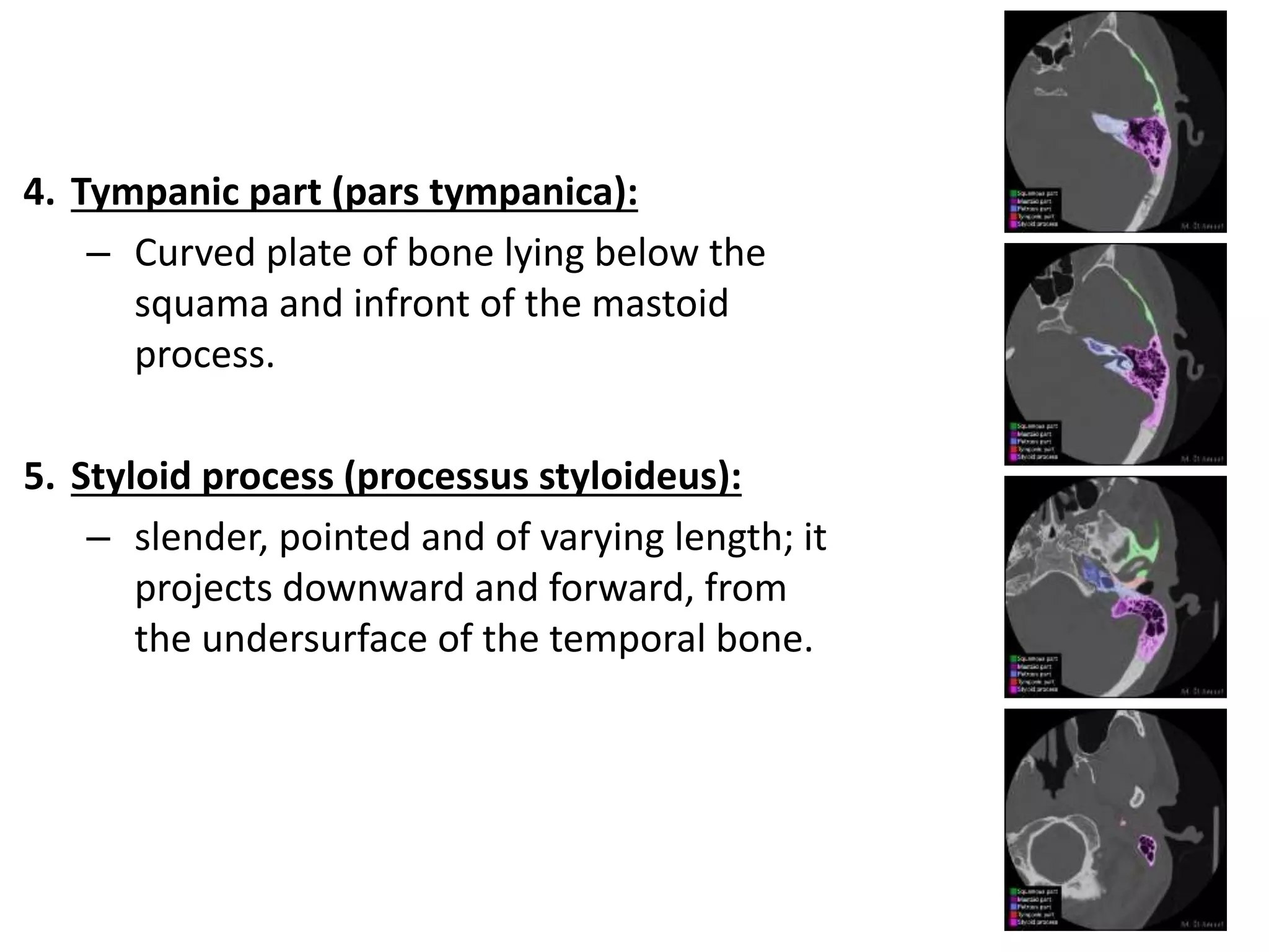
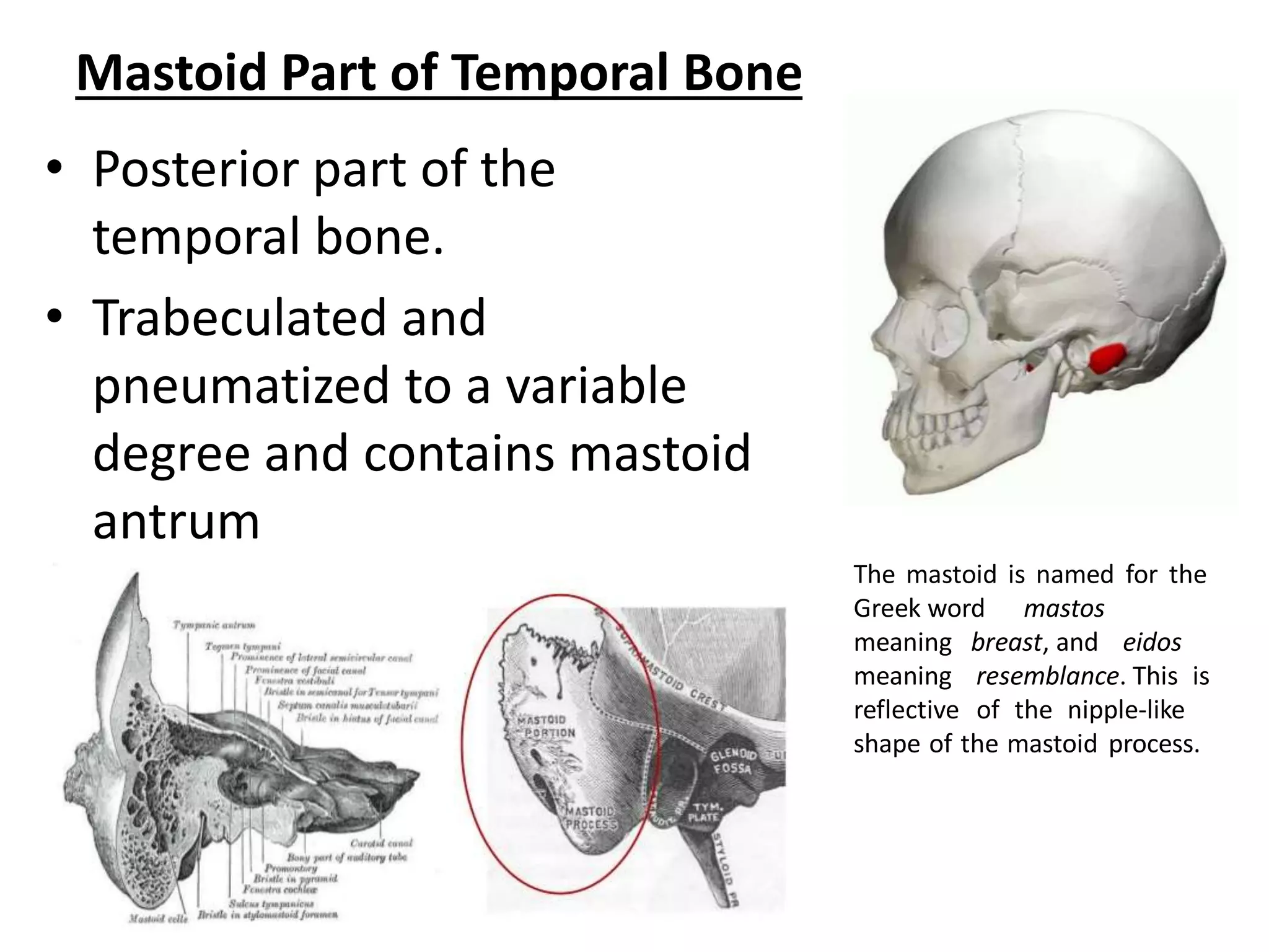
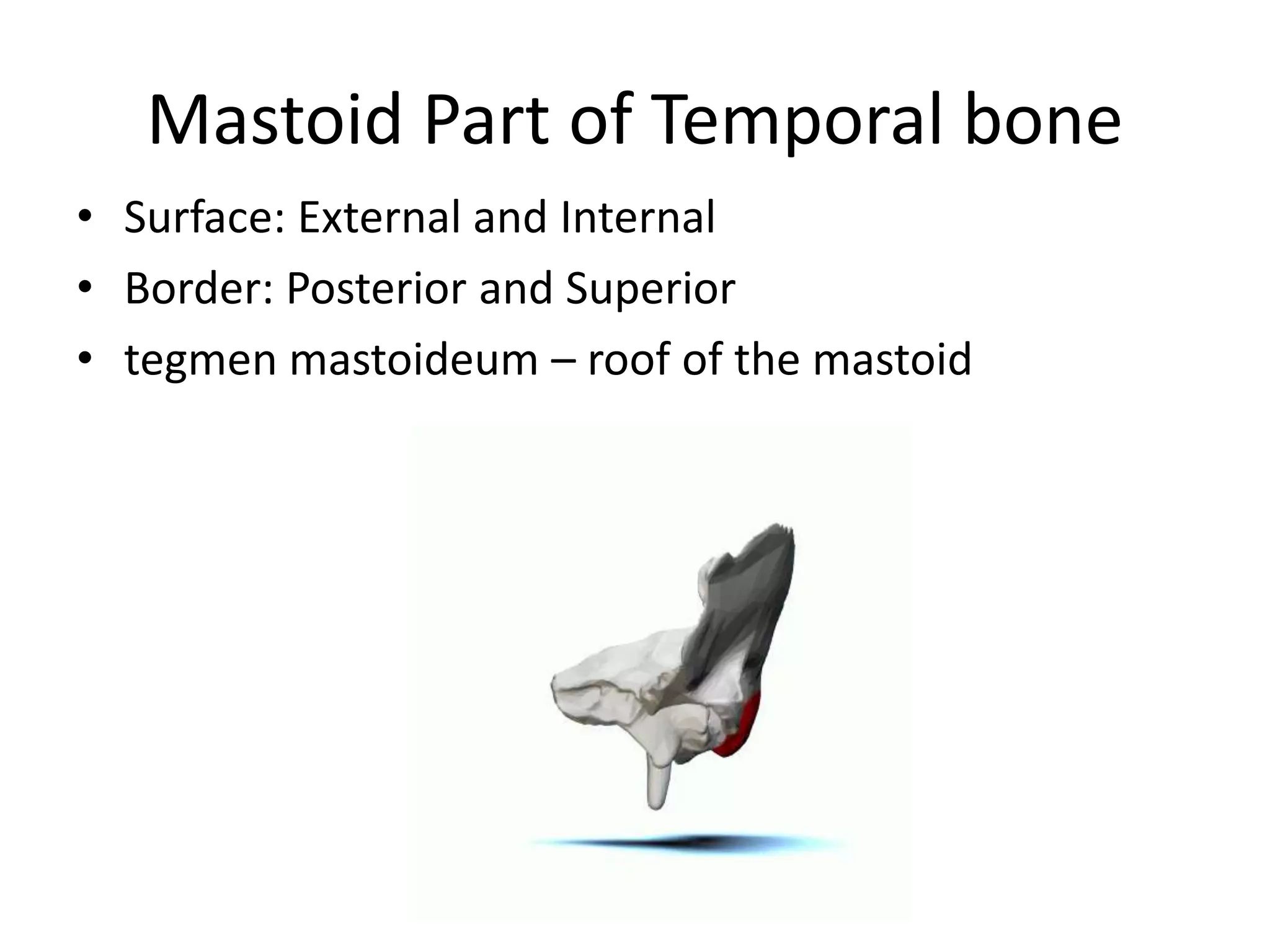
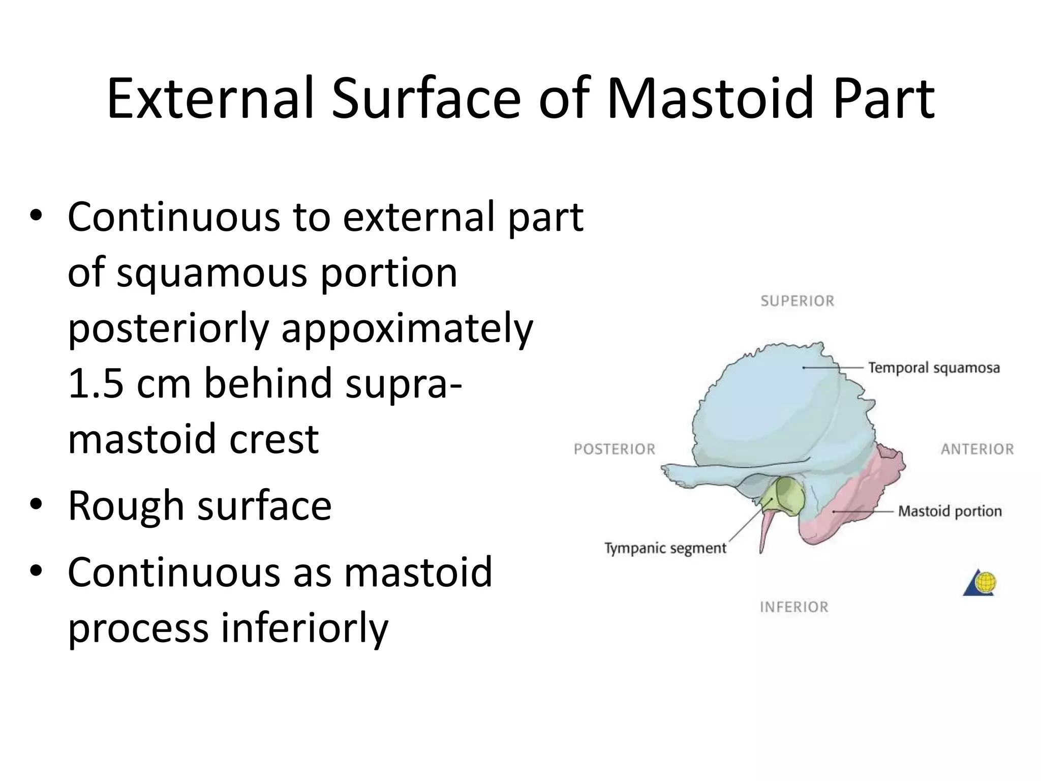
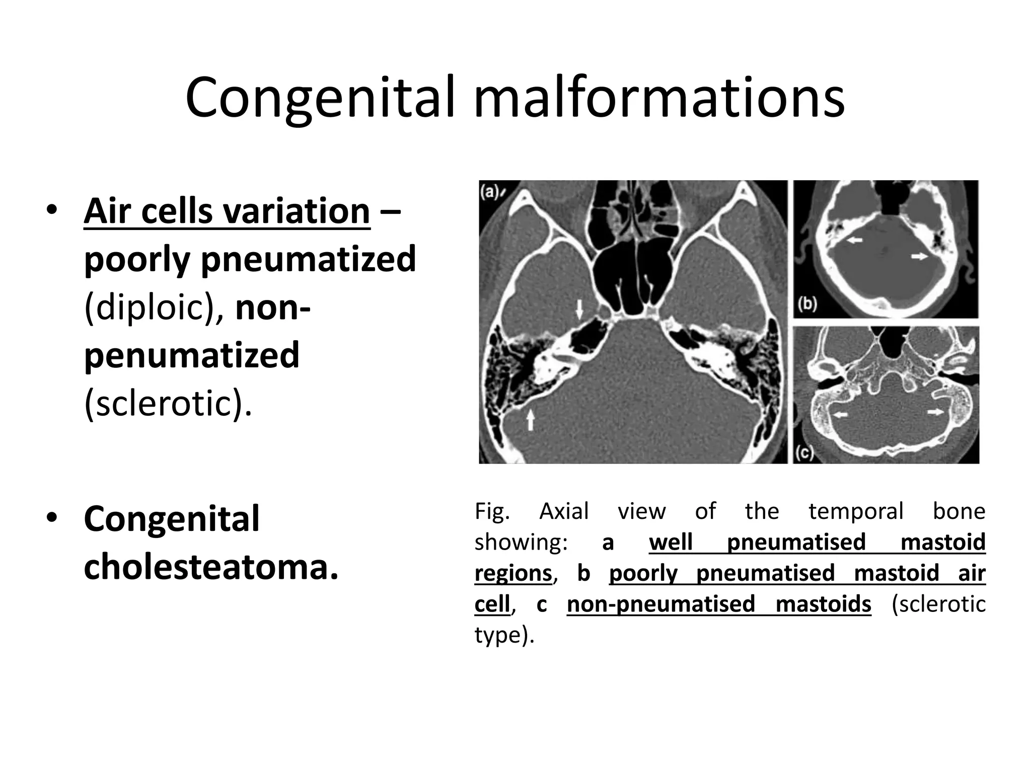
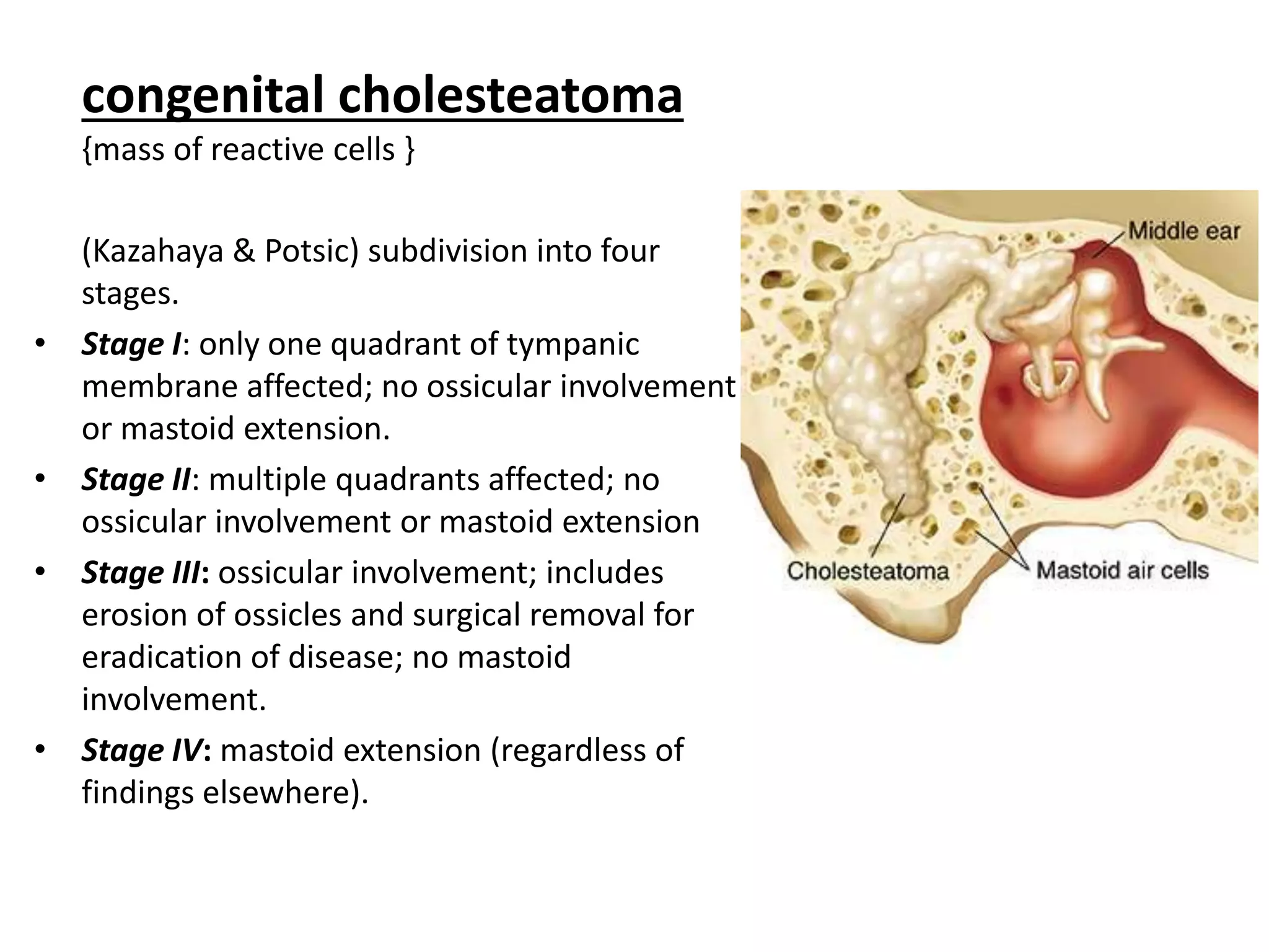
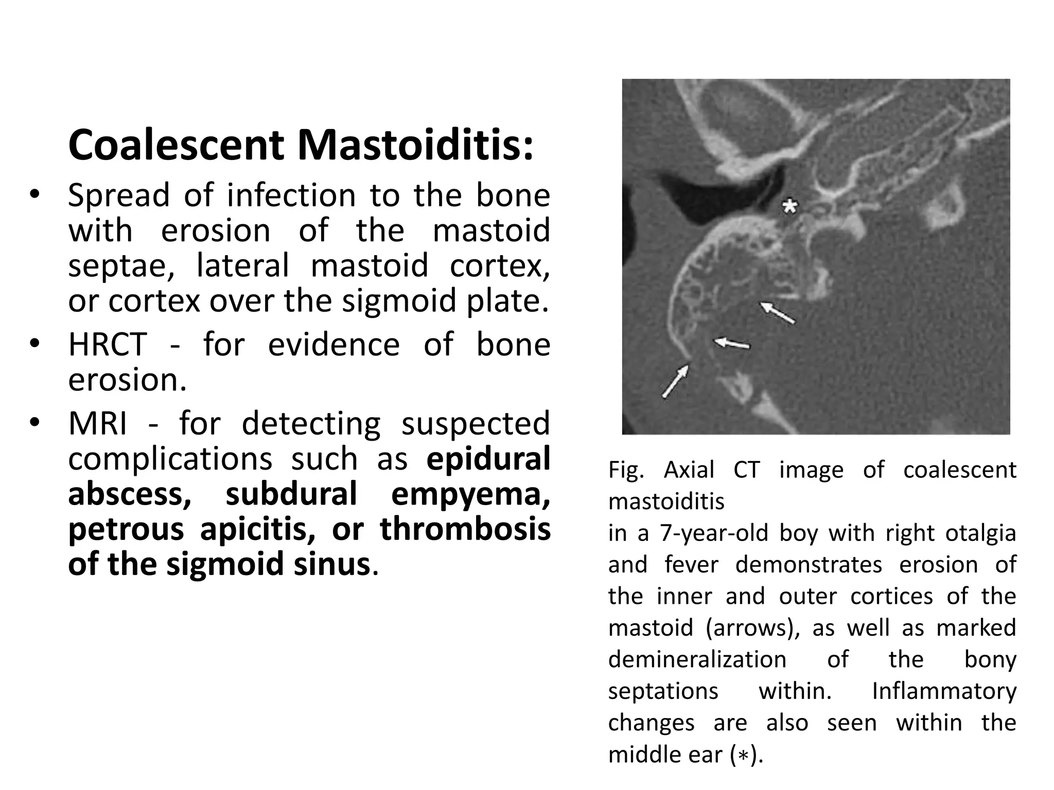
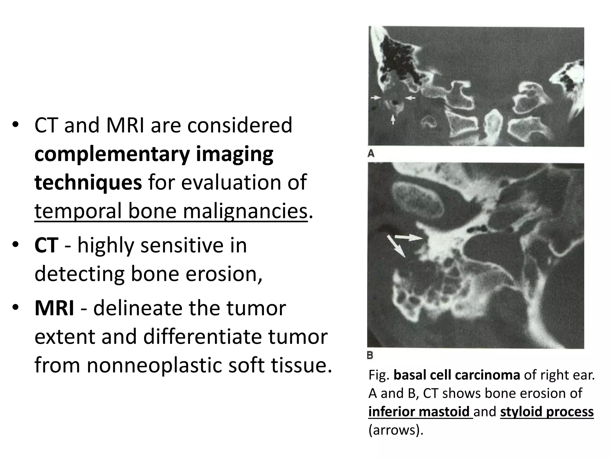
IMAGING IN
DISEASES OF MASTOID
Dr. Gobardhan Thapa
MD Radiodiagnosis
NAMS, Bir hospital
Presentation outlines
Normal anatomy
Imaging modalities
Common pathologies
conclusions
Temporal bone
anatomy
1. The squama (squama temporalis):
forms the anterior and upper part of
the bone; scale like thin and
translucent.
2. Mastoid portion (pars mastoidea):
forms the posterior portion of the
bone.
3. Petrous portion (pars petrosa
{pyramis}):
pyramidal and is wedged in at the
base of the skull between sphenoid
and occipital bone. Contains organ
of hearing.
Parts of the temporal bone
4. Tympanic part (pars tympanica):
– Curved plate of bone lying below the
squama and infront of the mastoid
process.
5. Styloid process (processus styloideus):
– slender, pointed and of varying length; it
projects downward and forward, from
the undersurface of the temporal bone.
Mastoid Part of Temporal Bone
• Posterior part of the
temporal bone.
• Trabeculated and
pneumatized to a variable
degree and contains mastoid
antrum
The mastoid is named for the
Greek word mastos
meaning breast, and eidos
meaning resemblance. This is
reflective of the nipple-like
shape of the mastoid process.
Embryology of mastoid
• The first pharyngeal pouch -> tubotympanic
recess.
• tubotympanic recess -> eustachian tube,
tympanic cavity, mastoid antrum, and their
epithelial lining.
• The middle ear cavity and mastoid antrum are
fluid filled until birth.
• The mastoid air cells develop as saclike
extensions from the mastoid antrum,
commencing around the time of birth and
continuing for several years.
Mastoid Part of Temporal bone
• Surface: External and Internal
• Border: Posterior and Superior
• tegmen mastoideum – roof of the mastoid
External Surface of Mastoid Part
• Continuous to external part
of squamous portion
posteriorly appoximately
1.5 cm behind supra-
mastoid crest
• Rough surface
• Continuous as mastoid
process inferiorly
Mastoid Process
• Bony prolongation at inferior
portion of mastoid part
• External aspect provides
attachment to
Sternocleidomastoid muscle
• Splenius capitis muscle and
Longissimus capitis muscle
are attached to it behind
sternocleidomastoid muscle
Mastoid Foramen
• Foramen present on external surface at the
junction of mastoid part and occipital bone.
• Opening to inner surface of mastoid part
• Lodges emissary vein which will communicate
posterior auricular vein exteriorly with
sigmoid sinus interiorly.
• Also allow passage of meningeal branch of
occipital artery.
Internal Surface of
Mastoid Process
• Consists of sigmoid
sulcus which lodges
sigmoid sinus
• Groove for Occipital
Artery
– Lodges occipital artery
Mastoid segment of the
facial nerve
• courses through the medial
portion of the mastoid bone
until it exits the skull through
the stylomastoid foramen
and enters the parotid gland.
• The chorda tympani
branches off of the mastoid
segment of the facial nerve
and returns to the middle ear
cavity via the canaliculus of
the chorda tympani.
Fig. mastoid segment of the facial nerve canal
(black arrowheads) is seen in its vertical course.
Parts of the lateral SCC and superior SCC are seen.
The mastoid antrum and jugular foramen are
seen.
Mastoid Antrum
• Air containing space in
antero-superior part of
mastoid.
• Variable size.
Mastoid Antrum:
Boundaries
– Roof: Tegmen antri, which
separates mastoid antrum
from middle cranial fossa
– Lateral Wall: Squamous part
by Macewen Triangle.
– Medial Wall: formed by
petrous bone and is related
to
• Posterior semicircular canal
• Endolymphatic sac
• Dura of posterior cranial fossa
Macewan/Supra-Meatal Triangle
Mastoid Antrum: Boundaries
– Anterior Wall: communicates with attic
(epitympanum) through aditus and is in relation
with facial nerve canal, middle ear and deep bony
external auditory canal
– Posterior Wall: communicates with mastoid air
cells; Sigmoid sinus curves downwards
– Floor: communicates with mastoid air cells; other
deeper relations from medial to lateral sides:
• Jugular bulb medial to facial canal
• Digastric ridge which gives origin to posterior belly of digastric
muscle
• Origin of sternocleidomastoid muscle
Korner’s/Petrosquamous Septum
• petrosquamosal suture – may persist as bony plate called
Korner’s septum.
• Separates superficial squamosal cells from deep petrosal cells.
• Mastoid antrum can be entered into only after removal of
Korner’s septum.
• If not recognized, it leads to incomplete removal of disease
during mastoidectomy
Radiography of the mastoid process
Imaging objectives:
• Are the mastoids cellular or acellular?
– If they are cellular, how are cells distributed and do they extend to their
normal anatomical limits? If the mastoid process is acellular, is it sclerotic
or diploetic?
• Is the cortical bone thin or thick?
• Are both the mastoid processes symmetrical?
– Asymmetry is usually the result of pathology early in life that interfered
with the normal anatomical extension of the mucosa of the antrum.
• Are there any signs of bone destruction?
• Is the mastoid cavity preserved?
• Can the sinus plate and the dural plate be visualized in their
normal positions?
• Is there any cholesteatoma formation?
Frontal-occipital 35
degrees caudad
Position of patient and
cassette:
• Head adjusted to bring the
external auditory meatuses
equidistant from the table, so
that the median sagittal plane is
at right-angles to, and in the
midline of, the table.
• Chin depressed so that the
orbito-meatal line is at right-
angles to the table.
Direction and centring of the X-
ray beam:
• A caudal angulation employed,
such that it makes an angle of
35 degrees to the orbito-meatal
plane.
• The beam is centred midway
between the external auditory
meatuses.
Submento-vertical
Supine
• Shoulders raised and the neck
hyperextended to bring the
vertex of the skull in contact
with the grid cassette or table.
• Head is adjusted to bring the
external auditory meatuses
equidistant from the cassette.
• The median sagittal plane
should be at right-angles to
the cassette along its midline.
Erect
• The patient sits a short distance away from a
vertical Bucky.
• neck hyperextended to allow the head to fall
back until the vertex of the skull makes
contact with the centre of the vertical Bucky.
• The remainder of the positioning is as
described for the supine technique.
Direction and centring of the
X-ray beam:
• Central ray is directed at
right-angles to the orbito-
meatal plane and centred
midway between the
external auditory meatuses.
Mastoid – lateral oblique
25 degrees caudad
Position of patient and
cassette:
• Sitting position facing the
erect Bucky. Head then
rotated, such that the median
sagittal plane is parallel to the
Bucky and the inter-orbital
line is perpendicular to the
Bucky.
Direction and centring of
the X-ray beam:
• A 25-degree caudal
angulation is employed
and centred 5 cm above
and 2.5 cm behind the
external auditory meatus
remote from the cassette.
Mastoid – profile
Position of patient and cassette:
• The patient lies supine on the
table, with the orbito-meatal
baseline perpendicular to the
table top.
• From a position with the median
sagittal plane perpendicular to
the table, the head is rotated
through an angle of 35 degrees
away from the side under
examination.
• The vertical tangent to the skull
should now be at the level of the
middle of the mastoid process
under examination, so that the
mastoid process is in profile.
Direction and centring of
the X-ray beam:
• The central ray is angled
caudally so that it makes
an angle of 25 degrees to
the orbito-meatal plane
and is centred to the
middle of the mastoid
process on the side under
examination.
• Collimate tightly around
the mastoid process.
CT
• High-spatial-resolution CT (HRCT) - best method for
evaluating bone and air space anatomy and disorders.
• MDCT - provides improved quality of the axial source
images as well as two- and three-dimensional (2D and
3D) reconstructions.
• Multiplanar CT reformations can display the complex
anatomy of the temporal bone in all planes while
exposing the patient to radiation only once.
• Usually acquired with thin sections (0.5-1 mm) and
special bone algorithms for high detail.
Fig. CT axial Mastoid antrum level. The aditus ad
antrum (aditus) is seen connecting the mastoid
antrum and epitympanum. Portions of the posterior
(PSSC) and superior (SSSC) semicircular canals and
their shared common crus are visualized
MRI
• Primarily for soft tissue entities.
• Much better than CT in characterizing the
CSF, brain, and cranial nerves.
Mastoid bone pathologies
Congenital malformations
Infection
Neoplasms
Fractures/Trauma
Post surgical changes
Congenital malformations
• Air cells variation –
poorly pneumatized
(diploic), non-
penumatized
(sclerotic).
• Congenital
cholesteatoma.
Fig. Axial view of the temporal bone
showing: a well pneumatised mastoid
regions, b poorly pneumatised mastoid air
cell, c non-pneumatised mastoids (sclerotic
type).
congenital cholesteatoma
{mass of reactive cells }
(Kazahaya & Potsic) subdivision into four
stages.
• Stage I: only one quadrant of tympanic
membrane affected; no ossicular involvement
or mastoid extension.
• Stage II: multiple quadrants affected; no
ossicular involvement or mastoid extension
• Stage III: ossicular involvement; includes
erosion of ossicles and surgical removal for
eradication of disease; no mastoid
involvement.
• Stage IV: mastoid extension (regardless of
findings elsewhere).
Infection:
Acute Otomastoiditis
• appears as middle ear and mastoid air cell
opacification with fluid and nonspecific
debris.
• Subacute otitis media shows similar findings
in the mastoid air cells and middle ear, with
the additional finding of focal or diffuse
mucosal thickening.
Coalescent Mastoiditis:
• Spread of infection to the bone
with erosion of the mastoid
septae, lateral mastoid cortex,
or cortex over the sigmoid plate.
• HRCT - for evidence of bone
erosion.
• MRI - for detecting suspected
complications such as epidural
abscess, subdural empyema,
petrous apicitis, or thrombosis
of the sigmoid sinus.
Fig. Axial CT image of coalescent
mastoiditis
in a 7-year-old boy with right otalgia
and fever demonstrates erosion of
the inner and outer cortices of the
mastoid (arrows), as well as marked
demineralization of the bony
septations within. Inflammatory
changes are also seen within the
middle ear (∗).
Complications
of otomastoiditis
Acute Mastoid Osteitis
• Destruction of the bone of the
mastoid air cells and a
subperiosteal abscess may
develop.
Fig. Otomastoiditis and
subperiosteal abscess. Axial HRCT of
the left temporal bone - fluid in the
middle ear cavity and mastoid air
cells, with osseous erosion.
Subperiosteal abscess:
• most commonly in the post-auricular region;
• zygomatic abscess, Bezold's abscess,
Retropharyngeal or parapharyngeal abscess.
Subacute ('masked') mastoiditis:
• incompletely treated AOM after 10-14 days of
infection.
Bezold abscess
• Bone defect at the
mastoid tip medial to the
insertion of the posterior
belly of the digastric
muscle (digastric notch).
• Inflammatory changes
then directed inferiorly
deep to the
sternocleidomastoid
muscle by the fascial
planes.
Fig. Coronal contrast-enhanced CT image of
Bezold abscess in a 7-year-old child with acute
ear infection. Image shows a rim-enhancing
collection below the mastoid tip compatible
with a Bezold abscess (∗∗). Also note thrombus
in the right sigmoid sinus (arrow).
Intracranial complications of acute
mastoiditis
• dural venous sinus thrombosis, most
commonly the sigmoid and transverse
sinuses,
• epidural abscess,
• subdural empyema,
• meningitis, and brain abscess
Petrous apicitis
• in pneumatized petrous apex (present in 30% of
the population).
• septal and cortical destruction, osteitis, and
adjacent meningeal inflammation.
• Close proximity of the fifth and sixth nerves to
the petrous apex - can develop sixth nerve palsy
and deep retro-orbital pain in the V1 distribution
in association with otomastoiditis and petrous
apicitis => Gradinego syndrome.
Imaging findings:
• include opacification
of an aerated petrous
apex with fluid levels
and destruction of the
inner septations or the
cortex.
• On MR images,
meningeal
enhancement is better
appreciated. Fig. left petrous apicitis. Contrast MRI –
irregular enhancement of left petrous apex
Fig. left petrous apicitis. A region of irregular lytic
destruction in the left petrous apex
Chronic oto-mastoiditis
• Follows repeated bouts of otitis media -
nonspecific opacification of the middle ear and
mastoid air cells by granulation tissue and
effusion.
• Reactive sclerosis results in thickening of the
bony septa of the mastoid.
• If chronic otomastoiditis occurs during
maturation of the mastoid during childhood,
there will be a gradual reduction in the number
of mastoid air cells, resulting in poorly developed
mastoids.
Fig. Right chronic otomastoiditis. Right
middle ear soft tissue opacity with
corresponding ossicular destruction.
Sclerosis and loss of aeration of mastoid air
cells.
Cholesteatoma
• conglomerate mass of reactive cells involved in
a localized chronic inflammatory process.
• lined by stratified keratinizing squamous
epithelium with subepithelial fibroconnective
or granulation tissue and keratin debris.
• clinically problematic because of their growth
over time, the osseous erosions they cause, and
their tendency to recur after resection.
• Most cholesteatomas are intratympanic.
• Very rarely they may occur in the mastoid.
• CT:
– rounded, expansile, soft tissue mass, with erosion
adjacent bones.
– Osseous erosion - may lead to facial nerve canal
dehiscence, labyrinthine fistulas, and dehiscence
of the tegmen tympani with or without
meningoencephalocele formation.
“automastoidectomy”
appearance
• mural cholesteatomas drain their
cystic contents through the
tympanic membrane into the
EAC, with only the matrix lining
remaining.
• Characteristic extensive bone
destructive process in the middle
ear/mastoid resembling a surgical
mastoidectomy cavity.
Axial CT image of automastoidectomy
in a 51-year-old man with a history of
longstanding chronic otitis media on the
left. Image demonstrates a large cavity (∗)
in the middle ear and mastoid antrum,
with nonvisualization of the ossicles.
There is a small amount of residual
inflammatory soft tissue. There is no
history of surgery.
• Differentiation of cholesteatoma from a chronic otitis
media may be impossible on CT owing to similar
appearance of the entities and lack of bony
destruction with small cholesteatomas.
• Standard MRI sequences cannot differentiate
cholesteatomas from other inflammatory lesions.
However, half-Fourier acquisition single-shot turbo-
spin echo (HASTE) diffusion-weighted sequences -
helpful in detecting cholesteatomas larger than 5 mm.
• Cholesteatomas will appear hyperintense on diffusion-
weighted imaging (DWI).
Ref: Neuroradiology of cholesteatomas. Barath et al, AJNR, 2011 Feb;32(2):221-9.
Neoplasms
Benign:
Osteomas
• rare benign tumors that are often
incidentally found on CT imaging.
• commonly located at the
tympanosquamous suture within
the EAC
• can also arise in the mastoid
process
• unilateral and solitary pedunculated
hyperdense masses on CT
• Often these lesions are clinically
asymptomatic and do not require
surgery
Fig. CT - pedunculated
osteoma (marked with an
arrow) arising from right
mastoid bone
Lipoma
Facial nerve lipoma. Incidental detection of
focal fat in the distal mastoid segment of the
left facial nerve (arrows), bright on T1- (A) and
T2-weighted (B) images in a patient being
evaluated for dizziness.
• Rare benign
neoplasms in this
region.
schwannoma
• Facial nerve schwannomas – rare.
• Can occur along the entire course of the facial
nerve but most commonly involve the
geniculate ganglion; rarely in the mastoid
segment.
• Classic MR features include a fusiform mass
with T1 hypo- to isointense signal, T2
hyperintense signal, and avid enhancement
along the facial nerve.
• Modern MR and CT techniques - variety of
appearances, including “dumbbell shape” in
the CPA-IAC, multilobular morphology in the
tympanic segment, and an “invasive”
appearance in the mastoid segment.
• mastoid-segment schwannomas may also
require following the nerve into the parotid
gland
Fig. facial nerve schwannoma involving the
right mastoid segment of the facial nerve canal.
axial T1-weighted postcontrast MR image (left)
shows homogeneous enhancement of the mass
(between arrows). The bone algorithm CT
(right) at the same level shows focal
enlargement of the descending segment.
(between arrows).
Ref: The Many Faces of Facial Nerve Schwannoma. Wiggins et al, American Journal of
Neuroradiology March 2006, 27 (3) 694-699;
Cholesterol granulomas
• usually in the middle ear and mastoid air cells
and less commonly in the petrous apex.
• Characteristically expansile with hyperintense
signal on T1- and T2-weighted images.
• slow-growing collections and are
nonneoplastic
• Mastoid granulomas: asymptomatic or
headache
Fig. cholesterol granuloma T1- (left) and T2- (right) weighed
axial MRI - well-defined, high-signal-intensity, soft-tissue
mass protruding to the left posterior fossa. The left sigmoid
sinus (arrow) was compressed by the cholesterol granuloma,
compared with the normal right sigmoid sinus (arrowhead).
Fig. cholesterol granuloma. CT -
large soft-tissue mass in the left
temporal region protruding to the
posterior fossa with bone erosion.
Fig. H&E stain reveals
cholesterol clefts,
hemosiderin deposits,
and granulomatous
inflammation with giant
cells.
Ref: A Large Mastoid Cholesterol Granuloma Eroding the Dura and Sigmoid Sinus. Hsieh et al, J Cytol Histol
5: 236.
Malignant Neoplasms
• Primary malignant neoplasms of the temporal bone are
relatively uncommon;
• Most common - squamous or basal cell carcinoma -
most commonly involve the EAC or mastoid region.
• aggressive when they occur, often extending to the
EAC, middle ear cavity, or mastoid air cells, with
involvement of the facial nerve and TMJ.
• Distant metastases are rare for these tumors.
• Adenocarcinoma can present locally with similar
features as squamous cell carcinoma, but lymph node
involvement is more common.
• CT and MRI are considered
complementary imaging
techniques for evaluation of
temporal bone malignancies.
• CT - highly sensitive in
detecting bone erosion,
• MRI - delineate the tumor
extent and differentiate tumor
from nonneoplastic soft tissue. Fig. basal cell carcinoma of right ear.
A and B, CT shows bone erosion of
inferior mastoid and styloid process
(arrows).
Metastases
• Metastases of the temporal bone are not
uncommon.
• can spread hematogenously from the breast,
lung, stomach, prostate gland, or kidney.
Fractures
(Traumatic Injuries)
Mechanisms
• Blunt trauma injuries.
• Penetrating and gun shot injuries.
• Sport injuries.
Battle sign: Postauricular ecchymosis from traumatic
rupture of a mastoid emissary vein.
Fig. 46-year-old man with head injury A) a subcutaneous hemorrhage in left
postauricular region, B) CT: acute epidural hematoma , C) left temporal bone fracture
(arrows).
Traditional classification:
by the direction it courses relative to the petrous portion of
the temporal bone.
Longitudinal temporal bone fractures
(80-90% of temporal bone fractures) - course parallel to
the long axis of the petrous pyramid, whereas
transverse fractures
course perpendicular to the long axis of the petrous
pyramid. Higher risk of facial nerve injury.
Complex, with both longitudinal and transverse components
Fig. Complex temporal bone fracture
in a 26-year-old male trauma patient.
Axial HRCT left temporal bone -
comminuted oblique fracture
involving the left mastoid and
petrous segments, left jugular
foramen (JF), and left carotid canal
(CC). Partial opacification of the left
mastoid air cells and middle ear
cavity (*) from posttraumatic
hemorrhage.
Axial CT (a) longitudinal fracture and (b) transverse
fracture in two different patients. The longitudinal
fracture is parallel to the long axis of the petrous
pyramid (arrow, a) and traverses the mastoid portion
of the temporal bone. The transverse fracture is
oriented perpendicular to the petrous pyramid
(arrow, b) and traverses the basal turn of the cochlea
(arrowhead, b).
a b
Newer classification: by Kelly and Tami
otic capsule–sparing Fracture:
• the otic capsule is spared.
• much more common (94%–97%), results from a
temporo-parietal blow, increased incidence of
conductive hearing loss due to ossicular injury.
otic capsule– violating fracture:
• otic capsule is violated
• (3%–6%); results from an occipital blow, higher
incidence of facial nerve paralysis (30%–50%),
sensorineural hearing loss, and CSF fistula (CSF
otorrhea and CSF rhinorrhea).
otic capsule - dense osseous labyrinth of the inner ear that surrounds the cochlea, the
vestibule and the semicircular canals.
Post operative imaging
Mastoidectomy
• Mastoiditis, cholesteatoma resection, cochlear
implantation, or endolymphatic surgery - resecting variable
portions of the mastoid air cells and adjacent structures.
• Canal wall- up mastoidectomy – exenteration of the
mastoid air cells with preservation of the posterior wall of
the external auditory canal, creating a mastoid bowl or
cavity.
• Canal wall- down mastoidectomy - performing the same
steps as in canal wall- up mastoidectomy, but in addition
the posterior wall of the external auditory canal is resected
to increase exposure to middle ear contents.
Fig. canal wall up mastoidectomy for
a patient with history of mastoiditis.
Axial CT – intact posterior wall of EAC
(arrow) and mastoidectomy bowl (*)
Fig. Canal-wall-down mastoidectomy with
ossicular preservation. both mastold air cells
and posterior wall of the EAC (canal wall
down). The ossicles are preserved in this
procedure. Postoperative mastoid bowl (M),
with preservation of ossicles (arrows).
• Radical mastoidectomy
– for extensive disease of the
middle ear cavity with ossicular
involvement.
– removal of the tympanic
membrane, malleus, and incus,
with attempted preservation of
the stapes, in addition to canal-
wall-down mastoidectomy.
• Tympanomastoidectomy
– mastoidectomy performed in
conjunction with a middle ear
procedure, such as
tympanoplasty and/or ossicular
reconstruction.
Fig. radical mastoidectomy for
extensive middle ear
cholesteatoma and ossicular
erosion. Axial CT shows a large
mastoidectomy bowl (*) with
absence of posterior wall of
external auditory canal and
ossicular chain.
• Occasionally, the mastoid bowl may be
obliterated using bone, cartilage, fat, or
hydroxyapatite to minimize the size of the
mastoidectomy cavity following canal-wall-down
procedures.
• Otherwise, the mastoid bowl - clear and presence
of soft-tissue attenuation material on CT images
=> may represent granulation tissue or, in the
appropriate scenario, recurrent cholesteatoma.
• Evaluation of recurrent cholesteatoma is an
important indication for imaging after
mastoidectomy.
• CT - recurrent cholesteatoma is suspected
when there is new bone erosion associated
with a soft-tissue focus, although the
attenuation characteristics are nonspecific and
it can be difficult to differentiate these lesions
from granulation tissue.
• On MR images, cholesteatoma - high T2 and
low T1 signal, without central enhancement,
while granulation tissue - internal
enhancement.
• Furthermore, cholesteatoma characteristically
shows reduced diffusion.
Fig. Recurrent cholesteatoma after surgery. A, Coronal HRCT scan shows the obliterated
mastoidectomy cavity (white arrow). B, Coronal FIESTA image distinguishes the slightly
hyperintense (to brain) cholesteatoma (thick white arrow on B, C, and D) from the strongly
hyperintense granulation tissue (dashed arrow on B and C). C, Coronal contrast-enhanced MR
image differentiates as well the nonenhancing cholesteatoma from the strongly enhancing
granulation tissue. D, Coronal DWI with the intensive intralesional diffusion restriction supports
the diagnosis.
Conclusions:
• Though a small portion of temporal bone,
mastoid can be affected by a variety of
pathologies.
• Conventional radiography may give a initial
clue to diagnosis, but rarely used these days.
• HRCT and MRI, with or without contrast are
extensively used to delineate the exquisitely
complex anatomy and related pathologies of
mastoid bone.
References
• Gray’s Anatomy for Students, 2/e
• Clark’s positioning in radiography, 12/e
• CT and MRI of the whole body, Haaga, 6/e.
• Imaging Review of the Temporal Bone: Part I. Anatomy and
Inflammatory and Neoplastic Processes. Juliano et al;
Radiology: Volume 269: Number 1—October 2013
• Imaging Review of the Temporal Bone: Part II. Traumatic,
Postoperative, and Noninflammatory Nonneoplastic
Conditions. Juliano et al; Radiology: Volume 276: Number 3—
September 2015
• Various internet sources.
Thank you

Mastoid diseases imaging

  • 1.
    IMAGING IN DISEASES OFMASTOID Dr. Gobardhan Thapa MD Radiodiagnosis NAMS, Bir hospital
  • 2.
    Presentation outlines Normal anatomy Imagingmodalities Common pathologies conclusions
  • 3.
  • 4.
    1. The squama(squama temporalis): forms the anterior and upper part of the bone; scale like thin and translucent. 2. Mastoid portion (pars mastoidea): forms the posterior portion of the bone. 3. Petrous portion (pars petrosa {pyramis}): pyramidal and is wedged in at the base of the skull between sphenoid and occipital bone. Contains organ of hearing. Parts of the temporal bone
  • 5.
    4. Tympanic part(pars tympanica): – Curved plate of bone lying below the squama and infront of the mastoid process. 5. Styloid process (processus styloideus): – slender, pointed and of varying length; it projects downward and forward, from the undersurface of the temporal bone.
  • 6.
    Mastoid Part ofTemporal Bone • Posterior part of the temporal bone. • Trabeculated and pneumatized to a variable degree and contains mastoid antrum The mastoid is named for the Greek word mastos meaning breast, and eidos meaning resemblance. This is reflective of the nipple-like shape of the mastoid process.
  • 7.
    Embryology of mastoid •The first pharyngeal pouch -> tubotympanic recess. • tubotympanic recess -> eustachian tube, tympanic cavity, mastoid antrum, and their epithelial lining. • The middle ear cavity and mastoid antrum are fluid filled until birth. • The mastoid air cells develop as saclike extensions from the mastoid antrum, commencing around the time of birth and continuing for several years.
  • 8.
    Mastoid Part ofTemporal bone • Surface: External and Internal • Border: Posterior and Superior • tegmen mastoideum – roof of the mastoid
  • 9.
    External Surface ofMastoid Part • Continuous to external part of squamous portion posteriorly appoximately 1.5 cm behind supra- mastoid crest • Rough surface • Continuous as mastoid process inferiorly
  • 10.
    Mastoid Process • Bonyprolongation at inferior portion of mastoid part • External aspect provides attachment to Sternocleidomastoid muscle • Splenius capitis muscle and Longissimus capitis muscle are attached to it behind sternocleidomastoid muscle
  • 11.
    Mastoid Foramen • Foramenpresent on external surface at the junction of mastoid part and occipital bone. • Opening to inner surface of mastoid part • Lodges emissary vein which will communicate posterior auricular vein exteriorly with sigmoid sinus interiorly. • Also allow passage of meningeal branch of occipital artery.
  • 12.
    Internal Surface of MastoidProcess • Consists of sigmoid sulcus which lodges sigmoid sinus • Groove for Occipital Artery – Lodges occipital artery
  • 13.
    Mastoid segment ofthe facial nerve • courses through the medial portion of the mastoid bone until it exits the skull through the stylomastoid foramen and enters the parotid gland. • The chorda tympani branches off of the mastoid segment of the facial nerve and returns to the middle ear cavity via the canaliculus of the chorda tympani. Fig. mastoid segment of the facial nerve canal (black arrowheads) is seen in its vertical course. Parts of the lateral SCC and superior SCC are seen. The mastoid antrum and jugular foramen are seen.
  • 14.
    Mastoid Antrum • Aircontaining space in antero-superior part of mastoid. • Variable size.
  • 15.
    Mastoid Antrum: Boundaries – Roof:Tegmen antri, which separates mastoid antrum from middle cranial fossa – Lateral Wall: Squamous part by Macewen Triangle. – Medial Wall: formed by petrous bone and is related to • Posterior semicircular canal • Endolymphatic sac • Dura of posterior cranial fossa Macewan/Supra-Meatal Triangle
  • 16.
    Mastoid Antrum: Boundaries –Anterior Wall: communicates with attic (epitympanum) through aditus and is in relation with facial nerve canal, middle ear and deep bony external auditory canal – Posterior Wall: communicates with mastoid air cells; Sigmoid sinus curves downwards – Floor: communicates with mastoid air cells; other deeper relations from medial to lateral sides: • Jugular bulb medial to facial canal • Digastric ridge which gives origin to posterior belly of digastric muscle • Origin of sternocleidomastoid muscle
  • 17.
    Korner’s/Petrosquamous Septum • petrosquamosalsuture – may persist as bony plate called Korner’s septum. • Separates superficial squamosal cells from deep petrosal cells. • Mastoid antrum can be entered into only after removal of Korner’s septum. • If not recognized, it leads to incomplete removal of disease during mastoidectomy
  • 18.
    Radiography of themastoid process Imaging objectives: • Are the mastoids cellular or acellular? – If they are cellular, how are cells distributed and do they extend to their normal anatomical limits? If the mastoid process is acellular, is it sclerotic or diploetic? • Is the cortical bone thin or thick? • Are both the mastoid processes symmetrical? – Asymmetry is usually the result of pathology early in life that interfered with the normal anatomical extension of the mucosa of the antrum. • Are there any signs of bone destruction? • Is the mastoid cavity preserved? • Can the sinus plate and the dural plate be visualized in their normal positions? • Is there any cholesteatoma formation?
  • 19.
    Frontal-occipital 35 degrees caudad Positionof patient and cassette: • Head adjusted to bring the external auditory meatuses equidistant from the table, so that the median sagittal plane is at right-angles to, and in the midline of, the table. • Chin depressed so that the orbito-meatal line is at right- angles to the table.
  • 20.
    Direction and centringof the X- ray beam: • A caudal angulation employed, such that it makes an angle of 35 degrees to the orbito-meatal plane. • The beam is centred midway between the external auditory meatuses.
  • 21.
    Submento-vertical Supine • Shoulders raisedand the neck hyperextended to bring the vertex of the skull in contact with the grid cassette or table. • Head is adjusted to bring the external auditory meatuses equidistant from the cassette. • The median sagittal plane should be at right-angles to the cassette along its midline.
  • 22.
    Erect • The patientsits a short distance away from a vertical Bucky. • neck hyperextended to allow the head to fall back until the vertex of the skull makes contact with the centre of the vertical Bucky. • The remainder of the positioning is as described for the supine technique.
  • 23.
    Direction and centringof the X-ray beam: • Central ray is directed at right-angles to the orbito- meatal plane and centred midway between the external auditory meatuses.
  • 24.
    Mastoid – lateraloblique 25 degrees caudad Position of patient and cassette: • Sitting position facing the erect Bucky. Head then rotated, such that the median sagittal plane is parallel to the Bucky and the inter-orbital line is perpendicular to the Bucky.
  • 25.
    Direction and centringof the X-ray beam: • A 25-degree caudal angulation is employed and centred 5 cm above and 2.5 cm behind the external auditory meatus remote from the cassette.
  • 26.
    Mastoid – profile Positionof patient and cassette: • The patient lies supine on the table, with the orbito-meatal baseline perpendicular to the table top. • From a position with the median sagittal plane perpendicular to the table, the head is rotated through an angle of 35 degrees away from the side under examination. • The vertical tangent to the skull should now be at the level of the middle of the mastoid process under examination, so that the mastoid process is in profile.
  • 27.
    Direction and centringof the X-ray beam: • The central ray is angled caudally so that it makes an angle of 25 degrees to the orbito-meatal plane and is centred to the middle of the mastoid process on the side under examination. • Collimate tightly around the mastoid process.
  • 28.
    CT • High-spatial-resolution CT(HRCT) - best method for evaluating bone and air space anatomy and disorders. • MDCT - provides improved quality of the axial source images as well as two- and three-dimensional (2D and 3D) reconstructions. • Multiplanar CT reformations can display the complex anatomy of the temporal bone in all planes while exposing the patient to radiation only once. • Usually acquired with thin sections (0.5-1 mm) and special bone algorithms for high detail.
  • 29.
    Fig. CT axialMastoid antrum level. The aditus ad antrum (aditus) is seen connecting the mastoid antrum and epitympanum. Portions of the posterior (PSSC) and superior (SSSC) semicircular canals and their shared common crus are visualized
  • 30.
    MRI • Primarily forsoft tissue entities. • Much better than CT in characterizing the CSF, brain, and cranial nerves.
  • 31.
    Mastoid bone pathologies Congenitalmalformations Infection Neoplasms Fractures/Trauma Post surgical changes
  • 32.
    Congenital malformations • Aircells variation – poorly pneumatized (diploic), non- penumatized (sclerotic). • Congenital cholesteatoma. Fig. Axial view of the temporal bone showing: a well pneumatised mastoid regions, b poorly pneumatised mastoid air cell, c non-pneumatised mastoids (sclerotic type).
  • 33.
    congenital cholesteatoma {mass ofreactive cells } (Kazahaya & Potsic) subdivision into four stages. • Stage I: only one quadrant of tympanic membrane affected; no ossicular involvement or mastoid extension. • Stage II: multiple quadrants affected; no ossicular involvement or mastoid extension • Stage III: ossicular involvement; includes erosion of ossicles and surgical removal for eradication of disease; no mastoid involvement. • Stage IV: mastoid extension (regardless of findings elsewhere).
  • 34.
    Infection: Acute Otomastoiditis • appearsas middle ear and mastoid air cell opacification with fluid and nonspecific debris. • Subacute otitis media shows similar findings in the mastoid air cells and middle ear, with the additional finding of focal or diffuse mucosal thickening.
  • 35.
    Coalescent Mastoiditis: • Spreadof infection to the bone with erosion of the mastoid septae, lateral mastoid cortex, or cortex over the sigmoid plate. • HRCT - for evidence of bone erosion. • MRI - for detecting suspected complications such as epidural abscess, subdural empyema, petrous apicitis, or thrombosis of the sigmoid sinus. Fig. Axial CT image of coalescent mastoiditis in a 7-year-old boy with right otalgia and fever demonstrates erosion of the inner and outer cortices of the mastoid (arrows), as well as marked demineralization of the bony septations within. Inflammatory changes are also seen within the middle ear (∗).
  • 36.
    Complications of otomastoiditis Acute MastoidOsteitis • Destruction of the bone of the mastoid air cells and a subperiosteal abscess may develop. Fig. Otomastoiditis and subperiosteal abscess. Axial HRCT of the left temporal bone - fluid in the middle ear cavity and mastoid air cells, with osseous erosion.
  • 37.
    Subperiosteal abscess: • mostcommonly in the post-auricular region; • zygomatic abscess, Bezold's abscess, Retropharyngeal or parapharyngeal abscess. Subacute ('masked') mastoiditis: • incompletely treated AOM after 10-14 days of infection.
  • 38.
    Bezold abscess • Bonedefect at the mastoid tip medial to the insertion of the posterior belly of the digastric muscle (digastric notch). • Inflammatory changes then directed inferiorly deep to the sternocleidomastoid muscle by the fascial planes. Fig. Coronal contrast-enhanced CT image of Bezold abscess in a 7-year-old child with acute ear infection. Image shows a rim-enhancing collection below the mastoid tip compatible with a Bezold abscess (∗∗). Also note thrombus in the right sigmoid sinus (arrow).
  • 39.
    Intracranial complications ofacute mastoiditis • dural venous sinus thrombosis, most commonly the sigmoid and transverse sinuses, • epidural abscess, • subdural empyema, • meningitis, and brain abscess
  • 40.
    Petrous apicitis • inpneumatized petrous apex (present in 30% of the population). • septal and cortical destruction, osteitis, and adjacent meningeal inflammation. • Close proximity of the fifth and sixth nerves to the petrous apex - can develop sixth nerve palsy and deep retro-orbital pain in the V1 distribution in association with otomastoiditis and petrous apicitis => Gradinego syndrome.
  • 41.
    Imaging findings: • includeopacification of an aerated petrous apex with fluid levels and destruction of the inner septations or the cortex. • On MR images, meningeal enhancement is better appreciated. Fig. left petrous apicitis. Contrast MRI – irregular enhancement of left petrous apex Fig. left petrous apicitis. A region of irregular lytic destruction in the left petrous apex
  • 42.
    Chronic oto-mastoiditis • Followsrepeated bouts of otitis media - nonspecific opacification of the middle ear and mastoid air cells by granulation tissue and effusion. • Reactive sclerosis results in thickening of the bony septa of the mastoid. • If chronic otomastoiditis occurs during maturation of the mastoid during childhood, there will be a gradual reduction in the number of mastoid air cells, resulting in poorly developed mastoids.
  • 43.
    Fig. Right chronicotomastoiditis. Right middle ear soft tissue opacity with corresponding ossicular destruction. Sclerosis and loss of aeration of mastoid air cells.
  • 44.
    Cholesteatoma • conglomerate massof reactive cells involved in a localized chronic inflammatory process. • lined by stratified keratinizing squamous epithelium with subepithelial fibroconnective or granulation tissue and keratin debris. • clinically problematic because of their growth over time, the osseous erosions they cause, and their tendency to recur after resection.
  • 45.
    • Most cholesteatomasare intratympanic. • Very rarely they may occur in the mastoid. • CT: – rounded, expansile, soft tissue mass, with erosion adjacent bones. – Osseous erosion - may lead to facial nerve canal dehiscence, labyrinthine fistulas, and dehiscence of the tegmen tympani with or without meningoencephalocele formation.
  • 46.
    “automastoidectomy” appearance • mural cholesteatomasdrain their cystic contents through the tympanic membrane into the EAC, with only the matrix lining remaining. • Characteristic extensive bone destructive process in the middle ear/mastoid resembling a surgical mastoidectomy cavity. Axial CT image of automastoidectomy in a 51-year-old man with a history of longstanding chronic otitis media on the left. Image demonstrates a large cavity (∗) in the middle ear and mastoid antrum, with nonvisualization of the ossicles. There is a small amount of residual inflammatory soft tissue. There is no history of surgery.
  • 47.
    • Differentiation ofcholesteatoma from a chronic otitis media may be impossible on CT owing to similar appearance of the entities and lack of bony destruction with small cholesteatomas. • Standard MRI sequences cannot differentiate cholesteatomas from other inflammatory lesions. However, half-Fourier acquisition single-shot turbo- spin echo (HASTE) diffusion-weighted sequences - helpful in detecting cholesteatomas larger than 5 mm. • Cholesteatomas will appear hyperintense on diffusion- weighted imaging (DWI). Ref: Neuroradiology of cholesteatomas. Barath et al, AJNR, 2011 Feb;32(2):221-9.
  • 48.
    Neoplasms Benign: Osteomas • rare benigntumors that are often incidentally found on CT imaging. • commonly located at the tympanosquamous suture within the EAC • can also arise in the mastoid process • unilateral and solitary pedunculated hyperdense masses on CT • Often these lesions are clinically asymptomatic and do not require surgery Fig. CT - pedunculated osteoma (marked with an arrow) arising from right mastoid bone
  • 49.
    Lipoma Facial nerve lipoma.Incidental detection of focal fat in the distal mastoid segment of the left facial nerve (arrows), bright on T1- (A) and T2-weighted (B) images in a patient being evaluated for dizziness. • Rare benign neoplasms in this region.
  • 50.
    schwannoma • Facial nerveschwannomas – rare. • Can occur along the entire course of the facial nerve but most commonly involve the geniculate ganglion; rarely in the mastoid segment. • Classic MR features include a fusiform mass with T1 hypo- to isointense signal, T2 hyperintense signal, and avid enhancement along the facial nerve. • Modern MR and CT techniques - variety of appearances, including “dumbbell shape” in the CPA-IAC, multilobular morphology in the tympanic segment, and an “invasive” appearance in the mastoid segment. • mastoid-segment schwannomas may also require following the nerve into the parotid gland Fig. facial nerve schwannoma involving the right mastoid segment of the facial nerve canal. axial T1-weighted postcontrast MR image (left) shows homogeneous enhancement of the mass (between arrows). The bone algorithm CT (right) at the same level shows focal enlargement of the descending segment. (between arrows). Ref: The Many Faces of Facial Nerve Schwannoma. Wiggins et al, American Journal of Neuroradiology March 2006, 27 (3) 694-699;
  • 51.
    Cholesterol granulomas • usuallyin the middle ear and mastoid air cells and less commonly in the petrous apex. • Characteristically expansile with hyperintense signal on T1- and T2-weighted images. • slow-growing collections and are nonneoplastic • Mastoid granulomas: asymptomatic or headache
  • 52.
    Fig. cholesterol granulomaT1- (left) and T2- (right) weighed axial MRI - well-defined, high-signal-intensity, soft-tissue mass protruding to the left posterior fossa. The left sigmoid sinus (arrow) was compressed by the cholesterol granuloma, compared with the normal right sigmoid sinus (arrowhead). Fig. cholesterol granuloma. CT - large soft-tissue mass in the left temporal region protruding to the posterior fossa with bone erosion. Fig. H&E stain reveals cholesterol clefts, hemosiderin deposits, and granulomatous inflammation with giant cells. Ref: A Large Mastoid Cholesterol Granuloma Eroding the Dura and Sigmoid Sinus. Hsieh et al, J Cytol Histol 5: 236.
  • 53.
    Malignant Neoplasms • Primarymalignant neoplasms of the temporal bone are relatively uncommon; • Most common - squamous or basal cell carcinoma - most commonly involve the EAC or mastoid region. • aggressive when they occur, often extending to the EAC, middle ear cavity, or mastoid air cells, with involvement of the facial nerve and TMJ. • Distant metastases are rare for these tumors. • Adenocarcinoma can present locally with similar features as squamous cell carcinoma, but lymph node involvement is more common.
  • 54.
    • CT andMRI are considered complementary imaging techniques for evaluation of temporal bone malignancies. • CT - highly sensitive in detecting bone erosion, • MRI - delineate the tumor extent and differentiate tumor from nonneoplastic soft tissue. Fig. basal cell carcinoma of right ear. A and B, CT shows bone erosion of inferior mastoid and styloid process (arrows).
  • 55.
    Metastases • Metastases ofthe temporal bone are not uncommon. • can spread hematogenously from the breast, lung, stomach, prostate gland, or kidney.
  • 56.
    Fractures (Traumatic Injuries) Mechanisms • Blunttrauma injuries. • Penetrating and gun shot injuries. • Sport injuries.
  • 57.
    Battle sign: Postauricularecchymosis from traumatic rupture of a mastoid emissary vein. Fig. 46-year-old man with head injury A) a subcutaneous hemorrhage in left postauricular region, B) CT: acute epidural hematoma , C) left temporal bone fracture (arrows).
  • 58.
    Traditional classification: by thedirection it courses relative to the petrous portion of the temporal bone. Longitudinal temporal bone fractures (80-90% of temporal bone fractures) - course parallel to the long axis of the petrous pyramid, whereas transverse fractures course perpendicular to the long axis of the petrous pyramid. Higher risk of facial nerve injury. Complex, with both longitudinal and transverse components
  • 59.
    Fig. Complex temporalbone fracture in a 26-year-old male trauma patient. Axial HRCT left temporal bone - comminuted oblique fracture involving the left mastoid and petrous segments, left jugular foramen (JF), and left carotid canal (CC). Partial opacification of the left mastoid air cells and middle ear cavity (*) from posttraumatic hemorrhage. Axial CT (a) longitudinal fracture and (b) transverse fracture in two different patients. The longitudinal fracture is parallel to the long axis of the petrous pyramid (arrow, a) and traverses the mastoid portion of the temporal bone. The transverse fracture is oriented perpendicular to the petrous pyramid (arrow, b) and traverses the basal turn of the cochlea (arrowhead, b). a b
  • 60.
    Newer classification: byKelly and Tami otic capsule–sparing Fracture: • the otic capsule is spared. • much more common (94%–97%), results from a temporo-parietal blow, increased incidence of conductive hearing loss due to ossicular injury. otic capsule– violating fracture: • otic capsule is violated • (3%–6%); results from an occipital blow, higher incidence of facial nerve paralysis (30%–50%), sensorineural hearing loss, and CSF fistula (CSF otorrhea and CSF rhinorrhea). otic capsule - dense osseous labyrinth of the inner ear that surrounds the cochlea, the vestibule and the semicircular canals.
  • 61.
    Post operative imaging Mastoidectomy •Mastoiditis, cholesteatoma resection, cochlear implantation, or endolymphatic surgery - resecting variable portions of the mastoid air cells and adjacent structures. • Canal wall- up mastoidectomy – exenteration of the mastoid air cells with preservation of the posterior wall of the external auditory canal, creating a mastoid bowl or cavity. • Canal wall- down mastoidectomy - performing the same steps as in canal wall- up mastoidectomy, but in addition the posterior wall of the external auditory canal is resected to increase exposure to middle ear contents.
  • 62.
    Fig. canal wallup mastoidectomy for a patient with history of mastoiditis. Axial CT – intact posterior wall of EAC (arrow) and mastoidectomy bowl (*) Fig. Canal-wall-down mastoidectomy with ossicular preservation. both mastold air cells and posterior wall of the EAC (canal wall down). The ossicles are preserved in this procedure. Postoperative mastoid bowl (M), with preservation of ossicles (arrows).
  • 63.
    • Radical mastoidectomy –for extensive disease of the middle ear cavity with ossicular involvement. – removal of the tympanic membrane, malleus, and incus, with attempted preservation of the stapes, in addition to canal- wall-down mastoidectomy. • Tympanomastoidectomy – mastoidectomy performed in conjunction with a middle ear procedure, such as tympanoplasty and/or ossicular reconstruction. Fig. radical mastoidectomy for extensive middle ear cholesteatoma and ossicular erosion. Axial CT shows a large mastoidectomy bowl (*) with absence of posterior wall of external auditory canal and ossicular chain.
  • 64.
    • Occasionally, themastoid bowl may be obliterated using bone, cartilage, fat, or hydroxyapatite to minimize the size of the mastoidectomy cavity following canal-wall-down procedures. • Otherwise, the mastoid bowl - clear and presence of soft-tissue attenuation material on CT images => may represent granulation tissue or, in the appropriate scenario, recurrent cholesteatoma.
  • 65.
    • Evaluation ofrecurrent cholesteatoma is an important indication for imaging after mastoidectomy. • CT - recurrent cholesteatoma is suspected when there is new bone erosion associated with a soft-tissue focus, although the attenuation characteristics are nonspecific and it can be difficult to differentiate these lesions from granulation tissue.
  • 66.
    • On MRimages, cholesteatoma - high T2 and low T1 signal, without central enhancement, while granulation tissue - internal enhancement. • Furthermore, cholesteatoma characteristically shows reduced diffusion.
  • 67.
    Fig. Recurrent cholesteatomaafter surgery. A, Coronal HRCT scan shows the obliterated mastoidectomy cavity (white arrow). B, Coronal FIESTA image distinguishes the slightly hyperintense (to brain) cholesteatoma (thick white arrow on B, C, and D) from the strongly hyperintense granulation tissue (dashed arrow on B and C). C, Coronal contrast-enhanced MR image differentiates as well the nonenhancing cholesteatoma from the strongly enhancing granulation tissue. D, Coronal DWI with the intensive intralesional diffusion restriction supports the diagnosis.
  • 68.
    Conclusions: • Though asmall portion of temporal bone, mastoid can be affected by a variety of pathologies. • Conventional radiography may give a initial clue to diagnosis, but rarely used these days. • HRCT and MRI, with or without contrast are extensively used to delineate the exquisitely complex anatomy and related pathologies of mastoid bone.
  • 69.
    References • Gray’s Anatomyfor Students, 2/e • Clark’s positioning in radiography, 12/e • CT and MRI of the whole body, Haaga, 6/e. • Imaging Review of the Temporal Bone: Part I. Anatomy and Inflammatory and Neoplastic Processes. Juliano et al; Radiology: Volume 269: Number 1—October 2013 • Imaging Review of the Temporal Bone: Part II. Traumatic, Postoperative, and Noninflammatory Nonneoplastic Conditions. Juliano et al; Radiology: Volume 276: Number 3— September 2015 • Various internet sources.
  • 70.

Editor's Notes

  • #7 Mastoid Part of Temporal Bone forms posterior part of the temporal bone. It is Trabeculated and pneumatized to a variable degree and contains mastoid antrum.
  • #9 It consists of two suface: External and internal and two borders: posterior and superior.
  • #10 External Surface of Mastoid Part is rough surface continuous to external part of squamous part posteriorly appoximately 1.5 cm behind supra-mastoid crest It is continuous as mastoid process inferiorly.
  • #11 Formed due to pulling action of sternocleidomastoid muscle when the child learns head holding.
  • #13 Internal Surface of Mastoid Process is present inferior to internal surface of mastoid part. It presents Digastric notch, also known as mastoid notch which provides attachment to posterior belly of digastric muscle. It also presents groove for Occipital Artery lodging occipital artery medial to digastric notch. POSTERIOR BELLY OF DIGASTRIC MUSCLE: SUPPLIED BY FACIAL NERVE ANTERIOR BELLY OF DIGASTRIC MUSCLE: SUPPLIED BY TRIGEMINAL NERVE
  • #18 In some cases, petrosquamosal suture persists as a bony plate called Korner’s septum It separates superficial squamosal cells from deep petrosal cells During mastoid surgery, it causes difficulty in locating antrum and deeper cells If not recognized, it leads to incomplete removal of disease during mastoidectomy Mastoid antrum can be entered into only after removal of Korner’s septum
  • #20 supine in the midline of the table or erect with back to an erect Bucky. A small (24-30 cm) cassette is placed transversely in the cassette tray and is centred to coincide with the angled central ray.
  • #21 The mastoid process can be palpated easily behind the ear. Collimate laterally to include the lateral margins of the skull and supra-inferiorly to include the mastoid and petrous parts of the temporal bone.
  • #22 The orbito-meatal plane should be as near as possible parallel to the cassette.
  • #25 Position the mastoid process in the middle of the Bucky. An 18 24-cm cassette is positioned longitudinally in the Bucky and is centred to coincide with the central ray and mastoid process. The shoulders may be rotated slightly to allow the correct position to be attained. The patient may grip the Bucky for stability. The auricle of the ear adjacent to the table is folded forward to ensure that its soft-tissue outline is not superimposed over the region of interest.
  • #26 Collimate to the area under examination.
  • #27 Finally, the head is moved transversely across the table so that the mastoid process being examined is in the midline of the table. , such that the median sagittal plane now makes an angle of 55 degrees to the table.
  • #29 The temporal bone has a high inherent contrast resolution, having both the densest bone in the body and air-filled spaces. provides improved quality of the axial source images as well as two- and three-dimensional (2D and 3D) reconstructions; this is due to a reduction in partial volume and motion artifacts and higher resolution along the z-axis with nearly isotropic voxel size.
  • #35 The fluid in serous otitis media cannot be distinguished from the pus seen with purulent otitis.
  • #39 In young children, the mastoid tip is usually not aerated; as a result, Bezold abscess in young children is uncommon.
  • #45 Congenital or acquired Primary and secondary
  • #46 M/C in in Prussak’s space
  • #48 cholesteatomas may or may not be accompanied by chronic otitis media.
  • #51 The presenting symptoms can provide insight on the location of the schwannoma. For example, sensorineural hearing loss is caused by compression of cranial nerve VIII at the CPA, whereas conductive hearing loss is caused by ossicular chain dysfunction from tympanic cavity schwannomas.
  • #58 Temporal bone fractures do not demonstrate normal bone healing, possibly related to low metabolic activity of the bone of the otic capsule
  • #61 CSF otorrhea and CSF rhinorrhea (leak of CSF through the Eustachian tube into the nasal cavity in the setting of an intact tympanic membrane).
  • #67 can potentially serve as an alternative to second-look surgery for the detection of recurrent cholesteatoma. Indeed, non– echo-planar imaging DWI confers a positive predictive value for cholesteatoma of 93%–100%. It is important to interpret the DWI studies in conjunction with the conventional pulse sequence images to avoid false-positive interpretations, which can result from the presence of fat grafts and hemorrhage.