Download to read offline
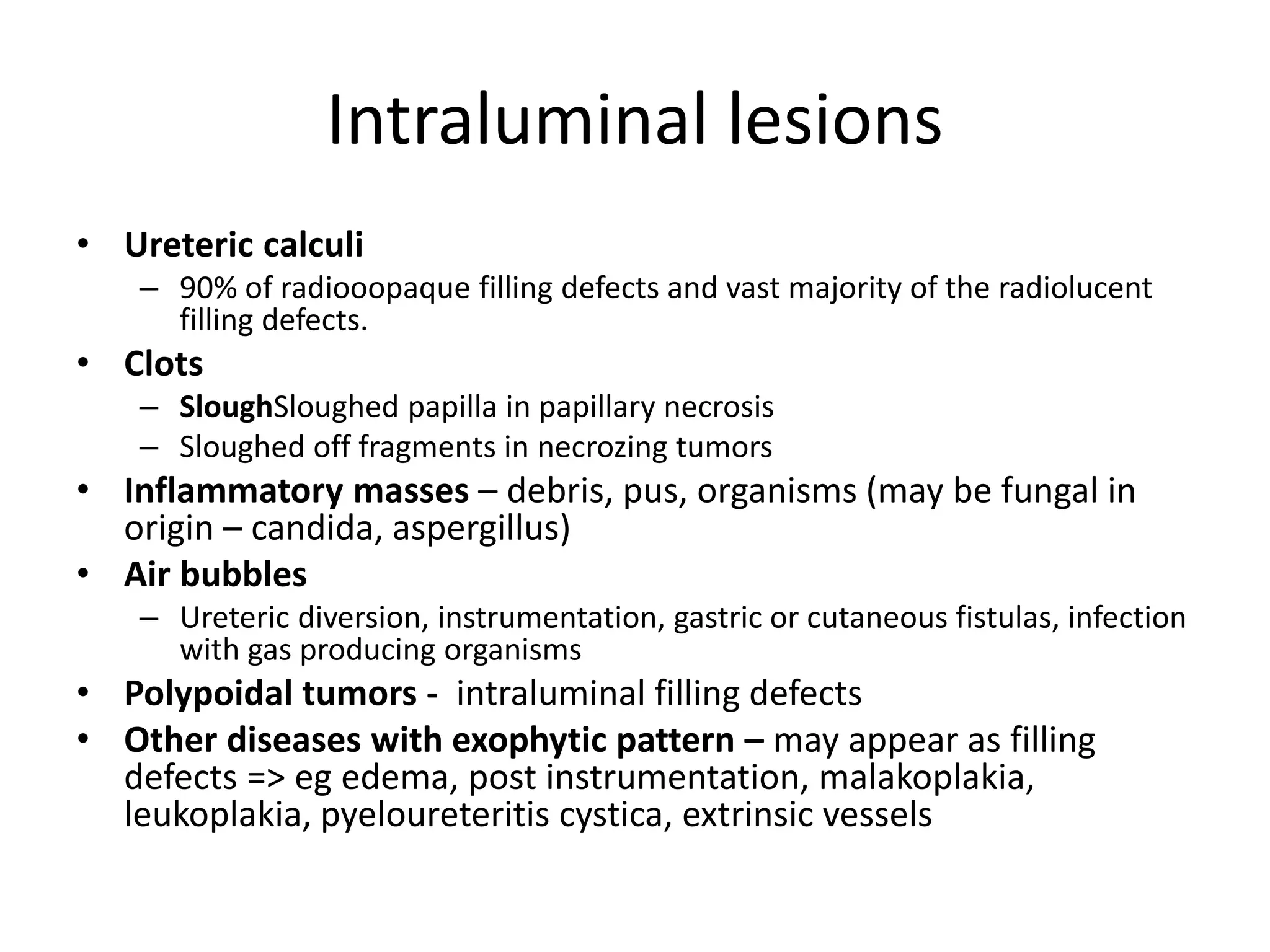
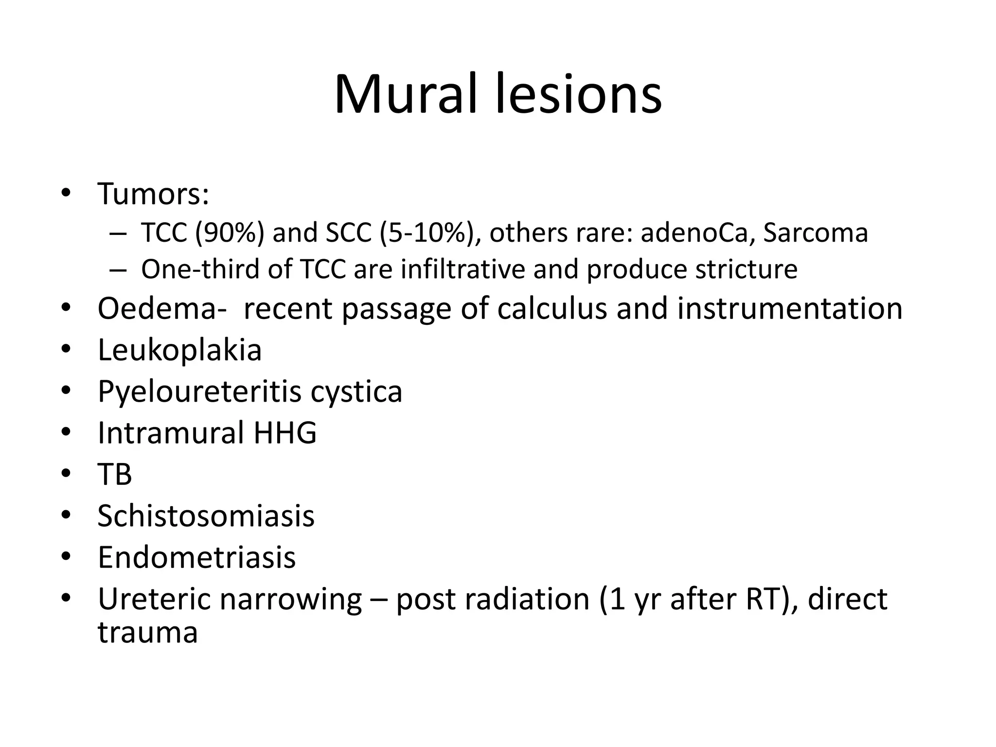
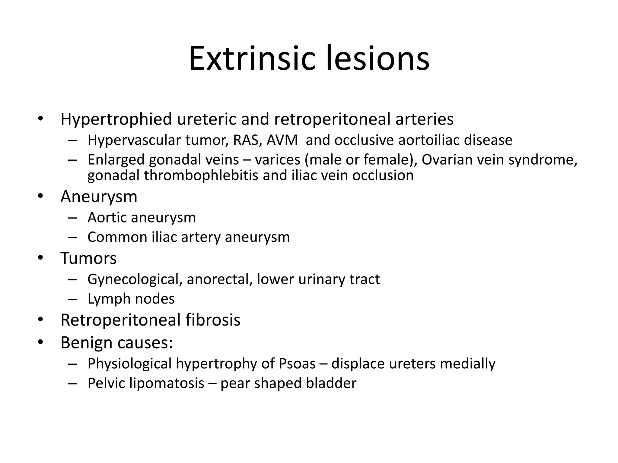


This document discusses lesions of the ureters categorized into intraluminal, mural, and extrinsic lesions. Intraluminal lesions include ureteric calculi, clots, inflammatory masses, air bubbles, and polypoidal tumors. Mural lesions include tumors like transitional cell carcinoma and squamous cell carcinoma, as well as edema, leukoplakia, and pyeloureteritis cystica. Extrinsic lesions compressing the ureters come from enlarged arteries, aneurysms, tumors, retroperitoneal fibrosis, and enlarged veins. Retroperitoneal fibrosis involves inflammatory fibrosis surrounding the ureters and aorta.


