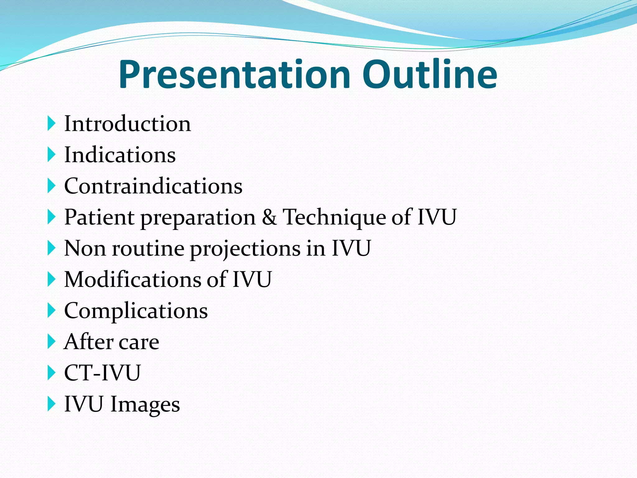
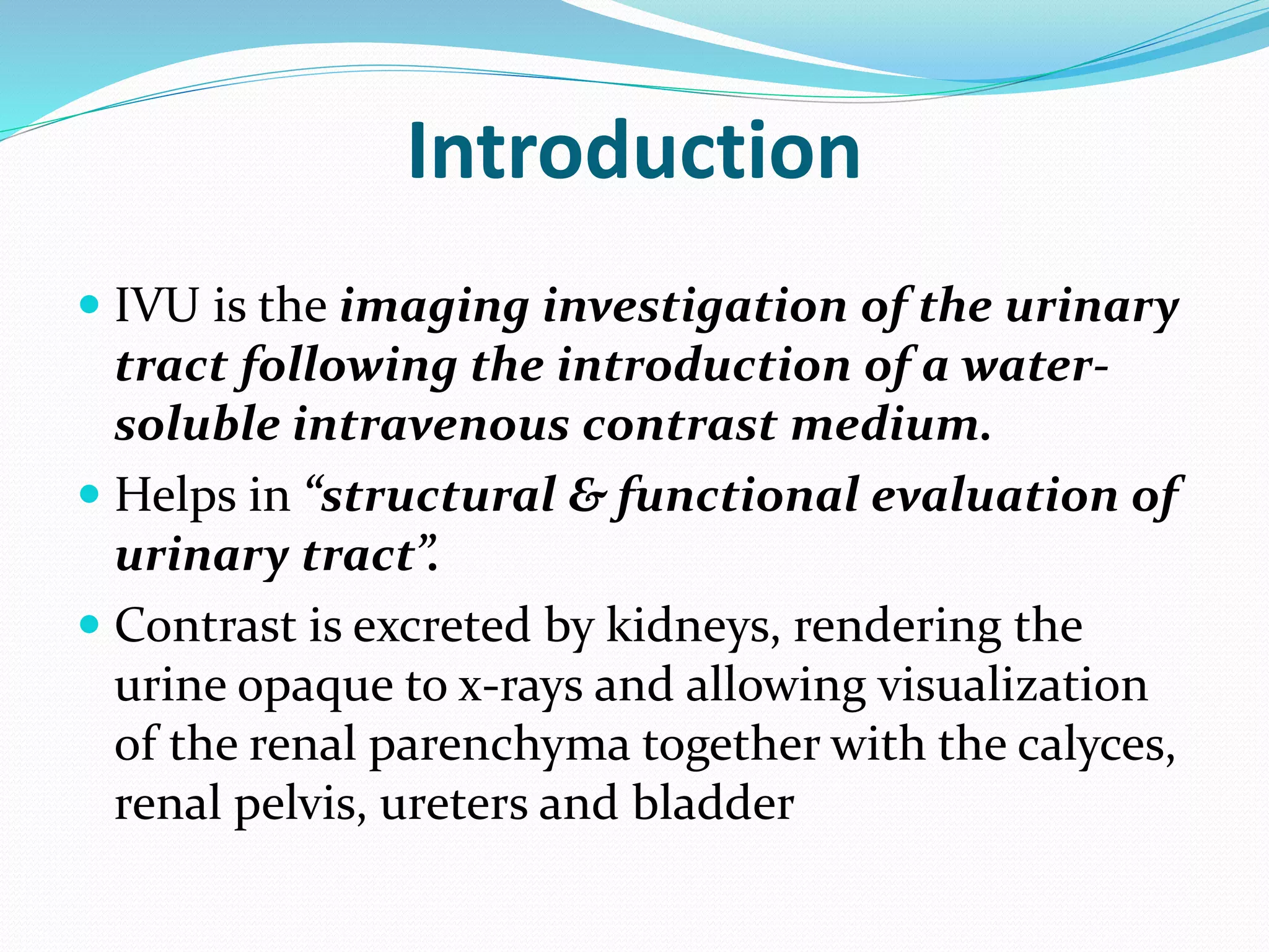
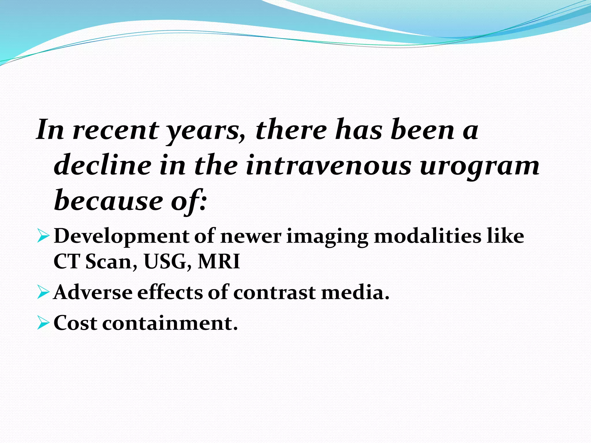
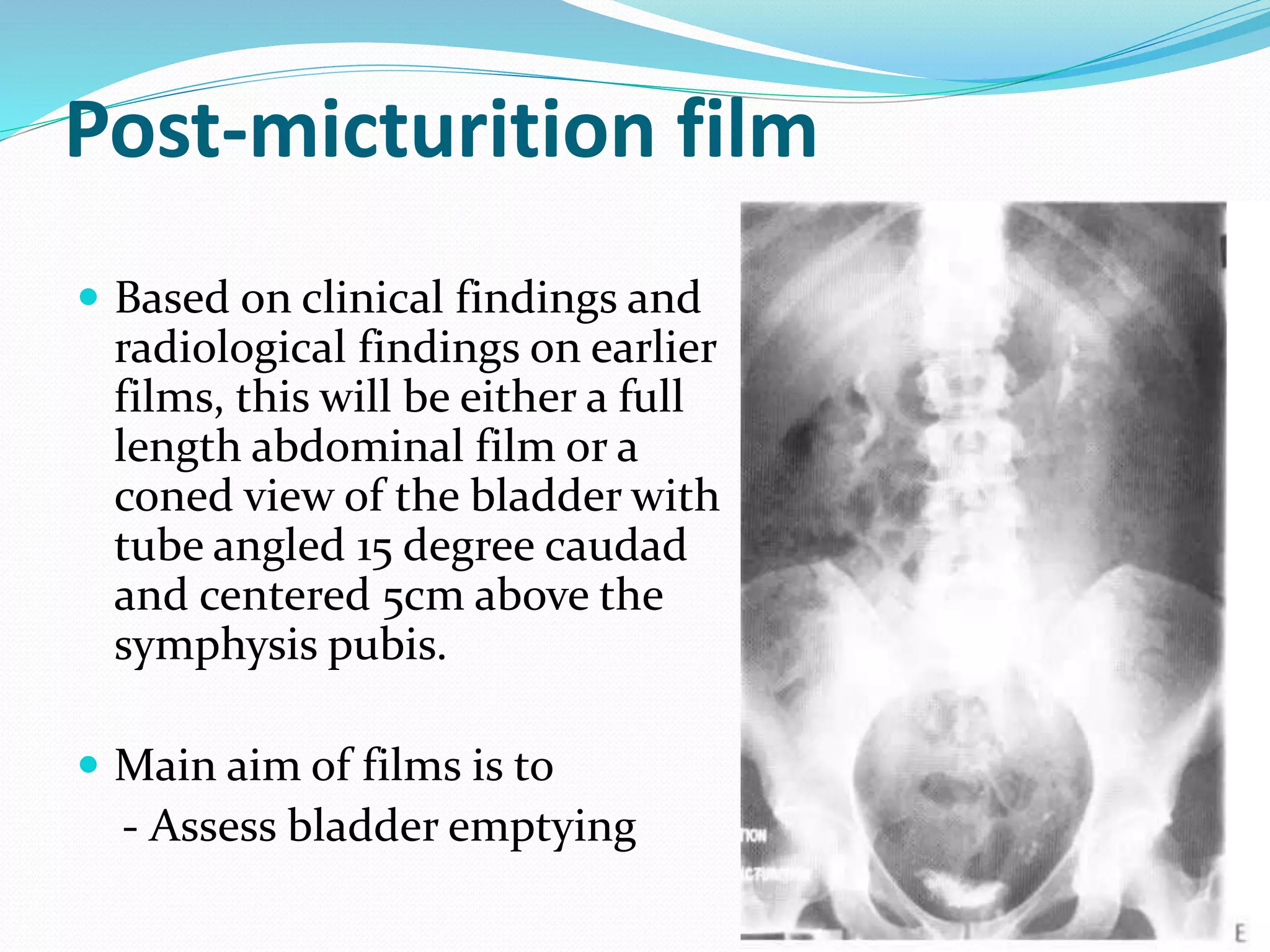
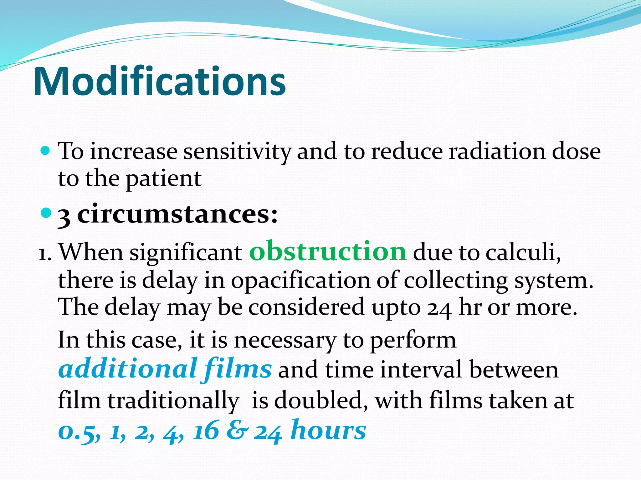
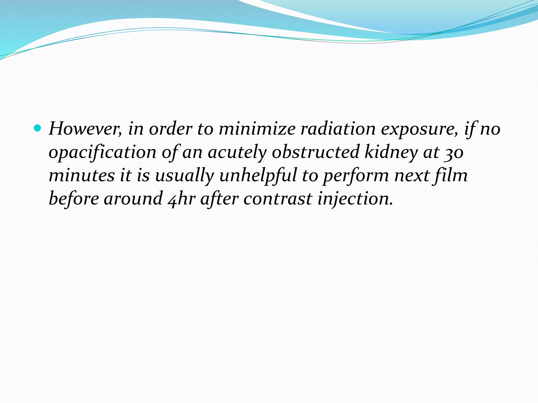
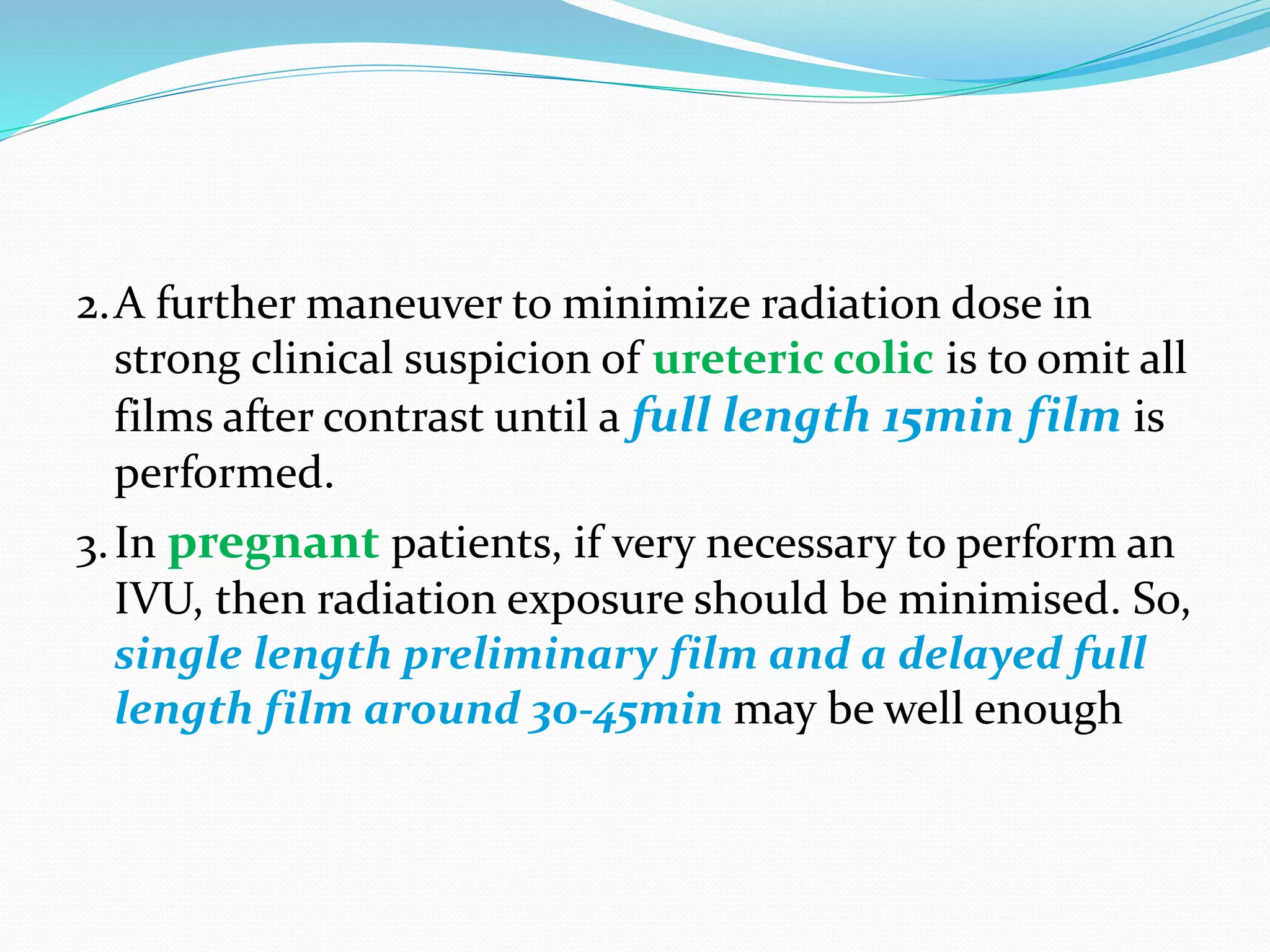
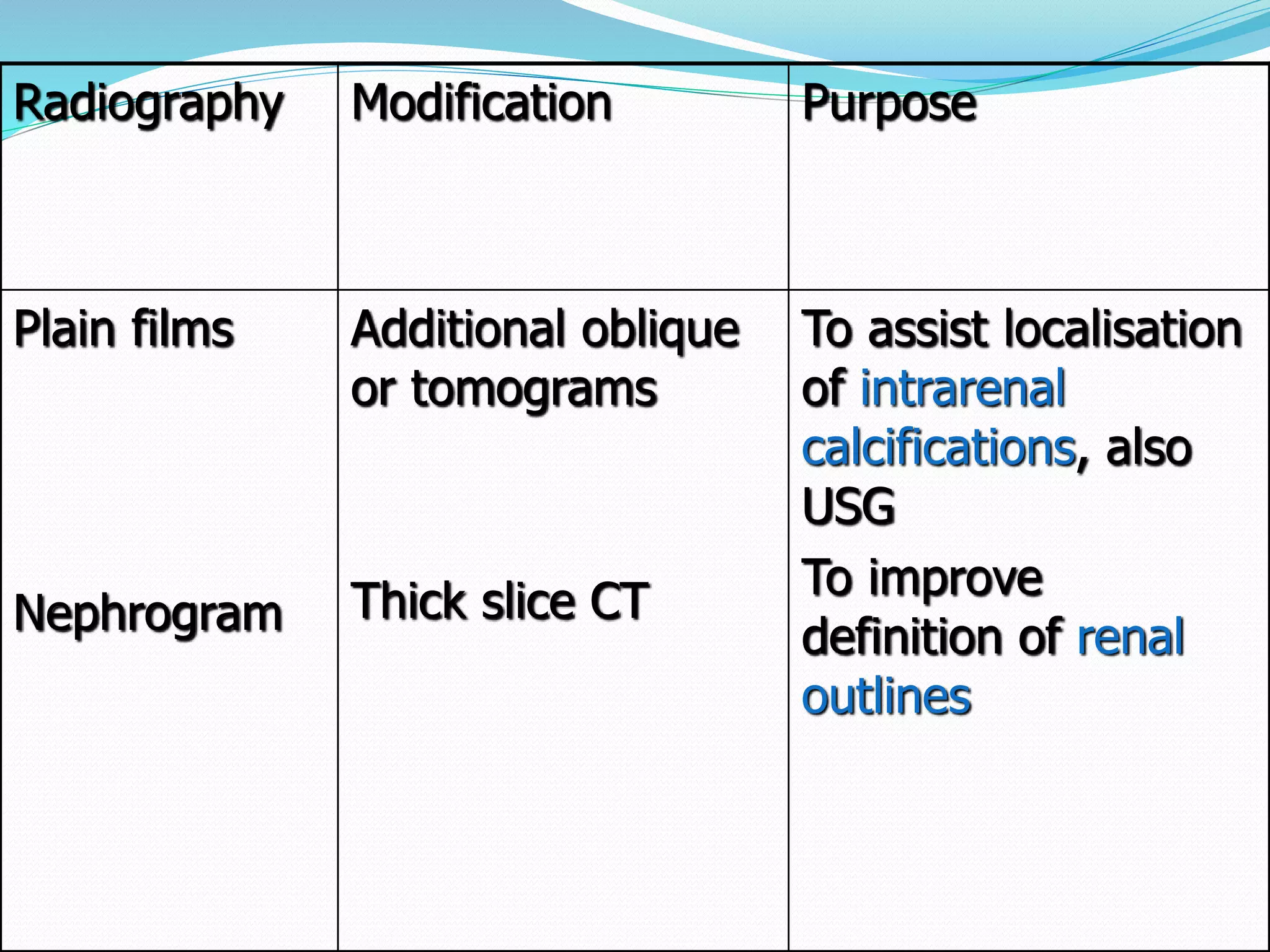
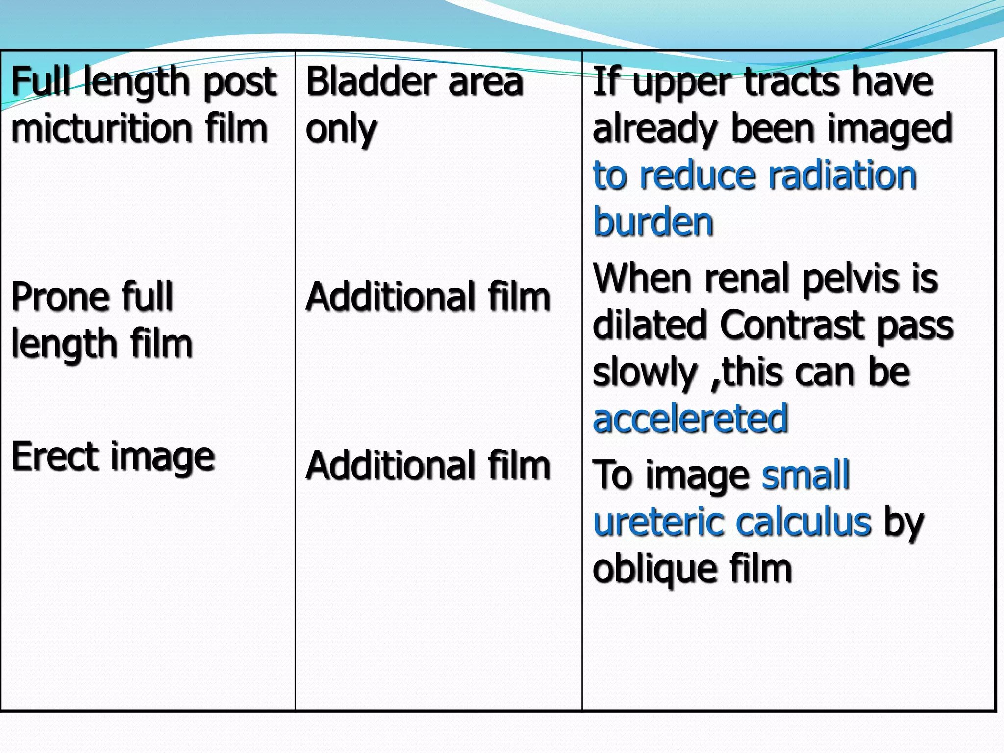
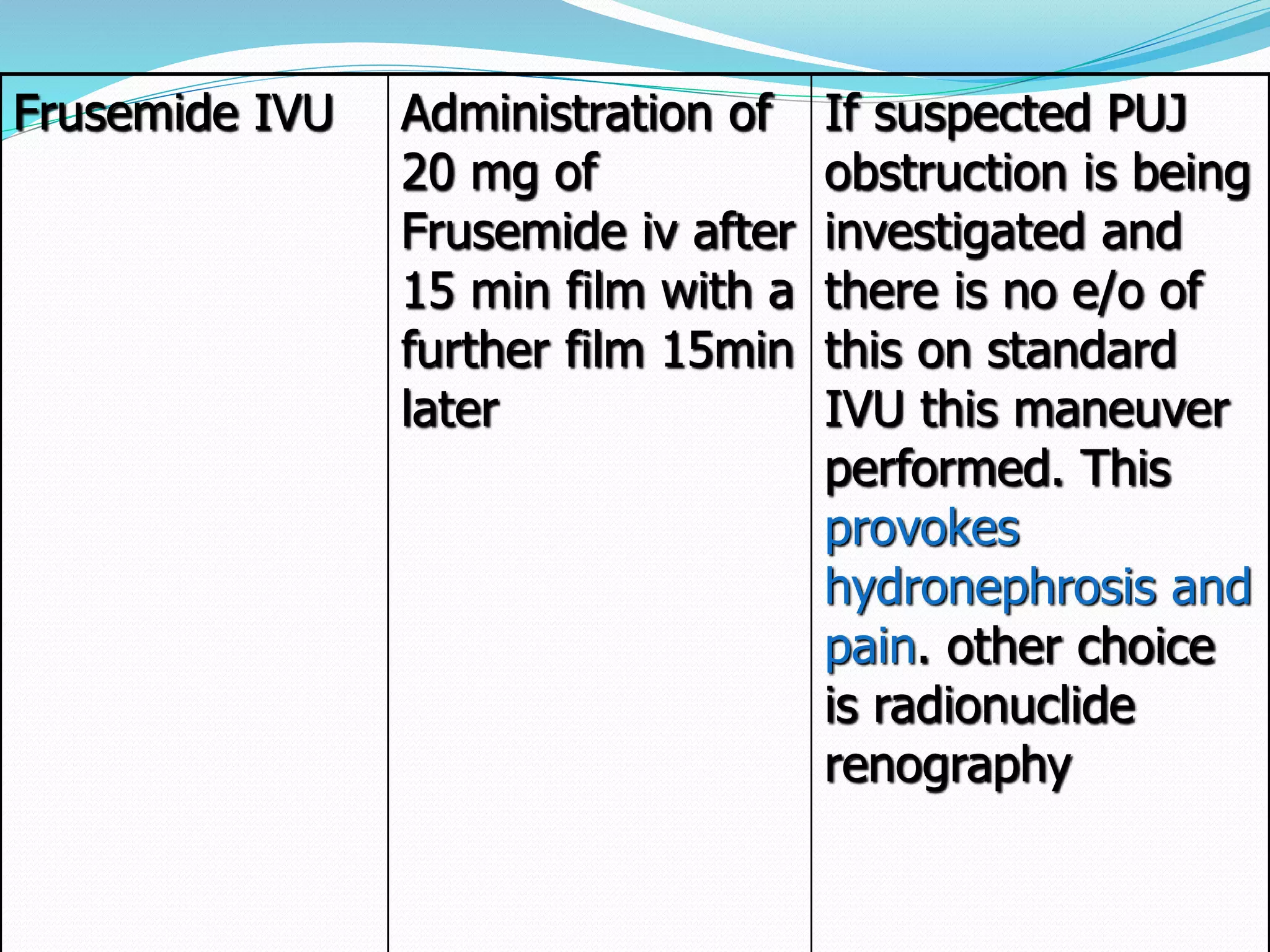
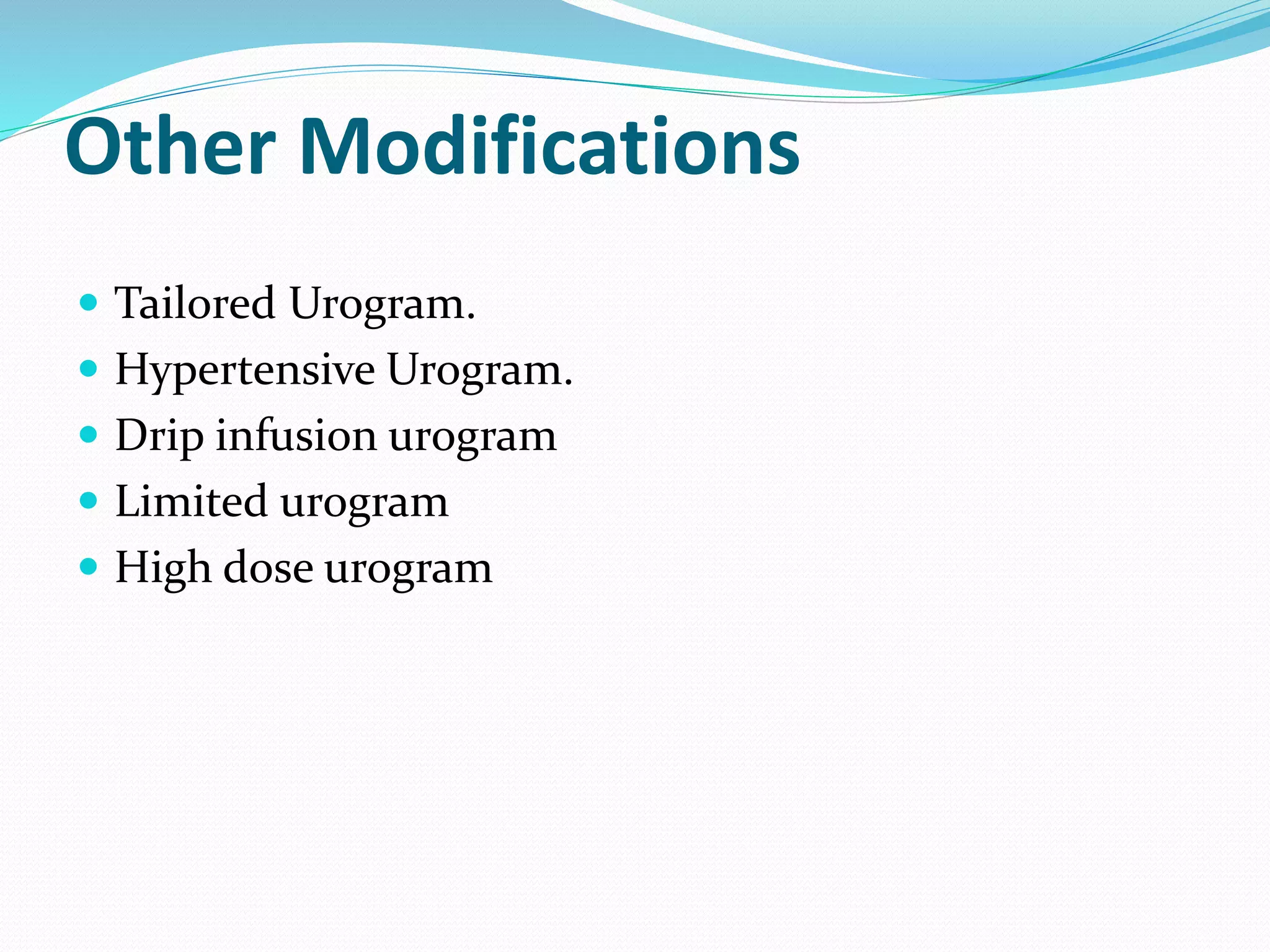
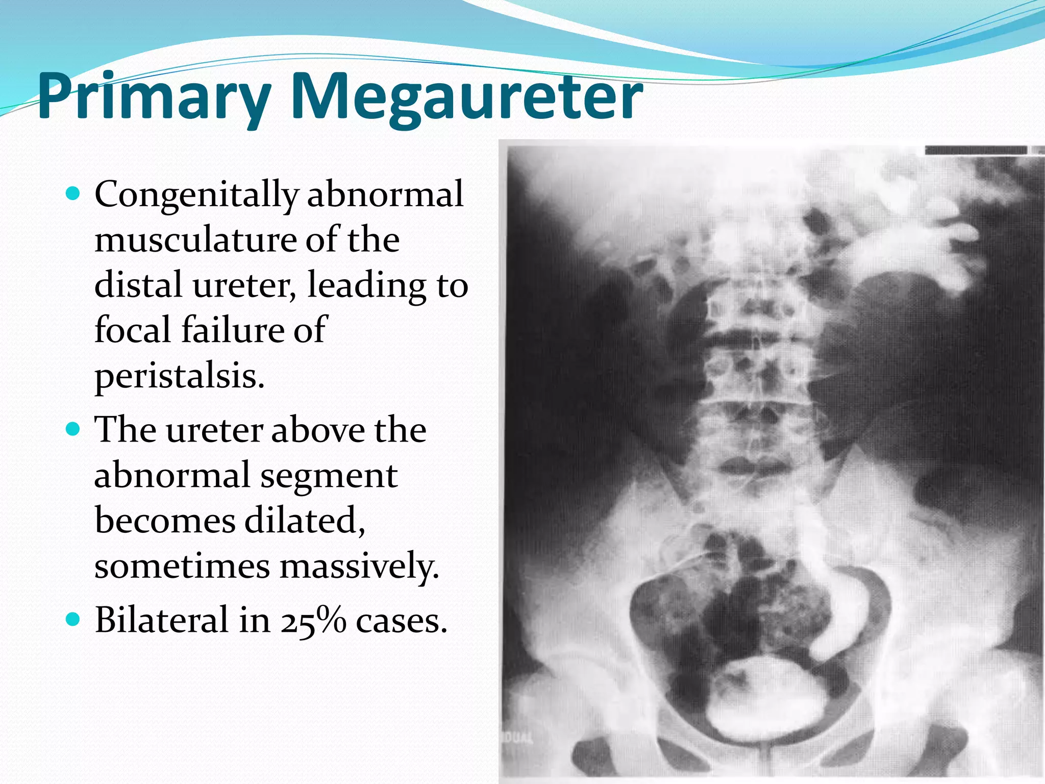
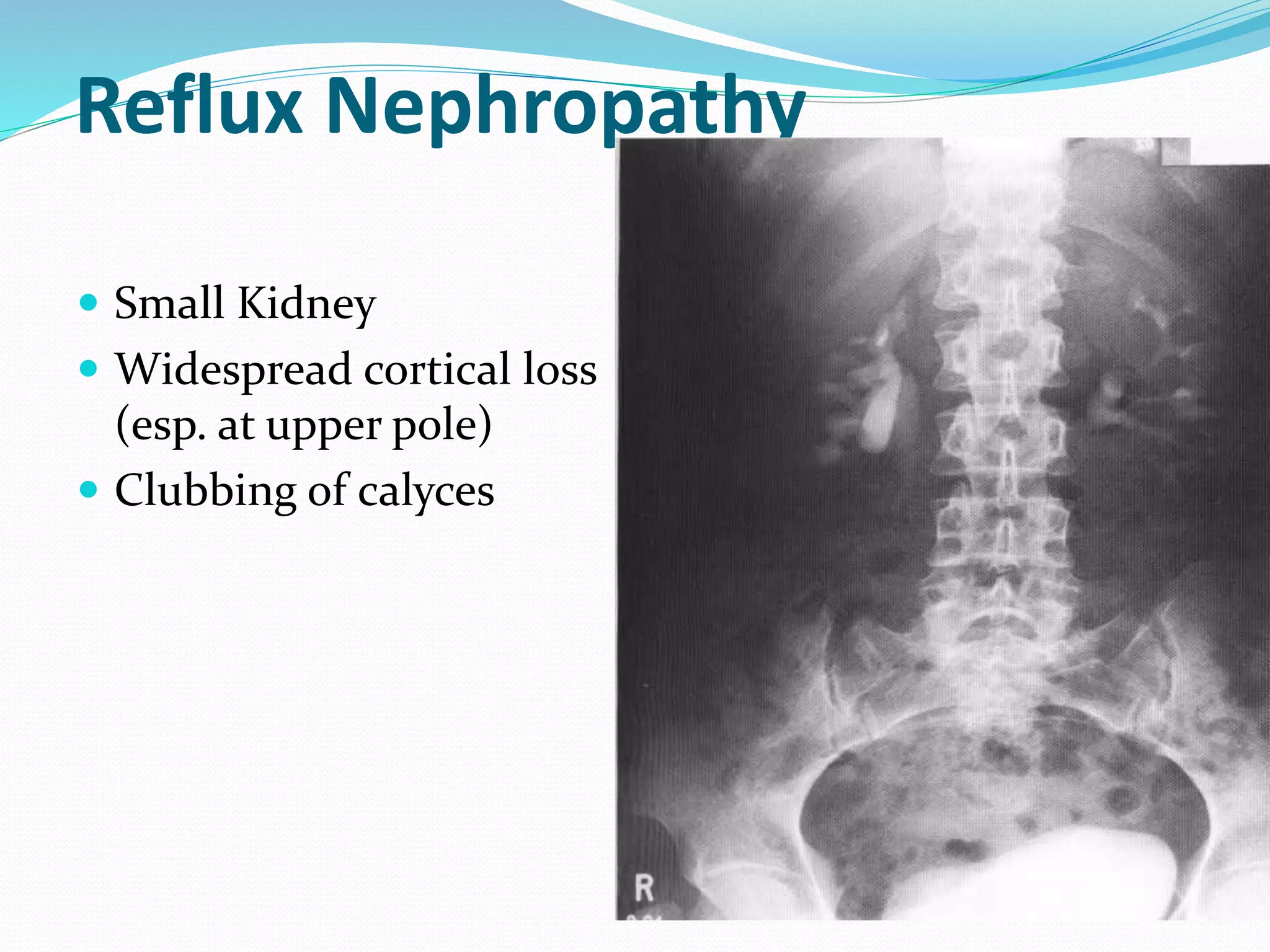
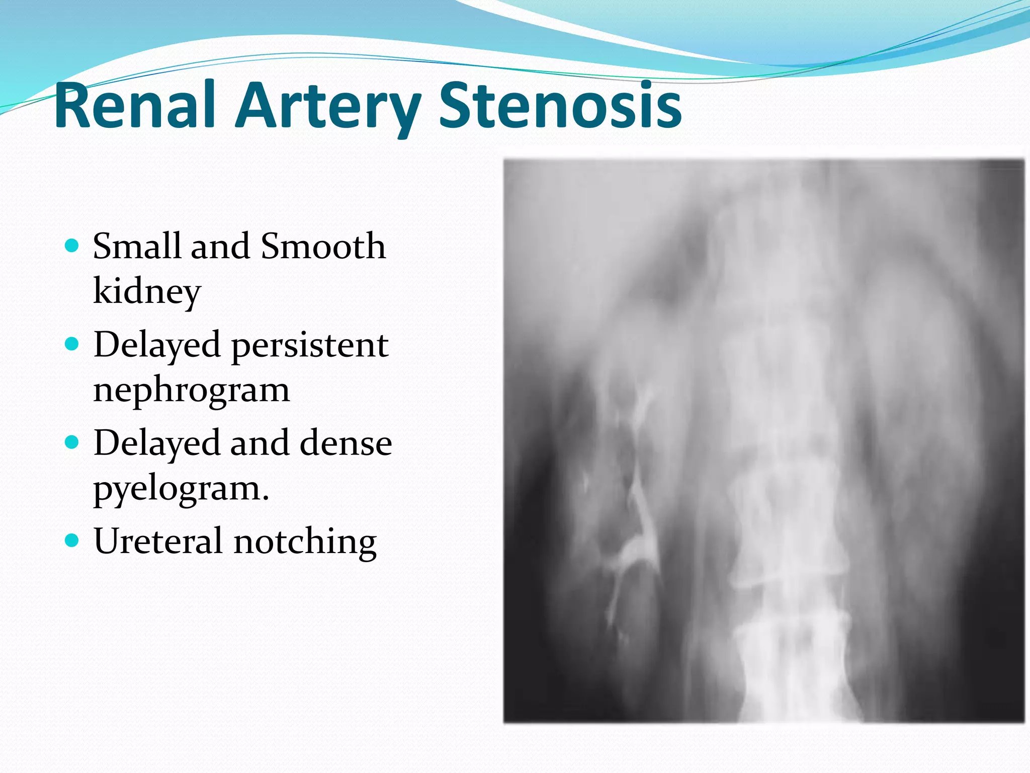
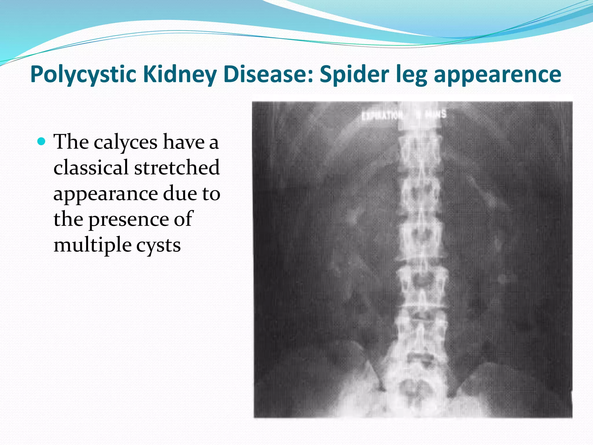
Ivu is a radiological investigation for visualization and assessment of the urinary tract.This presentation covers brief anatomy of urinary tract, indication and contraindication,contrast media dose and administration, routine and modified ivu procedure,its complication,ctivu and some abnormalities in the urinary tract.
Learn Barium Meal & Follow Through for the beginners from a Radiology Resident.For some image description please go through the text book "David Sutton" because i have described these image during my presentation Verbally..There are many animations used inside this presentation so to see all the pictures which are placed layer by layer with the help of animations you simple need to download this presentation first.... Thanx.
IVU is the radiographic examination of urinary tract including renal parenchyma, calyces and pelvis after intravenous injection of contrast media. Study was carried out at UCMS, Bhairawa, Nepal.
POSTERIOR URETHRAL VALVES- Pediatric Surgery
• Dear viewers,
• Greetings from “ Surgical Educator”
• Today I have uploaded one more video in Pediatric Surgery/Pediatric Urology- “ Posterior Urethral Valves”
• Posterior Urethral Valves is the congenital cause for Bladder Outlet Obstruction, resulting in abnormal development of the kidneys as well as the bladder.
• In this video, I talked about the learning outcomes, introduction, etiopathogenesis, clinical features, investigations, differential diagnosis, treatment, follow-up and prognosis of “ Posterior Urethral Valves”
• I hope you will enjoy the video for its educational value.
• You can watch all my teaching videos in the following links
• surgicaleducator.blogspot.com youtube.com/c/surgicaleducator
• Thank you for watching the video.
MANAGEMENT OF ATRIOVENTRICULAR CONDUCTION BLOCK.pdfJim Jacob Roy
Cardiac conduction defects can occur due to various causes.
Atrioventricular conduction blocks ( AV blocks ) are classified into 3 types.
This document describes the acute management of AV block.
Couples presenting to the infertility clinic- Do they really have infertility...Sujoy Dasgupta
Dr Sujoy Dasgupta presented the study on "Couples presenting to the infertility clinic- Do they really have infertility? – The unexplored stories of non-consummation" in the 13th Congress of the Asia Pacific Initiative on Reproduction (ASPIRE 2024) at Manila on 24 May, 2024.
New Directions in Targeted Therapeutic Approaches for Older Adults With Mantl...i3 Health
i3 Health is pleased to make the speaker slides from this activity available for use as a non-accredited self-study or teaching resource.
This slide deck presented by Dr. Kami Maddocks, Professor-Clinical in the Division of Hematology and
Associate Division Director for Ambulatory Operations
The Ohio State University Comprehensive Cancer Center, will provide insight into new directions in targeted therapeutic approaches for older adults with mantle cell lymphoma.
STATEMENT OF NEED
Mantle cell lymphoma (MCL) is a rare, aggressive B-cell non-Hodgkin lymphoma (NHL) accounting for 5% to 7% of all lymphomas. Its prognosis ranges from indolent disease that does not require treatment for years to very aggressive disease, which is associated with poor survival (Silkenstedt et al, 2021). Typically, MCL is diagnosed at advanced stage and in older patients who cannot tolerate intensive therapy (NCCN, 2022). Although recent advances have slightly increased remission rates, recurrence and relapse remain very common, leading to a median overall survival between 3 and 6 years (LLS, 2021). Though there are several effective options, progress is still needed towards establishing an accepted frontline approach for MCL (Castellino et al, 2022). Treatment selection and management of MCL are complicated by the heterogeneity of prognosis, advanced age and comorbidities of patients, and lack of an established standard approach for treatment, making it vital that clinicians be familiar with the latest research and advances in this area. In this activity chaired by Michael Wang, MD, Professor in the Department of Lymphoma & Myeloma at MD Anderson Cancer Center, expert faculty will discuss prognostic factors informing treatment, the promising results of recent trials in new therapeutic approaches, and the implications of treatment resistance in therapeutic selection for MCL.
Target Audience
Hematology/oncology fellows, attending faculty, and other health care professionals involved in the treatment of patients with mantle cell lymphoma (MCL).
Learning Objectives
1.) Identify clinical and biological prognostic factors that can guide treatment decision making for older adults with MCL
2.) Evaluate emerging data on targeted therapeutic approaches for treatment-naive and relapsed/refractory MCL and their applicability to older adults
3.) Assess mechanisms of resistance to targeted therapies for MCL and their implications for treatment selection
Title: Sense of Taste
Presenter: Dr. Faiza, Assistant Professor of Physiology
Qualifications:
MBBS (Best Graduate, AIMC Lahore)
FCPS Physiology
ICMT, CHPE, DHPE (STMU)
MPH (GC University, Faisalabad)
MBA (Virtual University of Pakistan)
Learning Objectives:
Describe the structure and function of taste buds.
Describe the relationship between the taste threshold and taste index of common substances.
Explain the chemical basis and signal transduction of taste perception for each type of primary taste sensation.
Recognize different abnormalities of taste perception and their causes.
Key Topics:
Significance of Taste Sensation:
Differentiation between pleasant and harmful food
Influence on behavior
Selection of food based on metabolic needs
Receptors of Taste:
Taste buds on the tongue
Influence of sense of smell, texture of food, and pain stimulation (e.g., by pepper)
Primary and Secondary Taste Sensations:
Primary taste sensations: Sweet, Sour, Salty, Bitter, Umami
Chemical basis and signal transduction mechanisms for each taste
Taste Threshold and Index:
Taste threshold values for Sweet (sucrose), Salty (NaCl), Sour (HCl), and Bitter (Quinine)
Taste index relationship: Inversely proportional to taste threshold
Taste Blindness:
Inability to taste certain substances, particularly thiourea compounds
Example: Phenylthiocarbamide
Structure and Function of Taste Buds:
Composition: Epithelial cells, Sustentacular/Supporting cells, Taste cells, Basal cells
Features: Taste pores, Taste hairs/microvilli, and Taste nerve fibers
Location of Taste Buds:
Found in papillae of the tongue (Fungiform, Circumvallate, Foliate)
Also present on the palate, tonsillar pillars, epiglottis, and proximal esophagus
Mechanism of Taste Stimulation:
Interaction of taste substances with receptors on microvilli
Signal transduction pathways for Umami, Sweet, Bitter, Sour, and Salty tastes
Taste Sensitivity and Adaptation:
Decrease in sensitivity with age
Rapid adaptation of taste sensation
Role of Saliva in Taste:
Dissolution of tastants to reach receptors
Washing away the stimulus
Taste Preferences and Aversions:
Mechanisms behind taste preference and aversion
Influence of receptors and neural pathways
Impact of Sensory Nerve Damage:
Degeneration of taste buds if the sensory nerve fiber is cut
Abnormalities of Taste Detection:
Conditions: Ageusia, Hypogeusia, Dysgeusia (parageusia)
Causes: Nerve damage, neurological disorders, infections, poor oral hygiene, adverse drug effects, deficiencies, aging, tobacco use, altered neurotransmitter levels
Neurotransmitters and Taste Threshold:
Effects of serotonin (5-HT) and norepinephrine (NE) on taste sensitivity
Supertasters:
25% of the population with heightened sensitivity to taste, especially bitterness
Increased number of fungiform papillae
Prix Galien International 2024 Forum ProgramLevi Shapiro
June 20, 2024, Prix Galien International and Jerusalem Ethics Forum in ROME. Detailed agenda including panels:
- ADVANCES IN CARDIOLOGY: A NEW PARADIGM IS COMING
- WOMEN’S HEALTH: FERTILITY PRESERVATION
- WHAT’S NEW IN THE TREATMENT OF INFECTIOUS,
ONCOLOGICAL AND INFLAMMATORY SKIN DISEASES?
- ARTIFICIAL INTELLIGENCE AND ETHICS
- GENE THERAPY
- BEYOND BORDERS: GLOBAL INITIATIVES FOR DEMOCRATIZING LIFE SCIENCE TECHNOLOGIES AND PROMOTING ACCESS TO HEALTHCARE
- ETHICAL CHALLENGES IN LIFE SCIENCES
- Prix Galien International Awards Ceremony
Factory Supply Best Quality Pmk Oil CAS 28578–16–7 PMK Powder in Stockrebeccabio
Factory Supply Best Quality Pmk Oil CAS 28578–16–7 PMK Powder in Stock
Telegram: bmksupplier
signal: +85264872720
threema: TUD4A6YC
You can contact me on Telegram or Threema
Communicate promptly and reply
Free of customs clearance, Double Clearance 100% pass delivery to USA, Canada, Spain, Germany, Netherland, Poland, Italy, Sweden, UK, Czech Republic, Australia, Mexico, Russia, Ukraine, Kazakhstan.Door to door service
Hot Selling Organic intermediates
Ethanol (CH3CH2OH), or beverage alcohol, is a two-carbon alcohol
that is rapidly distributed in the body and brain. Ethanol alters many
neurochemical systems and has rewarding and addictive properties. It
is the oldest recreational drug and likely contributes to more morbidity,
mortality, and public health costs than all illicit drugs combined. The
5th edition of the Diagnostic and Statistical Manual of Mental Disorders
(DSM-5) integrates alcohol abuse and alcohol dependence into a single
disorder called alcohol use disorder (AUD), with mild, moderate,
and severe subclassifications (American Psychiatric Association, 2013).
In the DSM-5, all types of substance abuse and dependence have been
combined into a single substance use disorder (SUD) on a continuum
from mild to severe. A diagnosis of AUD requires that at least two of
the 11 DSM-5 behaviors be present within a 12-month period (mild
AUD: 2–3 criteria; moderate AUD: 4–5 criteria; severe AUD: 6–11 criteria).
The four main behavioral effects of AUD are impaired control over
drinking, negative social consequences, risky use, and altered physiological
effects (tolerance, withdrawal). This chapter presents an overview
of the prevalence and harmful consequences of AUD in the U.S.,
the systemic nature of the disease, neurocircuitry and stages of AUD,
comorbidities, fetal alcohol spectrum disorders, genetic risk factors, and
pharmacotherapies for AUD.
Tom Selleck Health: A Comprehensive Look at the Iconic Actor’s Wellness Journeygreendigital
Tom Selleck, an enduring figure in Hollywood. has captivated audiences for decades with his rugged charm, iconic moustache. and memorable roles in television and film. From his breakout role as Thomas Magnum in Magnum P.I. to his current portrayal of Frank Reagan in Blue Bloods. Selleck's career has spanned over 50 years. But beyond his professional achievements. fans have often been curious about Tom Selleck Health. especially as he has aged in the public eye.
Follow us on: Pinterest
Introduction
Many have been interested in Tom Selleck health. not only because of his enduring presence on screen but also because of the challenges. and lifestyle choices he has faced and made over the years. This article delves into the various aspects of Tom Selleck health. exploring his fitness regimen, diet, mental health. and the challenges he has encountered as he ages. We'll look at how he maintains his well-being. the health issues he has faced, and his approach to ageing .
Early Life and Career
Childhood and Athletic Beginnings
Tom Selleck was born on January 29, 1945, in Detroit, Michigan, and grew up in Sherman Oaks, California. From an early age, he was involved in sports, particularly basketball. which played a significant role in his physical development. His athletic pursuits continued into college. where he attended the University of Southern California (USC) on a basketball scholarship. This early involvement in sports laid a strong foundation for his physical health and disciplined lifestyle.
Transition to Acting
Selleck's transition from an athlete to an actor came with its physical demands. His first significant role in "Magnum P.I." required him to perform various stunts and maintain a fit appearance. This role, which he played from 1980 to 1988. necessitated a rigorous fitness routine to meet the show's demands. setting the stage for his long-term commitment to health and wellness.
Fitness Regimen
Workout Routine
Tom Selleck health and fitness regimen has evolved. adapting to his changing roles and age. During his "Magnum, P.I." days. Selleck's workouts were intense and focused on building and maintaining muscle mass. His routine included weightlifting, cardiovascular exercises. and specific training for the stunts he performed on the show.
Selleck adjusted his fitness routine as he aged to suit his body's needs. Today, his workouts focus on maintaining flexibility, strength, and cardiovascular health. He incorporates low-impact exercises such as swimming, walking, and light weightlifting. This balanced approach helps him stay fit without putting undue strain on his joints and muscles.
Importance of Flexibility and Mobility
In recent years, Selleck has emphasized the importance of flexibility and mobility in his fitness regimen. Understanding the natural decline in muscle mass and joint flexibility with age. he includes stretching and yoga in his routine. These practices help prevent injuries, improve posture, and maintain mobilit
Explore natural remedies for syphilis treatment in Singapore. Discover alternative therapies, herbal remedies, and lifestyle changes that may complement conventional treatments. Learn about holistic approaches to managing syphilis symptoms and supporting overall health.
TEST BANK for Operations Management, 14th Edition by William J. Stevenson, Ve...kevinkariuki227
TEST BANK for Operations Management, 14th Edition by William J. Stevenson, Verified Chapters 1 - 19, Complete Newest Version.pdf
Pulmonary Thromboembolism - etilogy, types, medical- Surgical and nursing man...VarunMahajani
Disruption of blood supply to lung alveoli due to blockage of one or more pulmonary blood vessels is called as Pulmonary thromboembolism. In this presentation we will discuss its causes, types and its management in depth.
Flu Vaccine Alert in Bangalore Karnatakaaddon Scans
As flu season approaches, health officials in Bangalore, Karnataka, are urging residents to get their flu vaccinations. The seasonal flu, while common, can lead to severe health complications, particularly for vulnerable populations such as young children, the elderly, and those with underlying health conditions.
Dr. Vidisha Kumari, a leading epidemiologist in Bangalore, emphasizes the importance of getting vaccinated. "The flu vaccine is our best defense against the influenza virus. It not only protects individuals but also helps prevent the spread of the virus in our communities," he says.
This year, the flu season is expected to coincide with a potential increase in other respiratory illnesses. The Karnataka Health Department has launched an awareness campaign highlighting the significance of flu vaccinations. They have set up multiple vaccination centers across Bangalore, making it convenient for residents to receive their shots.
To encourage widespread vaccination, the government is also collaborating with local schools, workplaces, and community centers to facilitate vaccination drives. Special attention is being given to ensuring that the vaccine is accessible to all, including marginalized communities who may have limited access to healthcare.
Residents are reminded that the flu vaccine is safe and effective. Common side effects are mild and may include soreness at the injection site, mild fever, or muscle aches. These side effects are generally short-lived and far less severe than the flu itself.
Healthcare providers are also stressing the importance of continuing COVID-19 precautions. Wearing masks, practicing good hand hygiene, and maintaining social distancing are still crucial, especially in crowded places.
Protect yourself and your loved ones by getting vaccinated. Together, we can help keep Bangalore healthy and safe this flu season. For more information on vaccination centers and schedules, residents can visit the Karnataka Health Department’s official website or follow their social media pages.
Stay informed, stay safe, and get your flu shot today!
2. Presentation Outline
Introduction
Indications
Contraindications
Patient preparation & Technique of IVU
Non routine projections in IVU
Modifications of IVU
Complications
After care
CT-IVU
IVU Images
3. Introduction
IVU is the imaging investigation of the urinary
tract following the introduction of a water-
soluble intravenous contrast medium.
Helps in “structural & functional evaluation of
urinary tract”.
Contrast is excreted by kidneys, rendering the
urine opaque to x-rays and allowing visualization
of the renal parenchyma together with the calyces,
renal pelvis, ureters and bladder
4. In recent years, there has been a
decline in the intravenous urogram
because of:
Development of newer imaging modalities like
CT Scan, USG, MRI
Adverse effects of contrast media.
Cost containment.
5. Indications
In Adults
Suspected urinary tract pathology
Investigation of persistent or frank hematuria
Renal /ureteric calculi (prior to endourological
procedure)
Complex urinary tract infection (including Renal TB)
Ureteric fistulas and strictures
Suspected transitional cell carcinoma
6. In Children:
Evaluation of VATER anomalies- 90% has Renal
anomalies.
Malformation of genitalia –hypospadiasis
Enuresis
Constant or intermittent dampness in girls to rule out
ectopically inserted ureter.
7. Contraindications
Absolute CIs:
Past h/o severe adverse reaction to contrast media.
HOCM carries 20% risk and LOCM decrease risk to
5% , and in those cases radioisotope scan , USG, CT,
MRI provide alternative means of investigations
Proven hypersensitivity to iodine.
Relative CIs: (@ABCD MS)
Asthma /significant allergic history.
B-blockers
8. Chronic Renal insufficiency
Cardiac disease –Cardiac failure /arrhythmias may be
precipitated and in these cases lower risk with LOCM
Diabetes Mellitus
Dehydration
Multiple Myeloma
Metformin therapy: Co-administration of metformin
(glucophage)+ iv contrast to diabetics may lead to
acute alteration of renal function and lactic acidosis,
therefore metformin is withheld
Sickle cell anemia
9. Thyrotoxicosis
Pregnancy
A contrast material is excreted by a similar
mechanism to creatinine, a serum creatinine level
above 200micromol/l would indicate a patient who
would unlikely to excrete contrast satisfactorily.
So, cautions in diabetics and patients with severe
disturbances of liver and kidneys.
10. Contrast medium and injection data
Ionic and non-ionic are available, both of which are
excreted by different mechanisms. The ionic group is
excreted mainly by glomerular filtration causing a
peak concentration of iodine in the renal cortex faster
compared to nonionic which is mainly excreted by
proximal tubules
The timing for first radiograph to demonstrate
parenchymal phase best will thus differ.
11. HOCM or LOCM 370 are acceptable but the following
“high risk” group should receive LOCM.
Infants/small children/elderly.
Poorly hydrated patients
Those with renal /cardiac failure
Patients with diabetes, myelomatosis, sickle cell
disease
Patients with previous contrast medium reactions/
strong allergic history
12. Contrast agent: Ultravist ( Iopramide)
LOCM: 300-600mg Iodine meq/kg body weight
Standard Dose:
Adult Dose : 50-100 ml
Pediatric dose: 1 ml/kg
13. Patient Preparation
Bowel preparation is important as abdomen
should ideally be free of radio-opaque fecal matter
and gas
NPO (No food for 4-6 hr prior to examination)
Laxatives- Dulcolax 2-4 tabs at bed time for 2
days prior to procedure.
Bowel preparation is now generally regarded as
unhelpful and it is unpleasant to the patient.
14. Is fluid deprivation indicated?
Traditionally fluid was restricted prior to IVU in order
to improve opacification of collecting system.
However, dehydration increase risk of nephrotoxicity
which may be permanent in patients with DM,
Multiple Myeloma, Hyperuricemia, Sickle Cell Disease
and pre-existing renal disease.
15. Risk of irreversible renal damage to renal function in
previously healthy kidney due to contrast agent is very
low
Also, with the advent of modern non-ionic contrast
agents which do not provoke an osmotic diuresis,
degree of opacification is unlikely to be significantly
altered by dehydration.
So, fluid restriction should be avoided and if there
is a risk that the patient is dehydrated before the IVU,
this should be corrected first.
16. Radiation protection
“Pregnancy rule” should be applied.
If whole of renal tract is to be visualized, no gonad
shielding is possible for the females, but for males
the testis can be protected by placing a lead rubber
sheet over upper thighs below lower edge of
symphysis pubis.
When bladder and lower ureters are not included
then female can also be given gonad protection.
17. Technique
Informed consent
Median ante-cubital vein-preferred injection site.
19 G needle is advanced upto the vein and kept there
during entire procedure duration.
IV cannula in place
–provides ER treatment if required
-for further injection of contrast if opacification is
inadequate
18. Most adverse reactions are likely to occur within few
minutes after injection. So, Emergency drugs (eg.
Adrenaline), Oxygen and Resuscitation equipments
should also be readily available.
Doctor (radiologist) should be available in the
department.
19. Classic series of plain films
Preliminary post void full length film (control film).
Immediate film (Nephrogram)
5-min film
15-min compression film
15-min release film
Post-micturition film
20. Preliminary/Control film
Plain film is to demonstrate the urinary tract prior to
administration of contrast medium
kVp= 70-80 (low kVp), mAs= 60-70
Centering: the vertical central ray is directed to the
centre of the cassette
Supine full length AP view of the abdomen in
inspiration.
Pelvis should be adjusted so that the anterior
superior iliac spines are equidistant from table
top.
Lower border of cassette is at level of symphysis
pubis.
21.
22. Why to take preliminary/control film?
To check exposure factors, centering
State of bowel preparation
Obvious pre-existing pathology, particularly urinary
tract calculi/calcification.
23. Calcification on the KUB
In the Urinary tract
Renal: calculi, renal cell carcinoma, tuberculosis,
arterial (atheroma or aneurysm)
Ureter: calculi, tuberculosis, schistosomiasis
Bladder: calculi, tuberculosis, schistosomiasis,
transitional cell carcinoma
25. Immediate film (Nephrogram)
AP film of renal areas.
This film is exposed 10-14
seconds after contrast
injection (arm to kidney
time)
Renal parenchyma is
opacified by contrast
medium in the renal tubules.
Aim is to see Renal
outlines
26. Normal size: 9-13cm
cephalocaudally, left
is 0.5-1 cm larger
than right.
Normal kidney size
should not more
than 3 times the sum
of the height of L1
vertebra and height
of L1-L2
intervertebral disc.
Measurement of Kidney
27. Right kidney is more than 1.5cm larger than left kidney
Left kidney is more than 2cm larger than right kidney
Significant Discrepancies in size if
28. Average thickness 3-
3.5cm in polar region
and 2-2.5cm in
interpolar region
Decrease in
parenchymal thickness
seen in post
inflammatory or stone
related scarring.
Increase in parenchymal
thickness is seen in
renal mass.
Measurement of Parenchymal thickness
Interpapillary line
29. 5-min film
AP of Renal areas
Film is taken to
determine if excretion is
symmetrical and for
assessing if need to
modify technique e.g- a
further injection of
contrast medium if poor
opaification.
To see Pelvicalyceal
system
30. Compression band is now applied around the
patients abdomen and balloon is positioned
midway between the anterior superior iliac spine
i.e precisely over the ureters as they crosses pelvic
brim.
31. Why compression technique?
Compression inhibits ureteric drainage and
promote distension of pelvicalyceal system,
optimising their visualization
33. Contraindications of compression
Recent abdominal surgery
Abdominal Aortic Aneurysms
Acute painful abdomen/ renal colic
Large abdominal mass
Urinary tract trauma
Presence of Urinary diversion
Presence of Renal transplant
When 5-min film shows already distended calyces.
34. 15-min compression film
AP view of renal areas
There is usually adequate
distension of pelvicalyceal
system with opaque urine.
Compression removed
when satisfactory
demonstration of
pelvicalyceal system has
been achieved.
35.
36. 15-min Release film
Supine AP film
This film is taken to
show whole urinary
tract.
37.
38. Post-micturition film
Based on clinical findings and
radiological findings on earlier
films, this will be either a full
length abdominal film or a
coned view of the bladder with
tube angled 15 degree caudad
and centered 5cm above the
symphysis pubis.
Main aim of films is to
- Assess bladder emptying
39. To demonstrate return of dilated upper tracts with
relief of bladder pressure.
Aid diagnosis of VUJ calculi
Diagnosis of bladder tumors
Demonstrate urethral diverticulum.
40. Non-routine projections
Postero-anterior (prone)-abdomen
Projection is to promote emptying of contrast from the
pelvicalyceal system into the ureter.
Right or left posterior oblique
This is to show the relationship of the opacities to the
kidneys, ureters, and bladder.
41. Lateral Projection
May be used as an alternative to oblique projection in
relative position of the opacities near to or in the
kidneys.
Opacities in the kidneys will overshadow, or be very
near the vertebrae. Opacities outside the kidneys are
usually shown anterior to the vertebrae
42. Stereotypical appearances of normal
IVU are as follows
Takes 12-20 seconds for contrast to reach renal
arteries following iv injection
At this stage, its concentration is maximum in the
vascular compartment.
However, this falls rapidly as contrast medium begins
to escape into extracellular compartment and
undergoes rapid glomerular filtration and enters the
renal tubules
43. In first minute of IVU, healthy kidneys (assuming a
normal cardiovascular system) show diffuse
enhancement. This is referred as Nephrogram.
During this phase renal size (normally at least 3
vertebrae in length but no more than four) and outline
are seen.
44. In roughly 1st half minute – contrast in the
vascular compartment dominates and therefore
cortex is more enhanced than the medulla
This differentiation is sometimes visible in
immediate film of IVU series (but regularly visible
on CT performed at this stage)
In second half of minute - contrast in the
tubules increases and enhancement of kidneys is
more diffuse
45. At 1 minute: Contrast begins to appear in
calyces
After 1 minute: Contrast in the normal calyces
will begin to drain immediately into the pelvis and
ureter and this phased referred as Pyelogram
After compression is released ,there is transient
increase in flow down the ureters and release film
offers the best chance of demonstrating the
ureters.
Normal ureters exhibit peristalsis and on a single
film it is uncommon to demonstrate entire length
of both (or even either ) ureters.
46. Modifications
To increase sensitivity and to reduce radiation dose
to the patient
3 circumstances:
1. When significant obstruction due to calculi,
there is delay in opacification of collecting system.
The delay may be considered upto 24 hr or more.
In this case, it is necessary to perform
additional films and time interval between
film traditionally is doubled, with films taken at
0.5, 1, 2, 4, 16 & 24 hours
47. However, in order to minimize radiation exposure, if no
opacification of an acutely obstructed kidney at 30
minutes it is usually unhelpful to perform next film
before around 4hr after contrast injection.
48. 2.A further maneuver to minimize radiation dose in
strong clinical suspicion of ureteric colic is to omit all
films after contrast until a full length 15min film is
performed.
3.In pregnant patients, if very necessary to perform an
IVU, then radiation exposure should be minimised. So,
single length preliminary film and a delayed full
length film around 30-45min may be well enough
50. Radiography Modification Purpose
Plain films
Nephrogram
Additional oblique
or tomograms
Thick slice CT
To assist localisation
of intrarenal
calcifications, also
USG
To improve
definition of renal
outlines
51. 5min film
15 min
compression
film
15 min
release film
2nd injection
of contrast
Series of 1cm
thick
tomograms
Additional
bladder views
To improve opacification
of PCS.
To diff betn overlying
shadows and filling
defects within collecting
systems
When bladder poorly filled
in release film
When irregular filling
defects/calculus in distal
ureter seen oblique films
to be taken.
USG can be done to
reduce radiation dose
52. Full length post
micturition film
Prone full
length film
Erect image
Bladder area
only
Additional film
Additional film
If upper tracts have
already been imaged
to reduce radiation
burden
When renal pelvis is
dilated Contrast pass
slowly ,this can be
accelereted
To image small
ureteric calculus by
oblique film
53. Frusemide IVU Administration of
20 mg of
Frusemide iv after
15 min film with a
further film 15min
later
If suspected PUJ
obstruction is being
investigated and
there is no e/o of
this on standard
IVU this maneuver
performed. This
provokes
hydronephrosis and
pain. other choice
is radionuclide
renography
54. Tailored Urogram.
Hypertensive Urogram.
Drip infusion urogram
Limited urogram
High dose urogram
Other Modifications
55. Modifies the urogram to provide the information
needed to include or exclude the clinical problem.
Study is terminated as soon as the desired information
is available.
Tailored Urogram
56. Also called as minute sequence urogram.
Films are taken 1,2,3,5 minutes after injection of
contrast media.
Hypertensive urogram
57. Contrast is given in 500ml of normal saline.
Advantages
- Nephrogram persists for longer time.
- PCS and ureters are visualized for longer time.
- No significant increase in contrast reactions.
- Administration is easy.
Drip infusion urography
58. Overload the patient with more iodine than necessary.
Calyceal blunting may be produced suggesting
abnormal dilatation.
May precipitate CCF in patient with borderline
cardiac complaints.
Initial vascular nephrogram is not obtained.
Disadvantages
59. Useful for follow up for earlier pathology
Limited films are taken - KUB , 15 mins and post void.
@{limited phases are taken}
Limited Urography
60. Indications:
Renal impairment
Poor bowel preparation
Emergency urography
Vesical fistula
But should be very cautious in Diabetes, Dehydration
and in elderly patient.
High dose urography
61. Due to contrast
Minor reactions- Nausea, vomiting, mild rash,
headache, mild dyspnea
Intermediate reactions- Extensive urticaria, facial
edema, bronchospasm, laryngeal edema, hypotension.
Severe reactions- Circulatory collapse, pulmonary
edema, MI, cardiac and respiratory arrest
Complications
62. Due to Technique
Upper arm or shoulder pain.
Extravasation of contrast at injection site.
63. Observation for 6 hrs
Watch for late contrast reactions
Prevention of dehydration
In high risk patients – RFT should be done to watch
deterioration.
After care
65. Phase Timing Range
Slice
Thickne
ss
What to
Detect?
Nonenhanc
ed
Precontr
ast
Lung bases to
pubic
symphysis
5 mm Calculi,
calcifications,
hemorrhage/he
morrhagic
cysts
Nephrogra
m
100 sec Lung bases to
pubic
symphysis
3 mm Renal tumors,
renal vein
thrombosis
Excretory 5-8 min Lung bases to
base of
bladder
2 or 3
mm
Papillary
necrosis,
urothelial
carcinoma
67. Corticomedullary phase
Renal cortex can be differentiated from renal medulla
at this stage because (1) the vascularity of the cortex is
greater than that of the medulla, and (2) contrast
material has not yet reached the distal aspect of the
renal tubules
Useful for diagnosis of aneurysm or an arterio-
venous malformation or fistula
68. Nephrographic Phase
Offers the best opportunity for discrimination
between the normal renal medulla and a renal
mass.
Most valuable for detecting renal masses and
characterizing indeterminate lesions
69. Excretory phase
Helpful to better delineate the relationship of a
centrally located mass with the collecting system.
Also useful for evaluating urothelial masses.
70. CT-IVU
Progressively replacing conventional intravenous
urography (IVU).
Hybrid CT urography is a combination of CT and
IVU that uses projection radiographs along with
acquisition of CT images after intravenous contrast
injection
IVU abdominal compression is applied after the
intravenous contrast medium injection for better
opacification and distention of the intrarenal
collecting system and the ureter
71. Ten-minute decompressed film images help to
visualize almost the entire ureters. Twenty-minute and
postvoiding films are useful for bladder evaluation.
Role of MRI:
1. Determining the Renal vein thrombosis &
cephalic extent of an intracaval tumor in a
patient with renal cell carcinoma (RCC)
2. Characterization of small renal masses
3. Evaluation of donors & transplanted kidneys
74. Horseshoe Kidney: Flower Vase Appearence
In utero contact
between the
metanephric tissue of
the developing kidneys
results in a midline
connection (isthmus)
Often visible on the
plain film but is better
seen on the nephrogram
phase of an IVU
between the lower poles.
75. Ectopic Ureter: Drooping lily appearence
The lower pole moiety
is displaced
inferolaterally by an
upper pole
hydronephrosis.
This usually occurs due to
obstruction of the upper
pole moiety ureter at its
orifice associated with
ectopic insertion or a
ureterocele.
76. Ureterocele: Cobra/Adder Head appearence
On IVU, the ureterocele
can be seen as a non-
opacified structure
surrounded by opacified
urine in the bladder. This
has been described as a
cobra's head appearance.
77. Later, full length film
shows opacification of
the distended upper
moiety ureter running
down to the opacified
ureterocele
78. Medullary Sponge Kidney: Paint brush appearence
Ectasia (fusiform or
cystic) of the collecting
ducts within the renal
pyramids, seen in up to 1 in
200 IVUs.
Benign incidental finding
but there is a weak
association with some
tumours (Wilms' disease &
phaeochromocytomas),
horseshoe kidney, and
distal renal tubular acidosis
79. Hydronephrosis
During the acute episode, there are features on IVU of
severe acute obstruction, which include a delayed,
increasingly dense nephrogram and delayed
appearance (sometimes up to 24 h or more) of contrast
within the collecting system.
When opacification occurs, it demonstrates clubbed
calyces and a dilated pelvis.
Prior to opacification of the pelvicalyceal system, there
may be a negative pyelogram, i.e. dilated calyces
appearing as radiolucent areas surrounded by the
denser areas of the nephrogram
80. Crescent/Rim sign
Contrast may be seen with a
curvilinear configuration just
peripheral to the calyces.
This appearance has been
termed `crescents' and is
thought to represent contrast
stasis in collecting ducts
displaced around distended
calyces
81. Primary Megaureter
Congenitally abnormal
musculature of the
distal ureter, leading to
focal failure of
peristalsis.
The ureter above the
abnormal segment
becomes dilated,
sometimes massively.
Bilateral in 25% cases.
83. Small and Smooth
kidney
Delayed persistent
nephrogram
Delayed and dense
pyelogram.
Ureteral notching
Renal Artery Stenosis
84. Bladder diverticulum
Focal herniations of urothelium
and submucosa through the weak
sites in the bladder wall
In the early stages, multiple
(sometimes numerous) small
protrusions of the bladder lumen
appear between the trabeculae
(sacculations).
As they enlarge above 2 cm, they
become defined as diverticula
85. Polycystic Kidney Disease: Spider leg appearence
The calyces have a
classical stretched
appearance due to
the presence of
multiple cysts
86. Take Home messages
Indications of IVU
Contraindications of IVU
Patient preparation for
IVU
Classic series of films.
Non routine projections in
IVU
Modifications of IVU
Role of compression ?
Contraindications of
compression films ?
Advantage of IVU over
CT-IVU
Advantages of CT-IVU over
IVU
Ureteric calculus Vs
Phlebolith (4)
Ureteric calculus Vs Blood
clot (2)
Flower vase appearance,
Cobra head appearance,
Drooping lily sign,
Paint brush appearance,
Rim/Crescent sign,
87. References
Textbook of Radiology, David Sutton, 7th edition
Fundamentals of Radiology, Brynts and Helms
CT and MRI of Whole Body, John R. Haaga, 5th edition
Clark’s positioning, Stewart Whitley, 12th edition
*to include urethra (prostatic urethra) in the film*
From lung base to pubic symphysis, kVp= 120, mAs= 250, Field of View= 300mm, Slice Thickness= 5mm, 3mm, 2mm, Radiation dose= 14.8 mSv+-3.1, 1.5 times more than Conventional IVU
Abdominal radiograph, a nonenhanced renal CT, and a multiphasic contrast-enhanced renal CT scan, followed by overhead excretory urographic and postvoid radiographs, are obtained
Gd-enhanced MRI: Risk of Nephrogenic Systemic fibrosis (NSF) in renal insufficiency (GFR<30), Advantage: can be used in Iodine allergies, Pregnancy & Children, Disadv: Insensitive for calculus compared to CT
Ectatic DCTs contain microcalcifications (bunch of grapes appearance or Bouquet of flowers appearence)
B/L PUJ obstruction, LK more severely affected
DD: Severe HN, Acute ureteric obstruction, Dehydration
Usually do not enlarge beyond this as they act to moderate the intravesical pressure by accomodating urine
IVU- higher spatial resolution & better evaluation of urothelium, CT-IVU- Higher contrast resolution & additional evaluation of soft tissues