SlideShare a Scribd company logo
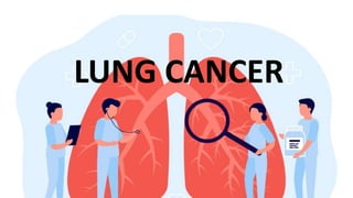
LUNG CANCER
Lung cancer Types
The most common type of lung cancer
In the UK Squamous cell cancer
In the USA Adenocarcinoma
Lung cancer : Non-Small Cell Carcinoma
• There are three main subtypes of non-small cell lung cancer:
1. Squamous cell cancer 2. Adenocarcinoma 3. Large cell lung carcinoma
Squamous cell cancer
Typically central (Squamous= Sentral)
Associated with:
• Parathyroid hormone-related protein (PTHrP) secretion hypercalcaemia
• Strongly associated with Finger clubbing
• Hypertrophic pulmonary osteoarthropathy (HPOA)
The presence of clubbing and tender wrists without synovitis makes pulmonary
osteoarthropathy the most likely diagnosis.
It is usually associated with underlying carcinoma of the lung.
Associated with bronchogenic carcinoma in 90% of cases.
The most sensitive diagnostic investigation is isotope bone scan: increase in the uptake in long
bones, around periarticular surfaces, and also mandible and scapulae
Regression of the pain has been reported with successful resection of the tumor and after vagotomy
• Hyperthyroidism due to Ectopic TSH
Most squamous-cell carcinomas present as obstructive lesions, which can manifest as
Infection.
Life threatening haemoptysis
is a medical emergency that requires prompt action.
Pulmonary angiography will identify the blood supply to the tumor and embolization of this vessel(s) will
immediately stem the bleeding.
Histology
will show clusters of lightly stained cells, often associated with groups of partially
keratinized, acidophilic cell clusters.
Pleomorphic cells in cluster with keratin pearls and intercellular bridges
Adenocarcinoma
• most common type of lung cancer in non-smokers, although the majority of patients who
develop lung adenocarcinoma are smokers
• Typically located on the lung periphery Normal bronchoscopy.
• May associate with Gynecomastia.
• PET/CT scan offers the best imaging modality to determine LN involvement in bronchial
adenocarcinoma
Histology will show:
• Malignant cells more often arranged in small clusters with an obvious lumen
and duct-like structures.
• Mucin-containing tumor cells with glandular differentiation
Bronchioloalveolar cell carcinoma
• It is an adenocarcinoma.
• accounts for around 5% of all primary lung carcinomas.
• 1% of all bronchial carcinomas.
• Growth along the alveolar walls without actually destroying them.
• The classic massive clear frothy sputum (bronchorrhoea) can be up to one liter a
day.
• Other symptoms are dyspnea, weight loss and chest pain.
• Almost a half of patients are diagnosed on routine CXR, usually demonstrating a
peripheral lesion.
• The tumor spreads using the alveolar walls as a frame and the alveoli are often
filled with mucin.
• In those whose tumor is not resect able, prognosis is poor.
Management of non-small cell lung cancer
Surgery
• only 20% suitable for surgery
• Stage I (cT1N0 and cT2N0) and stage II (cT1N1, cT2N1 and cT3N0) tumors
should be considered operable.
• Stage IIIA (cT3N1 and cT1-3N2) tumors have a low chance of being cured by
surgery alone, but it can be used in combination with chemotherapy.
• Stage IIIB and IV tumors considered inoperable.
 mediastinoscopy performed prior to surgery as CT does not always show mediastinal
lymph node involvement
The functional criteria for pneumonectomy are:
• Forced expiratory volume in 1 second (FEV-1) of >1.5 litres
• FEV-1 > 50% of the observed forced vital capacity, and
• Normal partial pressure of arterial CO2 (Paco2) with the patient at rest
• Prognosis after surgery is about 50-67% at 5 years with stage 1disease
Curative and palliative radiotherapy
Contraindications for surgery include:
• Patient refusal
• Asses general health (age 70+rs IHD,MI recent 6wks,increased PCO2
• FEV1 < 1.5 liters is considered a general cut-off point
• If the tumor necessitates a pneumonectomy, the post-bronchodilator FEV should be more than 2 liters.
• Metastases.
• stage III b or IV (i.e. metastases present)
• tumor near hilum
• vocal cord paralysis (implies extracapsular spread to mediastinal L.N)
• SVC obstruction
• Malignant pleural effusion
• Most pleural effusions associated with lung carcinoma are due to the tumor (and results in classification as a
T4 tumor).
• Spread to involve the C8, T1 and T2 nerve roots occurs by rib erosion by tumor to involve the lower roots of
the brachial plexus and is known as a Pancoast tumor.
• This causes severe pain in the shoulder and down the inner aspect of the
arm on the affected side and is a contraindication to surgery
Pancoast tumor
• Neoplasm apex of the lung that typically invades the chest wall and brachial plexus causing a Horner's
syndrome (ptosis and constriction of the pupil).
 compresses the sympathetic fibers (from T1 and T2) as they travel upwards to
superior cervical ganglion and then to the dilator pupillae as the long ciliary
nerve.
• The tumor causes pain in the C8 and T1 distribution and Horner's syndrome.
 pain in the shoulder and the medial surface of the arm.
 Patients may initially present to rheumatologists or orthopedic surgeons.
• It may cause small muscle wasting of the hands and erosion of the first rib
 the investigation of choice CT of chest
 CT is essential to locate the tumor and the extent of rib, vertebral and muscle
involvement.
 Chest x-ray may be normal
• Tumors are most commonly squamous cell and are usually inoperable on
presentation
Staging lung carcinoma
• It is important to remember the criteria for staging carcinoma of the lung. TNM staging
takes into account;
The size and position of the tumor (T)
Whether the cancer cells have spread into the lymph nodes (N)
Whether the tumor has spread anywhere else in the body - secondary cancer or
metastases (M)
• Computed tomographic (CT) scanning is recommended as a staging procedure. Where
available, PET scanning may be superior
• CT can be performed with or without contrast enhancement, but Contrast enhancement
makes it easier for inexperienced observers to interpret the CT.
• Overall, preoperative CT staging has been shown to overstage or understage when
compared with operative findings in 40% of patients. So biopsy is also needed.
TNM System
The International Association for the Study of Lung Cancer (IASLC) developed a SEVENTH EDITION
OF THE TNM SYSTEM in 2010 replaced earlier editions: as fellow
Primary tumor — (T)
T0 – No evidence of primary tumor.
Tis – Carcinoma in situ.
T1 – Tumor that is ≤3 cm
T1a: Tumor is ≤2 cm
T1b: Tumor is >2 cm, but ≤3 cm
T2 – Tumor that is >3 cm but ≤7 cm
T2a: Tumor is >3 cm, but ≤5 cm
T2b: Tumor is >5 cm, but ≤7 cm
T3 – Tumor that is >7 cm ; invades the chest wall (including superior sulcus tumors),
diaphragm, phrenic nerve, mediastinal pleura, parietal pericardium
T4 – Tumor of any size that invades the mediastinum, heart, great vessels, trachea,
recurrent laryngeal nerve, esophagus, vertebral body, or carina; or separate tumor
nodule(s) located in a different lobe of the ipsilateral lung.
Regional lymph nodes — (N)
• N0 – No regional lymph node involvement.
• N1 – Involvement of ipsilateral intrapulmonary, peri bronchial, or hilar lymph
nodes.
• N2 – Involvement of ipsilateral mediastinal or sub carinal lymph nodes.
• N3 – Involvement of contralateral mediastinal or hilar lymph nodes.
Metastasis — (M)
• M0 – No distant metastasis
• M1a – Malignant pleural effusion, pericardial effusion, pleural nodules, or metastatic
nodules in the contralateral lung
• M1b – Distant (extra-thoracic) metastasis
_____________________________
Carcinoid lung
Overview
• Carcinoid tumors are neuroendocrine
• Originate from Kulchitsky (K) cells in the lung
• Slow growing
• Smoking is NOT a risk factor
Incidence: Lung carcinoid accounts
• 1% of lung tumors
• 10% of carcinoid tumors.
• typical age = 40-50 years
• The incidence is equal between men and women
FEATURES
• Often asymptomatic
• Long history of cough,
• Recurrent hemoptysis
• Recurrent infections
Carcinoid tumors (80-90%) develops in a bronchus cause bronchial obstruction Lower
respiratory tract infection.
• Carcinoid syndrome (rare)
• Associated conditions with carcinoid tumor in the lung
Carcinoid syndrome (rare)
• Depends on associated liver metastases
• Occurs in less than 10% of patients with carcinoid tumors, but occurs
most commonly in GIT tumors.
• Can secrete a number of vasoactive compounds (including serotonin and
bradykinin), which result in:
bronchospasm, diarrhea, skin flushing, right sided valvular heart lesions
ACTH secretion and subsequent Cushing's syndrome.
Ectopic growth hormone-releasing hormone [GHRH] and subsequent acromegaly
Multiple endocrine neoplasia (MEN) type 1 where pancreatic neuroendocrine
Investigation
CXR
• Often centrally located and not seen on CXR.
• Usually occur in the major bronchi, 85% can be seen Bronchoscopically.
• Most often located in the main bronchi, and occur most frequency in the right
middle lobe.
• A carcinoid tumor in the left lower lobe bronchus could cause distal collapse of
the left lower lobe.
Bronchoscopy
• identifies up to 80% of carcinoid tumors in the main bronchi.
• seen as a highly vascular 'cherry-like' tumor ('cherry red ball')Biopsy is usually followed with brisk
bleeding and should be done via rigid bronchoscopy.
• The histological picture of granular eosinophilic staining of the cytoplasm, is highly suggestive of a
carcinoid tumor.
• Histologically, these tumors consist of compact nests of epithelial cells surrounded by neat, delicate
connective tissue capsules.
• Plasma chromograffin A is an effective screening test for carcinoid as it is
very
sensitive, but it is not specific.
• 24 hour urinary excretion of 5-hydroxyindoleacetic acid is more specific for
the
diagnosis, but false positives and negatives are present.
• Scintigraphic imaging with labelled somatostatin increases the ability to
diagnose a carcinoid tumor, but biopsy is required to confirm.
Management
• Surgical resection
• A person with an isolated pulmonary carcinoid should be referred for
tumor resection,
• Somatostatin analogues can be used where there is no possibility
Prognosis
• If no metastases then 90% survival at 5 years
Small cell Lung CA
• Also known as "oat-cell carcinoma“ usually central
• arise from APUD* cells an acronym for
• Amine - high amine content
• Precursor Uptake - high uptake of amine precursors
• Decarboxylase - high content of the enzyme decarboxylase
• Most aggressive cancer which typically presents with a short history and 80–90% will have metastases at the time of presentation.
Features
• Ectopic ADH hyponatremia (SIADH )occurs in 5–10% of cases
• Ectopic ACTH Cushing's syndrome
• Due to the short natural history of this type of cancer, Cushing syndrome in small cell ca does not manifest classically by buffalo hump, striae
or central obesity.
• Its presence is suspected by arterial hypertension, hyperglycemia,
hypokalemia, alkalosis and muscle weakness.
• ACTH secretion can cause bilateral adrenal hyperplasia,
• The high levels of cortisol can lead to hypokalemic alkalosis
• In case of chronic heavy smoker with chest infection +hyperglycaemia + Ļow K + increase HCO3? Suspect SCLC with ectopic ACTH secretion.
But if with Low Na and increased K think of Adrenal metastasis MRI adrenals
• Lambert-Eaton syndrome: antibodies to voltage gated calcium channels causing myasthenia like syndrome
• Neuron-specific enolase (NSE) is a tumor marker indicative of small cell carcinoma
• Associated with L-myc amplification.
• Histologically, Small cell carcinoma has clusters of small, basophilic cells and
Management
Usually metastatic disease by time of diagnosis and surgery done only for de bulking
Stage
Early stage (T1-
2a,N0,M0)
Surgery
Early stage (T1-
2a,N0,M0)-
Limited disease
(T1-4,N0-3,M0)
4-6 cycles cisplatin based
chemotherapy, carboplatin if poor
renal function/poor performance
status +/- radiotherapy
Extensive disease
(T1-4, N0-3,
M1a/b)
6 cycles platinum based
combination chemotherapy +
thoracic
radiotherapy if good response
Prognosis
Very poor and survival beyond two 2 years is exceptional
Adverse prognostic factors in small cell lung cancer:
• Serum Na < 132 mmol/l
• Weight loss > 10%
• WHO performance status > 2
• Alkaline phosphatase > 1.5 times upper limit of normal
• LDH > 1.5 times upper limit of normal
• Extensive disease (occurring outside one hemi-thorax and ipsilateral supraclavicular fossa
nodes).
______________________
___
Bronchial Carcinoma
• 20-30% of cases with bronchial carcinoma are of small (oat) cell type from endocrine Kcells (Kulchitsky) cells
• Primary bronchial cancer the tumour edge may have a fluffy or spiked
appearance.
• Paraneoplastic manifestations:
 SIADH (5 - 10%)
 ACTH (5 %)
 ANP
• The most appropriate tool to confirm the diagnosis of bronchial carcinoma is
Bronchoscopy ± Trans bronchial biopsy (NOT CT-guided FNA biopsy).
• CT-guided FNA would be useful; however, there is a high risk of pneumothorax
in patients with FEV1 < 1.
________________________________________________
Lung cancer: paraneoplastic features
• Paraneoplastic syndromes are a result of antibody generation from or against malignant cells attacking
normal tissue.
• Both non-small cell and small cell lung cancers are associated with Paraneoplastic syndromes, although they
are more common with the small cell due to its neuroendocrine cell origin.
Squamous cell
• Parathyroid hormone-related protein (PTH-rp) secretion causing hypercalcemia
• Occurs in about 15%
• Best treated with intravenous fluids and bisphosphonates
• Clubbing
• Hypertrophic pulmonary osteoarthropathy (HPOA)
(HPOA) is a proliferative peri-ostisis typically involves the long bones.
It is often painful.
• Hyperthyroidism due to ectopic TSH
Adenocarcinoma
• Gynecomastia
• Bronchial carcinoma can presents with ataxia and bilateral Gynecomastia
• It can be painful and may be associated with testicular atrophy.
• Ataxia can occur as a result of a paraneoplastic cerebellar degeneration associated
with the malignancy
Small cell
• ADH : Symptomatic hyponatremia due to SIADH is treated with demeclocycline which induces nephrogenic diabetes insipidus leading to
excretion of excess water.
• ACTH : not typical, hypertension, hyperglycemia, hypokalemia, alkalosis and muscle weakness are more common than buffalo hump etc.
 May manifest by Cushingoid facieses and hyperpigmentation of the skin
• Lambert-Eaton syndrome
70% occur in small cell carcinoma
 is a pre-synaptic disorder of auto-antibody IgG directed against the pre-synaptic voltage gated calcium
channel (VGCC) leading to impaired acetylcholine release.
 Characterized by:
• Proximal muscle weakness (the cranial nerves and respiratory muscles are usually spared)
• Depressed or absent tendon reflexes and
• Autonomic features (for example, dry mouth, impotence, etc.).
Weakness and fatigability can be improved with guanidine hydrochloride
Unlike myasthenia gravis exercise is associated with increasing muscle strength and there is a negative
response to Tensilon.
Electromyography is useful in confirming the diagnosis where repeated nerve stimulations cause a
progressive increase in the size of the muscle action potential.
Cerebellar ataxia:
 Paraneoplastic syndromes are a result of antibody generation from or against malignant cells attacking normal tissue.
 Example: anti neuronal antibodies (anti-Hu, anti-Yo, anti-Ri ) directed against the Purkinje cells of the cerebellum
leading to the cerebellar syndrome (truncal ataxia).
 To diagnose this : Anti-Purkinje cell antibody levels.
Lungs Cancer etiology sign symtom causes.pptx

More Related Content

Similar to Lungs Cancer etiology sign symtom causes.pptx

Lung maligncy updated 27 04-18 dr jarisha miot radiology
Lung maligncy updated 27 04-18 dr jarisha miot radiologyLung maligncy updated 27 04-18 dr jarisha miot radiology
Lung maligncy updated 27 04-18 dr jarisha miot radiology
Jarisha Vj
 
Presentation1.pptx. radiological imaging of bronchogenic carcinom.
Presentation1.pptx. radiological imaging of bronchogenic carcinom.Presentation1.pptx. radiological imaging of bronchogenic carcinom.
Presentation1.pptx. radiological imaging of bronchogenic carcinom.Abdellah Nazeer
 
Atypical lung neoplasms1
Atypical lung neoplasms1Atypical lung neoplasms1
Atypical lung neoplasms1
Jayanth Hiremagalur
 
Malignant lung tumours
Malignant lung tumoursMalignant lung tumours
Malignant lung tumours
farranajwa
 
lung cancer.pptx
lung cancer.pptxlung cancer.pptx
lung cancer.pptx
ssuser1ba639
 
bronchogenic.pptx
bronchogenic.pptxbronchogenic.pptx
bronchogenic.pptx
MdRaihan58
 
Lung cancer
Lung cancerLung cancer
Lung cancer
Abhay Rajpoot
 
Locally advanced lung ca
Locally advanced lung caLocally advanced lung ca
Locally advanced lung ca
amanuelasefa1
 
testicular tumors
testicular tumorstesticular tumors
testicular tumors
DrAyush Garg
 
Lung cancer.
Lung cancer.Lung cancer.
Lung cancer.
YusufSiddiq
 
Carcinoma of esophagus
Carcinoma of esophagusCarcinoma of esophagus
Carcinoma of esophagus
prabhanjan chakravarthy
 
LUNG CANCER vandana..pptx
LUNG CANCER vandana..pptxLUNG CANCER vandana..pptx
LUNG CANCER vandana..pptx
VandanaKoranga
 
Ca esophagus 12th
Ca esophagus 12thCa esophagus 12th
Ca esophagus 12th
Gowtham Manimaran
 
Staging of bronchogenic carcinoma
Staging of bronchogenic carcinomaStaging of bronchogenic carcinoma
Staging of bronchogenic carcinoma
Dr. Kaliprasanna chatterjee
 
Radiological imaging of pulmonary neoplasms
Radiological imaging of pulmonary neoplasmsRadiological imaging of pulmonary neoplasms
Radiological imaging of pulmonary neoplasms
Pankaj Kaira
 
Lung cancer
Lung cancerLung cancer
Lung cancer
Mahesh Dayyala
 
Lung ca &amp; staging
Lung ca &amp; stagingLung ca &amp; staging
Lung ca &amp; staging
Balaram Prasad
 
LUNG CANCER
LUNG CANCERLUNG CANCER
LUNG CANCER
Vijay Sal
 
Radiological approach to lung neoplasms
Radiological approach to lung neoplasmsRadiological approach to lung neoplasms
Radiological approach to lung neoplasms
SnehaMandal5
 
lymphangitis carcinomatosis
lymphangitis carcinomatosislymphangitis carcinomatosis
lymphangitis carcinomatosis
mbito1
 

Similar to Lungs Cancer etiology sign symtom causes.pptx (20)

Lung maligncy updated 27 04-18 dr jarisha miot radiology
Lung maligncy updated 27 04-18 dr jarisha miot radiologyLung maligncy updated 27 04-18 dr jarisha miot radiology
Lung maligncy updated 27 04-18 dr jarisha miot radiology
 
Presentation1.pptx. radiological imaging of bronchogenic carcinom.
Presentation1.pptx. radiological imaging of bronchogenic carcinom.Presentation1.pptx. radiological imaging of bronchogenic carcinom.
Presentation1.pptx. radiological imaging of bronchogenic carcinom.
 
Atypical lung neoplasms1
Atypical lung neoplasms1Atypical lung neoplasms1
Atypical lung neoplasms1
 
Malignant lung tumours
Malignant lung tumoursMalignant lung tumours
Malignant lung tumours
 
lung cancer.pptx
lung cancer.pptxlung cancer.pptx
lung cancer.pptx
 
bronchogenic.pptx
bronchogenic.pptxbronchogenic.pptx
bronchogenic.pptx
 
Lung cancer
Lung cancerLung cancer
Lung cancer
 
Locally advanced lung ca
Locally advanced lung caLocally advanced lung ca
Locally advanced lung ca
 
testicular tumors
testicular tumorstesticular tumors
testicular tumors
 
Lung cancer.
Lung cancer.Lung cancer.
Lung cancer.
 
Carcinoma of esophagus
Carcinoma of esophagusCarcinoma of esophagus
Carcinoma of esophagus
 
LUNG CANCER vandana..pptx
LUNG CANCER vandana..pptxLUNG CANCER vandana..pptx
LUNG CANCER vandana..pptx
 
Ca esophagus 12th
Ca esophagus 12thCa esophagus 12th
Ca esophagus 12th
 
Staging of bronchogenic carcinoma
Staging of bronchogenic carcinomaStaging of bronchogenic carcinoma
Staging of bronchogenic carcinoma
 
Radiological imaging of pulmonary neoplasms
Radiological imaging of pulmonary neoplasmsRadiological imaging of pulmonary neoplasms
Radiological imaging of pulmonary neoplasms
 
Lung cancer
Lung cancerLung cancer
Lung cancer
 
Lung ca &amp; staging
Lung ca &amp; stagingLung ca &amp; staging
Lung ca &amp; staging
 
LUNG CANCER
LUNG CANCERLUNG CANCER
LUNG CANCER
 
Radiological approach to lung neoplasms
Radiological approach to lung neoplasmsRadiological approach to lung neoplasms
Radiological approach to lung neoplasms
 
lymphangitis carcinomatosis
lymphangitis carcinomatosislymphangitis carcinomatosis
lymphangitis carcinomatosis
 

Recently uploaded

Alcohol_Dr. Jeenal Mistry MD Pharmacology.pdf
Alcohol_Dr. Jeenal Mistry MD Pharmacology.pdfAlcohol_Dr. Jeenal Mistry MD Pharmacology.pdf
Alcohol_Dr. Jeenal Mistry MD Pharmacology.pdf
Dr Jeenal Mistry
 
Pulmonary Thromboembolism - etilogy, types, medical- Surgical and nursing man...
Pulmonary Thromboembolism - etilogy, types, medical- Surgical and nursing man...Pulmonary Thromboembolism - etilogy, types, medical- Surgical and nursing man...
Pulmonary Thromboembolism - etilogy, types, medical- Surgical and nursing man...
VarunMahajani
 
HOT NEW PRODUCT! BIG SALES FAST SHIPPING NOW FROM CHINA!! EU KU DB BK substit...
HOT NEW PRODUCT! BIG SALES FAST SHIPPING NOW FROM CHINA!! EU KU DB BK substit...HOT NEW PRODUCT! BIG SALES FAST SHIPPING NOW FROM CHINA!! EU KU DB BK substit...
HOT NEW PRODUCT! BIG SALES FAST SHIPPING NOW FROM CHINA!! EU KU DB BK substit...
GL Anaacs
 
Non-respiratory Functions of the Lungs.pdf
Non-respiratory Functions of the Lungs.pdfNon-respiratory Functions of the Lungs.pdf
Non-respiratory Functions of the Lungs.pdf
MedicoseAcademics
 
263778731218 Abortion Clinic /Pills In Harare ,
263778731218 Abortion Clinic /Pills In Harare ,263778731218 Abortion Clinic /Pills In Harare ,
263778731218 Abortion Clinic /Pills In Harare ,
sisternakatoto
 
heat stroke and heat exhaustion in children
heat stroke and heat exhaustion in childrenheat stroke and heat exhaustion in children
heat stroke and heat exhaustion in children
SumeraAhmad5
 
24 Upakrama.pptx class ppt useful in all
24 Upakrama.pptx class ppt useful in all24 Upakrama.pptx class ppt useful in all
24 Upakrama.pptx class ppt useful in all
DrSathishMS1
 
Ocular injury ppt Upendra pal optometrist upums saifai etawah
Ocular injury  ppt  Upendra pal  optometrist upums saifai etawahOcular injury  ppt  Upendra pal  optometrist upums saifai etawah
Ocular injury ppt Upendra pal optometrist upums saifai etawah
pal078100
 
NVBDCP.pptx Nation vector borne disease control program
NVBDCP.pptx Nation vector borne disease control programNVBDCP.pptx Nation vector borne disease control program
NVBDCP.pptx Nation vector borne disease control program
Sapna Thakur
 
Ophthalmology Clinical Tests for OSCE exam
Ophthalmology Clinical Tests for OSCE examOphthalmology Clinical Tests for OSCE exam
Ophthalmology Clinical Tests for OSCE exam
KafrELShiekh University
 
Report Back from SGO 2024: What’s the Latest in Cervical Cancer?
Report Back from SGO 2024: What’s the Latest in Cervical Cancer?Report Back from SGO 2024: What’s the Latest in Cervical Cancer?
Report Back from SGO 2024: What’s the Latest in Cervical Cancer?
bkling
 
How STIs Influence the Development of Pelvic Inflammatory Disease.pptx
How STIs Influence the Development of Pelvic Inflammatory Disease.pptxHow STIs Influence the Development of Pelvic Inflammatory Disease.pptx
How STIs Influence the Development of Pelvic Inflammatory Disease.pptx
FFragrant
 
Hemodialysis: Chapter 3, Dialysis Water Unit - Dr.Gawad
Hemodialysis: Chapter 3, Dialysis Water Unit - Dr.GawadHemodialysis: Chapter 3, Dialysis Water Unit - Dr.Gawad
Hemodialysis: Chapter 3, Dialysis Water Unit - Dr.Gawad
NephroTube - Dr.Gawad
 
Flu Vaccine Alert in Bangalore Karnataka
Flu Vaccine Alert in Bangalore KarnatakaFlu Vaccine Alert in Bangalore Karnataka
Flu Vaccine Alert in Bangalore Karnataka
addon Scans
 
Physiology of Chemical Sensation of smell.pdf
Physiology of Chemical Sensation of smell.pdfPhysiology of Chemical Sensation of smell.pdf
Physiology of Chemical Sensation of smell.pdf
MedicoseAcademics
 
BRACHYTHERAPY OVERVIEW AND APPLICATORS
BRACHYTHERAPY OVERVIEW  AND  APPLICATORSBRACHYTHERAPY OVERVIEW  AND  APPLICATORS
BRACHYTHERAPY OVERVIEW AND APPLICATORS
Krishan Murari
 
Superficial & Deep Fascia of the NECK.pptx
Superficial & Deep Fascia of the NECK.pptxSuperficial & Deep Fascia of the NECK.pptx
Superficial & Deep Fascia of the NECK.pptx
Dr. Rabia Inam Gandapore
 
ACUTE SCROTUM.....pdf. ACUTE SCROTAL CONDITIOND
ACUTE SCROTUM.....pdf. ACUTE SCROTAL CONDITIONDACUTE SCROTUM.....pdf. ACUTE SCROTAL CONDITIOND
ACUTE SCROTUM.....pdf. ACUTE SCROTAL CONDITIOND
DR SETH JOTHAM
 
Charaka Samhita Sutra sthana Chapter 15 Upakalpaniyaadhyaya
Charaka Samhita Sutra sthana Chapter 15 UpakalpaniyaadhyayaCharaka Samhita Sutra sthana Chapter 15 Upakalpaniyaadhyaya
Charaka Samhita Sutra sthana Chapter 15 Upakalpaniyaadhyaya
Dr KHALID B.M
 
Couples presenting to the infertility clinic- Do they really have infertility...
Couples presenting to the infertility clinic- Do they really have infertility...Couples presenting to the infertility clinic- Do they really have infertility...
Couples presenting to the infertility clinic- Do they really have infertility...
Sujoy Dasgupta
 

Recently uploaded (20)

Alcohol_Dr. Jeenal Mistry MD Pharmacology.pdf
Alcohol_Dr. Jeenal Mistry MD Pharmacology.pdfAlcohol_Dr. Jeenal Mistry MD Pharmacology.pdf
Alcohol_Dr. Jeenal Mistry MD Pharmacology.pdf
 
Pulmonary Thromboembolism - etilogy, types, medical- Surgical and nursing man...
Pulmonary Thromboembolism - etilogy, types, medical- Surgical and nursing man...Pulmonary Thromboembolism - etilogy, types, medical- Surgical and nursing man...
Pulmonary Thromboembolism - etilogy, types, medical- Surgical and nursing man...
 
HOT NEW PRODUCT! BIG SALES FAST SHIPPING NOW FROM CHINA!! EU KU DB BK substit...
HOT NEW PRODUCT! BIG SALES FAST SHIPPING NOW FROM CHINA!! EU KU DB BK substit...HOT NEW PRODUCT! BIG SALES FAST SHIPPING NOW FROM CHINA!! EU KU DB BK substit...
HOT NEW PRODUCT! BIG SALES FAST SHIPPING NOW FROM CHINA!! EU KU DB BK substit...
 
Non-respiratory Functions of the Lungs.pdf
Non-respiratory Functions of the Lungs.pdfNon-respiratory Functions of the Lungs.pdf
Non-respiratory Functions of the Lungs.pdf
 
263778731218 Abortion Clinic /Pills In Harare ,
263778731218 Abortion Clinic /Pills In Harare ,263778731218 Abortion Clinic /Pills In Harare ,
263778731218 Abortion Clinic /Pills In Harare ,
 
heat stroke and heat exhaustion in children
heat stroke and heat exhaustion in childrenheat stroke and heat exhaustion in children
heat stroke and heat exhaustion in children
 
24 Upakrama.pptx class ppt useful in all
24 Upakrama.pptx class ppt useful in all24 Upakrama.pptx class ppt useful in all
24 Upakrama.pptx class ppt useful in all
 
Ocular injury ppt Upendra pal optometrist upums saifai etawah
Ocular injury  ppt  Upendra pal  optometrist upums saifai etawahOcular injury  ppt  Upendra pal  optometrist upums saifai etawah
Ocular injury ppt Upendra pal optometrist upums saifai etawah
 
NVBDCP.pptx Nation vector borne disease control program
NVBDCP.pptx Nation vector borne disease control programNVBDCP.pptx Nation vector borne disease control program
NVBDCP.pptx Nation vector borne disease control program
 
Ophthalmology Clinical Tests for OSCE exam
Ophthalmology Clinical Tests for OSCE examOphthalmology Clinical Tests for OSCE exam
Ophthalmology Clinical Tests for OSCE exam
 
Report Back from SGO 2024: What’s the Latest in Cervical Cancer?
Report Back from SGO 2024: What’s the Latest in Cervical Cancer?Report Back from SGO 2024: What’s the Latest in Cervical Cancer?
Report Back from SGO 2024: What’s the Latest in Cervical Cancer?
 
How STIs Influence the Development of Pelvic Inflammatory Disease.pptx
How STIs Influence the Development of Pelvic Inflammatory Disease.pptxHow STIs Influence the Development of Pelvic Inflammatory Disease.pptx
How STIs Influence the Development of Pelvic Inflammatory Disease.pptx
 
Hemodialysis: Chapter 3, Dialysis Water Unit - Dr.Gawad
Hemodialysis: Chapter 3, Dialysis Water Unit - Dr.GawadHemodialysis: Chapter 3, Dialysis Water Unit - Dr.Gawad
Hemodialysis: Chapter 3, Dialysis Water Unit - Dr.Gawad
 
Flu Vaccine Alert in Bangalore Karnataka
Flu Vaccine Alert in Bangalore KarnatakaFlu Vaccine Alert in Bangalore Karnataka
Flu Vaccine Alert in Bangalore Karnataka
 
Physiology of Chemical Sensation of smell.pdf
Physiology of Chemical Sensation of smell.pdfPhysiology of Chemical Sensation of smell.pdf
Physiology of Chemical Sensation of smell.pdf
 
BRACHYTHERAPY OVERVIEW AND APPLICATORS
BRACHYTHERAPY OVERVIEW  AND  APPLICATORSBRACHYTHERAPY OVERVIEW  AND  APPLICATORS
BRACHYTHERAPY OVERVIEW AND APPLICATORS
 
Superficial & Deep Fascia of the NECK.pptx
Superficial & Deep Fascia of the NECK.pptxSuperficial & Deep Fascia of the NECK.pptx
Superficial & Deep Fascia of the NECK.pptx
 
ACUTE SCROTUM.....pdf. ACUTE SCROTAL CONDITIOND
ACUTE SCROTUM.....pdf. ACUTE SCROTAL CONDITIONDACUTE SCROTUM.....pdf. ACUTE SCROTAL CONDITIOND
ACUTE SCROTUM.....pdf. ACUTE SCROTAL CONDITIOND
 
Charaka Samhita Sutra sthana Chapter 15 Upakalpaniyaadhyaya
Charaka Samhita Sutra sthana Chapter 15 UpakalpaniyaadhyayaCharaka Samhita Sutra sthana Chapter 15 Upakalpaniyaadhyaya
Charaka Samhita Sutra sthana Chapter 15 Upakalpaniyaadhyaya
 
Couples presenting to the infertility clinic- Do they really have infertility...
Couples presenting to the infertility clinic- Do they really have infertility...Couples presenting to the infertility clinic- Do they really have infertility...
Couples presenting to the infertility clinic- Do they really have infertility...
 

Lungs Cancer etiology sign symtom causes.pptx

  • 2.
  • 3.
  • 4. Lung cancer Types The most common type of lung cancer In the UK Squamous cell cancer In the USA Adenocarcinoma
  • 5.
  • 6. Lung cancer : Non-Small Cell Carcinoma • There are three main subtypes of non-small cell lung cancer: 1. Squamous cell cancer 2. Adenocarcinoma 3. Large cell lung carcinoma Squamous cell cancer Typically central (Squamous= Sentral) Associated with: • Parathyroid hormone-related protein (PTHrP) secretion hypercalcaemia • Strongly associated with Finger clubbing • Hypertrophic pulmonary osteoarthropathy (HPOA) The presence of clubbing and tender wrists without synovitis makes pulmonary osteoarthropathy the most likely diagnosis. It is usually associated with underlying carcinoma of the lung. Associated with bronchogenic carcinoma in 90% of cases. The most sensitive diagnostic investigation is isotope bone scan: increase in the uptake in long bones, around periarticular surfaces, and also mandible and scapulae Regression of the pain has been reported with successful resection of the tumor and after vagotomy • Hyperthyroidism due to Ectopic TSH
  • 7. Most squamous-cell carcinomas present as obstructive lesions, which can manifest as Infection. Life threatening haemoptysis is a medical emergency that requires prompt action. Pulmonary angiography will identify the blood supply to the tumor and embolization of this vessel(s) will immediately stem the bleeding. Histology will show clusters of lightly stained cells, often associated with groups of partially keratinized, acidophilic cell clusters. Pleomorphic cells in cluster with keratin pearls and intercellular bridges
  • 8. Adenocarcinoma • most common type of lung cancer in non-smokers, although the majority of patients who develop lung adenocarcinoma are smokers • Typically located on the lung periphery Normal bronchoscopy. • May associate with Gynecomastia. • PET/CT scan offers the best imaging modality to determine LN involvement in bronchial adenocarcinoma Histology will show: • Malignant cells more often arranged in small clusters with an obvious lumen and duct-like structures. • Mucin-containing tumor cells with glandular differentiation
  • 9. Bronchioloalveolar cell carcinoma Bronchioloalveolar cell carcinoma • It is an adenocarcinoma. • accounts for around 5% of all primary lung carcinomas. • 1% of all bronchial carcinomas. • Growth along the alveolar walls without actually destroying them. • The classic massive clear frothy sputum (bronchorrhoea) can be up to one liter a day. • Other symptoms are dyspnea, weight loss and chest pain. • Almost a half of patients are diagnosed on routine CXR, usually demonstrating a peripheral lesion. • The tumor spreads using the alveolar walls as a frame and the alveoli are often filled with mucin. • In those whose tumor is not resect able, prognosis is poor.
  • 10. Management of non-small cell lung cancer Surgery • only 20% suitable for surgery • Stage I (cT1N0 and cT2N0) and stage II (cT1N1, cT2N1 and cT3N0) tumors should be considered operable. • Stage IIIA (cT3N1 and cT1-3N2) tumors have a low chance of being cured by surgery alone, but it can be used in combination with chemotherapy. • Stage IIIB and IV tumors considered inoperable.  mediastinoscopy performed prior to surgery as CT does not always show mediastinal lymph node involvement The functional criteria for pneumonectomy are: • Forced expiratory volume in 1 second (FEV-1) of >1.5 litres • FEV-1 > 50% of the observed forced vital capacity, and • Normal partial pressure of arterial CO2 (Paco2) with the patient at rest • Prognosis after surgery is about 50-67% at 5 years with stage 1disease Curative and palliative radiotherapy
  • 11. Contraindications for surgery include: • Patient refusal • Asses general health (age 70+rs IHD,MI recent 6wks,increased PCO2 • FEV1 < 1.5 liters is considered a general cut-off point • If the tumor necessitates a pneumonectomy, the post-bronchodilator FEV should be more than 2 liters. • Metastases. • stage III b or IV (i.e. metastases present) • tumor near hilum • vocal cord paralysis (implies extracapsular spread to mediastinal L.N) • SVC obstruction • Malignant pleural effusion • Most pleural effusions associated with lung carcinoma are due to the tumor (and results in classification as a T4 tumor). • Spread to involve the C8, T1 and T2 nerve roots occurs by rib erosion by tumor to involve the lower roots of the brachial plexus and is known as a Pancoast tumor. • This causes severe pain in the shoulder and down the inner aspect of the arm on the affected side and is a contraindication to surgery
  • 12. Pancoast tumor • Neoplasm apex of the lung that typically invades the chest wall and brachial plexus causing a Horner's syndrome (ptosis and constriction of the pupil).  compresses the sympathetic fibers (from T1 and T2) as they travel upwards to superior cervical ganglion and then to the dilator pupillae as the long ciliary nerve. • The tumor causes pain in the C8 and T1 distribution and Horner's syndrome.  pain in the shoulder and the medial surface of the arm.  Patients may initially present to rheumatologists or orthopedic surgeons. • It may cause small muscle wasting of the hands and erosion of the first rib  the investigation of choice CT of chest  CT is essential to locate the tumor and the extent of rib, vertebral and muscle involvement.  Chest x-ray may be normal • Tumors are most commonly squamous cell and are usually inoperable on presentation
  • 13. Staging lung carcinoma • It is important to remember the criteria for staging carcinoma of the lung. TNM staging takes into account; The size and position of the tumor (T) Whether the cancer cells have spread into the lymph nodes (N) Whether the tumor has spread anywhere else in the body - secondary cancer or metastases (M) • Computed tomographic (CT) scanning is recommended as a staging procedure. Where available, PET scanning may be superior • CT can be performed with or without contrast enhancement, but Contrast enhancement makes it easier for inexperienced observers to interpret the CT. • Overall, preoperative CT staging has been shown to overstage or understage when compared with operative findings in 40% of patients. So biopsy is also needed.
  • 14. TNM System The International Association for the Study of Lung Cancer (IASLC) developed a SEVENTH EDITION OF THE TNM SYSTEM in 2010 replaced earlier editions: as fellow Primary tumor — (T) T0 – No evidence of primary tumor. Tis – Carcinoma in situ. T1 – Tumor that is ≤3 cm T1a: Tumor is ≤2 cm T1b: Tumor is >2 cm, but ≤3 cm T2 – Tumor that is >3 cm but ≤7 cm T2a: Tumor is >3 cm, but ≤5 cm T2b: Tumor is >5 cm, but ≤7 cm T3 – Tumor that is >7 cm ; invades the chest wall (including superior sulcus tumors), diaphragm, phrenic nerve, mediastinal pleura, parietal pericardium T4 – Tumor of any size that invades the mediastinum, heart, great vessels, trachea, recurrent laryngeal nerve, esophagus, vertebral body, or carina; or separate tumor nodule(s) located in a different lobe of the ipsilateral lung.
  • 15. Regional lymph nodes — (N) • N0 – No regional lymph node involvement. • N1 – Involvement of ipsilateral intrapulmonary, peri bronchial, or hilar lymph nodes. • N2 – Involvement of ipsilateral mediastinal or sub carinal lymph nodes. • N3 – Involvement of contralateral mediastinal or hilar lymph nodes. Metastasis — (M) • M0 – No distant metastasis • M1a – Malignant pleural effusion, pericardial effusion, pleural nodules, or metastatic nodules in the contralateral lung • M1b – Distant (extra-thoracic) metastasis _____________________________
  • 16.
  • 17. Carcinoid lung Overview • Carcinoid tumors are neuroendocrine • Originate from Kulchitsky (K) cells in the lung • Slow growing • Smoking is NOT a risk factor Incidence: Lung carcinoid accounts • 1% of lung tumors • 10% of carcinoid tumors. • typical age = 40-50 years • The incidence is equal between men and women
  • 18. FEATURES • Often asymptomatic • Long history of cough, • Recurrent hemoptysis • Recurrent infections Carcinoid tumors (80-90%) develops in a bronchus cause bronchial obstruction Lower respiratory tract infection. • Carcinoid syndrome (rare) • Associated conditions with carcinoid tumor in the lung Carcinoid syndrome (rare) • Depends on associated liver metastases • Occurs in less than 10% of patients with carcinoid tumors, but occurs most commonly in GIT tumors. • Can secrete a number of vasoactive compounds (including serotonin and bradykinin), which result in: bronchospasm, diarrhea, skin flushing, right sided valvular heart lesions ACTH secretion and subsequent Cushing's syndrome. Ectopic growth hormone-releasing hormone [GHRH] and subsequent acromegaly Multiple endocrine neoplasia (MEN) type 1 where pancreatic neuroendocrine
  • 19. Investigation CXR • Often centrally located and not seen on CXR. • Usually occur in the major bronchi, 85% can be seen Bronchoscopically. • Most often located in the main bronchi, and occur most frequency in the right middle lobe. • A carcinoid tumor in the left lower lobe bronchus could cause distal collapse of the left lower lobe. Bronchoscopy • identifies up to 80% of carcinoid tumors in the main bronchi. • seen as a highly vascular 'cherry-like' tumor ('cherry red ball')Biopsy is usually followed with brisk bleeding and should be done via rigid bronchoscopy. • The histological picture of granular eosinophilic staining of the cytoplasm, is highly suggestive of a carcinoid tumor. • Histologically, these tumors consist of compact nests of epithelial cells surrounded by neat, delicate connective tissue capsules.
  • 20. • Plasma chromograffin A is an effective screening test for carcinoid as it is very sensitive, but it is not specific. • 24 hour urinary excretion of 5-hydroxyindoleacetic acid is more specific for the diagnosis, but false positives and negatives are present. • Scintigraphic imaging with labelled somatostatin increases the ability to diagnose a carcinoid tumor, but biopsy is required to confirm. Management • Surgical resection • A person with an isolated pulmonary carcinoid should be referred for tumor resection, • Somatostatin analogues can be used where there is no possibility Prognosis • If no metastases then 90% survival at 5 years
  • 21. Small cell Lung CA • Also known as "oat-cell carcinoma“ usually central • arise from APUD* cells an acronym for • Amine - high amine content • Precursor Uptake - high uptake of amine precursors • Decarboxylase - high content of the enzyme decarboxylase • Most aggressive cancer which typically presents with a short history and 80–90% will have metastases at the time of presentation. Features • Ectopic ADH hyponatremia (SIADH )occurs in 5–10% of cases • Ectopic ACTH Cushing's syndrome • Due to the short natural history of this type of cancer, Cushing syndrome in small cell ca does not manifest classically by buffalo hump, striae or central obesity. • Its presence is suspected by arterial hypertension, hyperglycemia, hypokalemia, alkalosis and muscle weakness. • ACTH secretion can cause bilateral adrenal hyperplasia, • The high levels of cortisol can lead to hypokalemic alkalosis • In case of chronic heavy smoker with chest infection +hyperglycaemia + Ļow K + increase HCO3? Suspect SCLC with ectopic ACTH secretion. But if with Low Na and increased K think of Adrenal metastasis MRI adrenals • Lambert-Eaton syndrome: antibodies to voltage gated calcium channels causing myasthenia like syndrome • Neuron-specific enolase (NSE) is a tumor marker indicative of small cell carcinoma • Associated with L-myc amplification. • Histologically, Small cell carcinoma has clusters of small, basophilic cells and
  • 22. Management Usually metastatic disease by time of diagnosis and surgery done only for de bulking Stage Early stage (T1- 2a,N0,M0) Surgery Early stage (T1- 2a,N0,M0)- Limited disease (T1-4,N0-3,M0) 4-6 cycles cisplatin based chemotherapy, carboplatin if poor renal function/poor performance status +/- radiotherapy Extensive disease (T1-4, N0-3, M1a/b) 6 cycles platinum based combination chemotherapy + thoracic radiotherapy if good response Prognosis Very poor and survival beyond two 2 years is exceptional Adverse prognostic factors in small cell lung cancer: • Serum Na < 132 mmol/l • Weight loss > 10% • WHO performance status > 2 • Alkaline phosphatase > 1.5 times upper limit of normal • LDH > 1.5 times upper limit of normal • Extensive disease (occurring outside one hemi-thorax and ipsilateral supraclavicular fossa nodes). ______________________
  • 23. ___ Bronchial Carcinoma • 20-30% of cases with bronchial carcinoma are of small (oat) cell type from endocrine Kcells (Kulchitsky) cells • Primary bronchial cancer the tumour edge may have a fluffy or spiked appearance. • Paraneoplastic manifestations:  SIADH (5 - 10%)  ACTH (5 %)  ANP • The most appropriate tool to confirm the diagnosis of bronchial carcinoma is Bronchoscopy ± Trans bronchial biopsy (NOT CT-guided FNA biopsy). • CT-guided FNA would be useful; however, there is a high risk of pneumothorax in patients with FEV1 < 1. ________________________________________________
  • 24. Lung cancer: paraneoplastic features • Paraneoplastic syndromes are a result of antibody generation from or against malignant cells attacking normal tissue. • Both non-small cell and small cell lung cancers are associated with Paraneoplastic syndromes, although they are more common with the small cell due to its neuroendocrine cell origin. Squamous cell • Parathyroid hormone-related protein (PTH-rp) secretion causing hypercalcemia • Occurs in about 15% • Best treated with intravenous fluids and bisphosphonates • Clubbing • Hypertrophic pulmonary osteoarthropathy (HPOA) (HPOA) is a proliferative peri-ostisis typically involves the long bones. It is often painful. • Hyperthyroidism due to ectopic TSH Adenocarcinoma • Gynecomastia • Bronchial carcinoma can presents with ataxia and bilateral Gynecomastia • It can be painful and may be associated with testicular atrophy. • Ataxia can occur as a result of a paraneoplastic cerebellar degeneration associated with the malignancy
  • 25. Small cell • ADH : Symptomatic hyponatremia due to SIADH is treated with demeclocycline which induces nephrogenic diabetes insipidus leading to excretion of excess water. • ACTH : not typical, hypertension, hyperglycemia, hypokalemia, alkalosis and muscle weakness are more common than buffalo hump etc.  May manifest by Cushingoid facieses and hyperpigmentation of the skin • Lambert-Eaton syndrome 70% occur in small cell carcinoma  is a pre-synaptic disorder of auto-antibody IgG directed against the pre-synaptic voltage gated calcium channel (VGCC) leading to impaired acetylcholine release.  Characterized by: • Proximal muscle weakness (the cranial nerves and respiratory muscles are usually spared) • Depressed or absent tendon reflexes and • Autonomic features (for example, dry mouth, impotence, etc.). Weakness and fatigability can be improved with guanidine hydrochloride Unlike myasthenia gravis exercise is associated with increasing muscle strength and there is a negative response to Tensilon. Electromyography is useful in confirming the diagnosis where repeated nerve stimulations cause a progressive increase in the size of the muscle action potential. Cerebellar ataxia:  Paraneoplastic syndromes are a result of antibody generation from or against malignant cells attacking normal tissue.  Example: anti neuronal antibodies (anti-Hu, anti-Yo, anti-Ri ) directed against the Purkinje cells of the cerebellum leading to the cerebellar syndrome (truncal ataxia).  To diagnose this : Anti-Purkinje cell antibody levels.