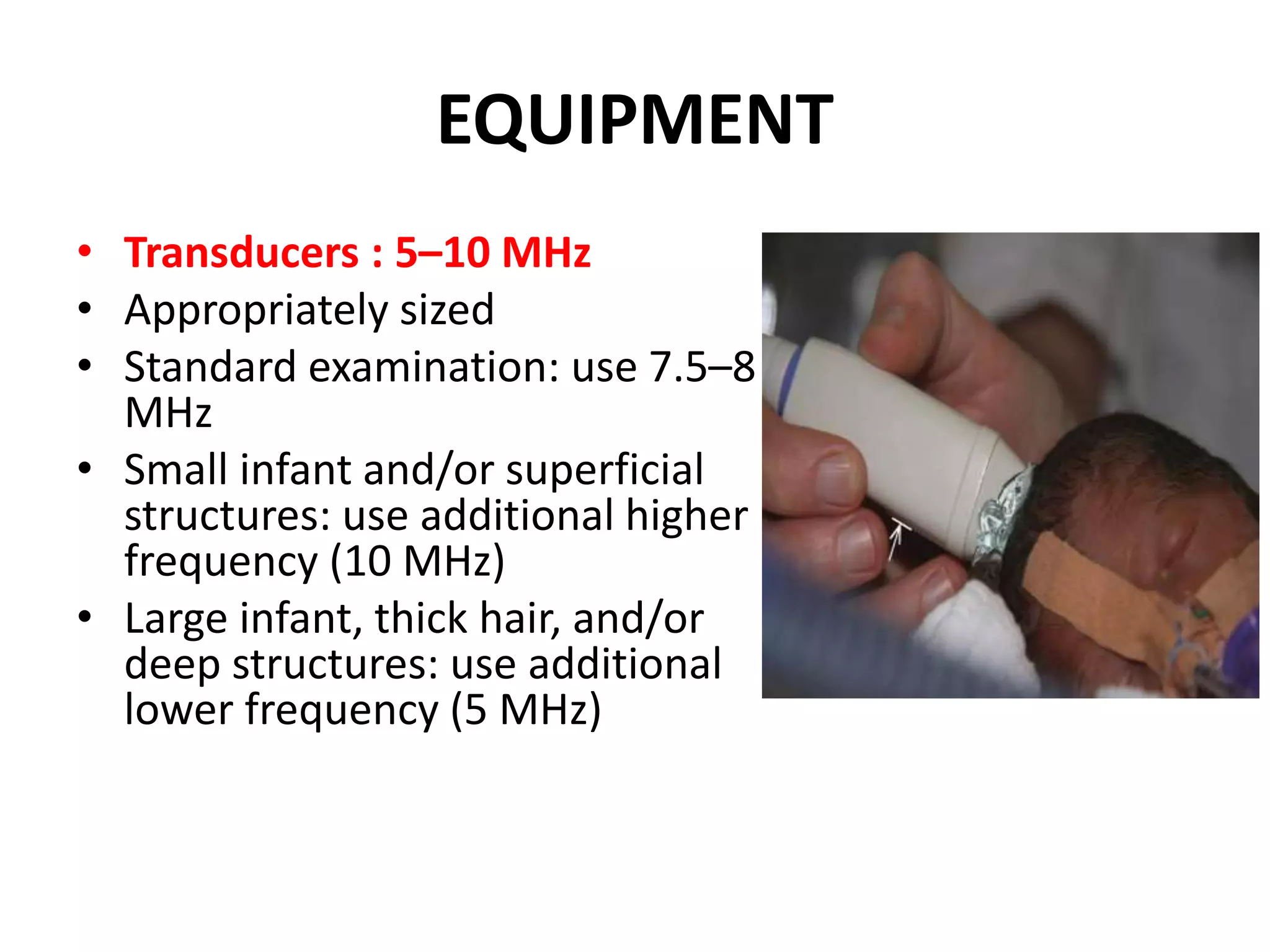
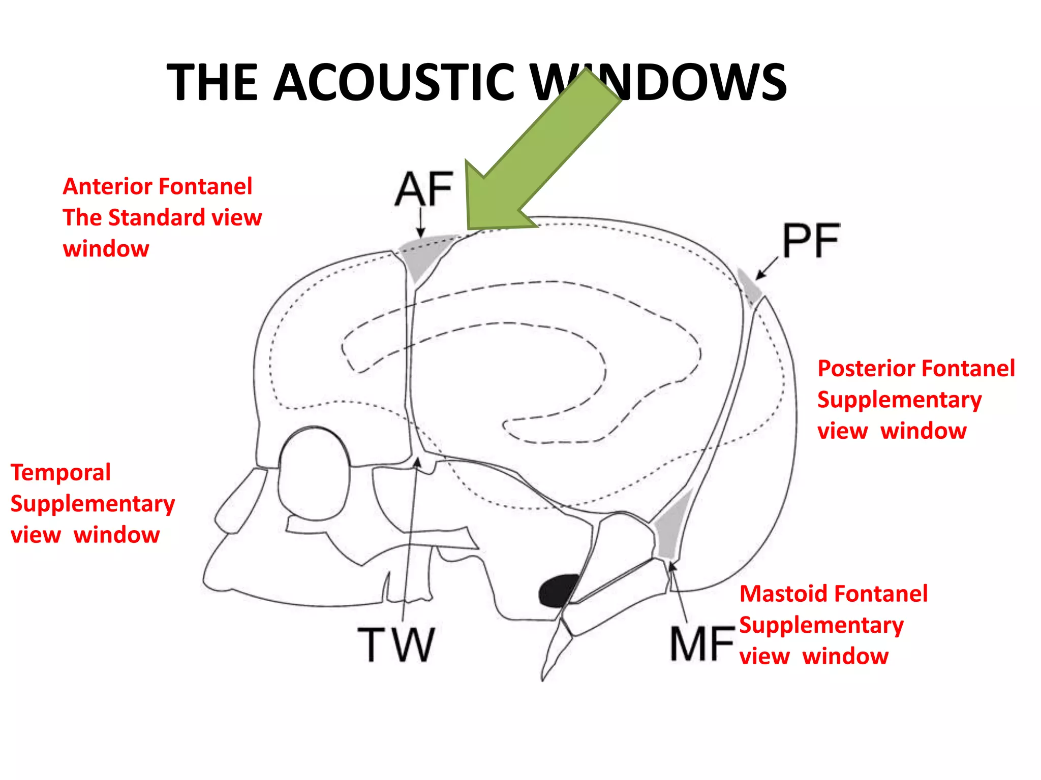
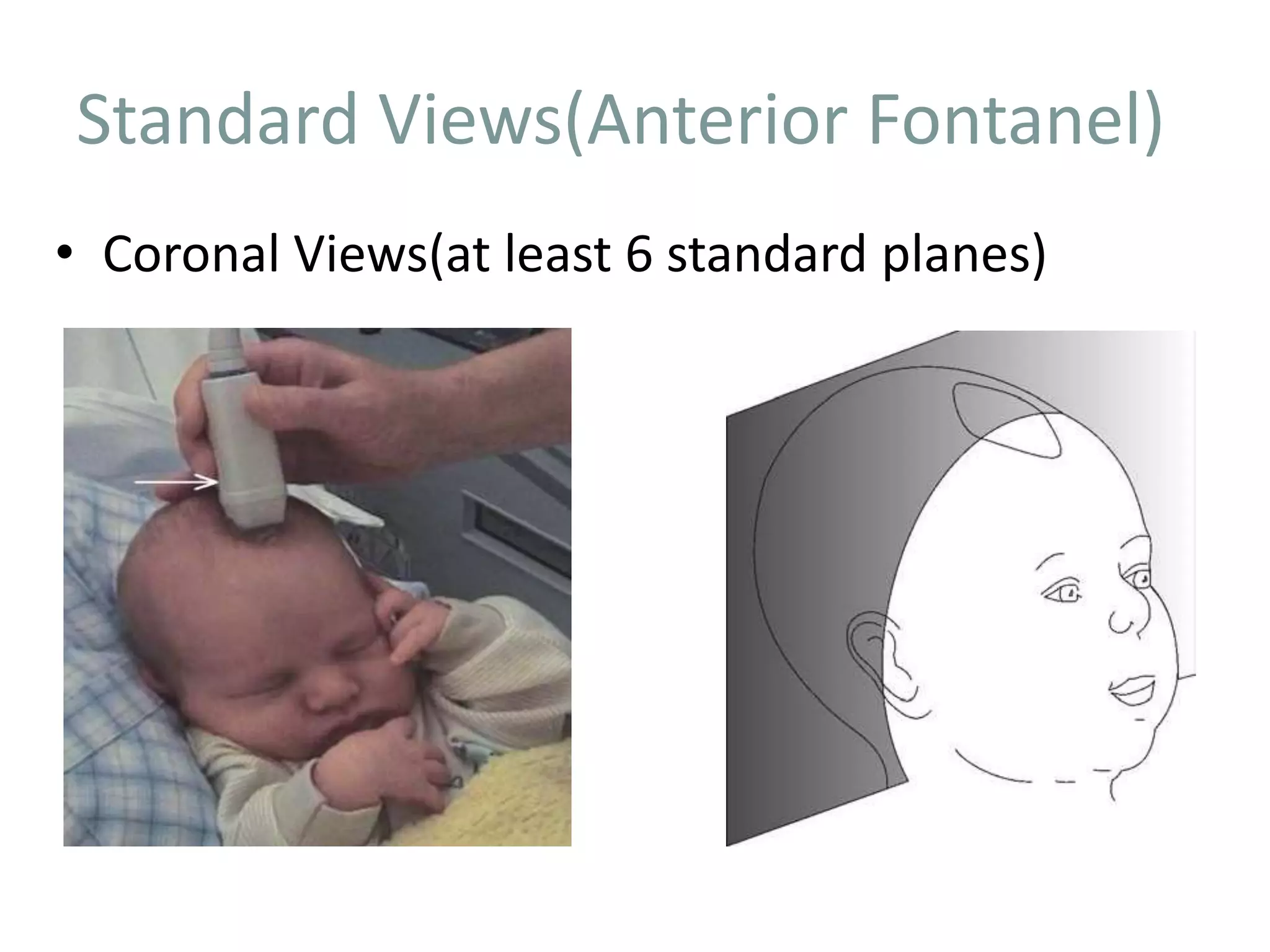
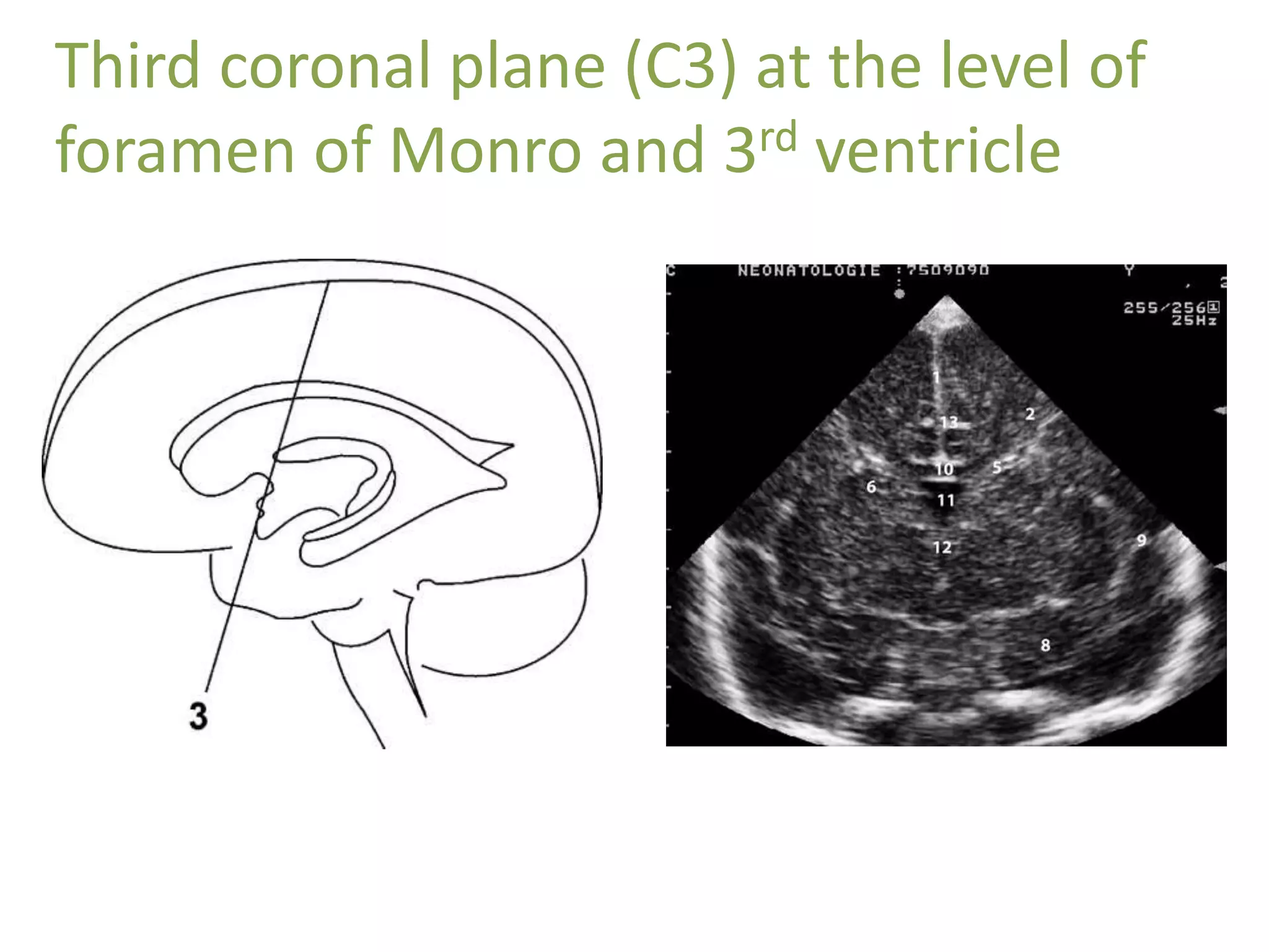
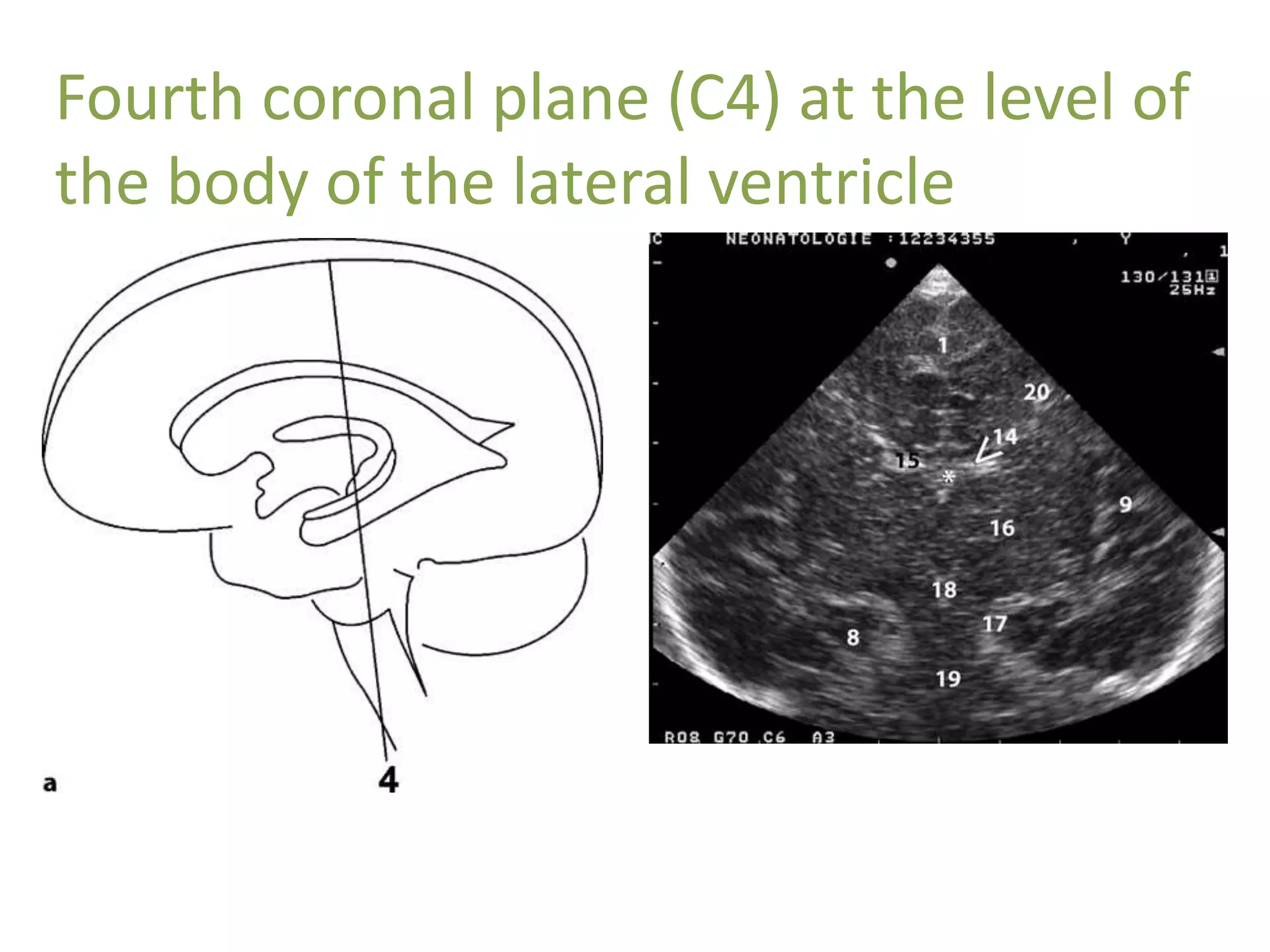
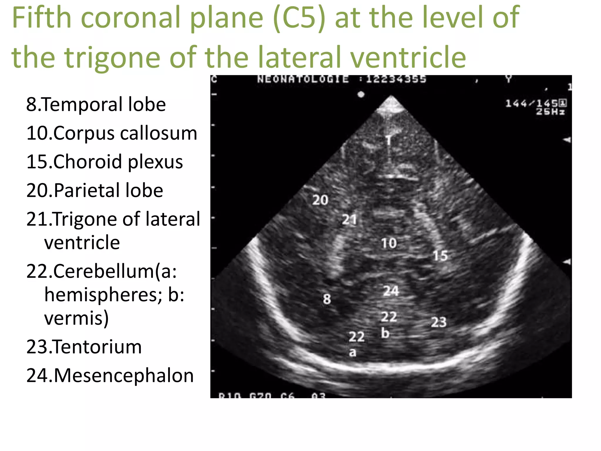
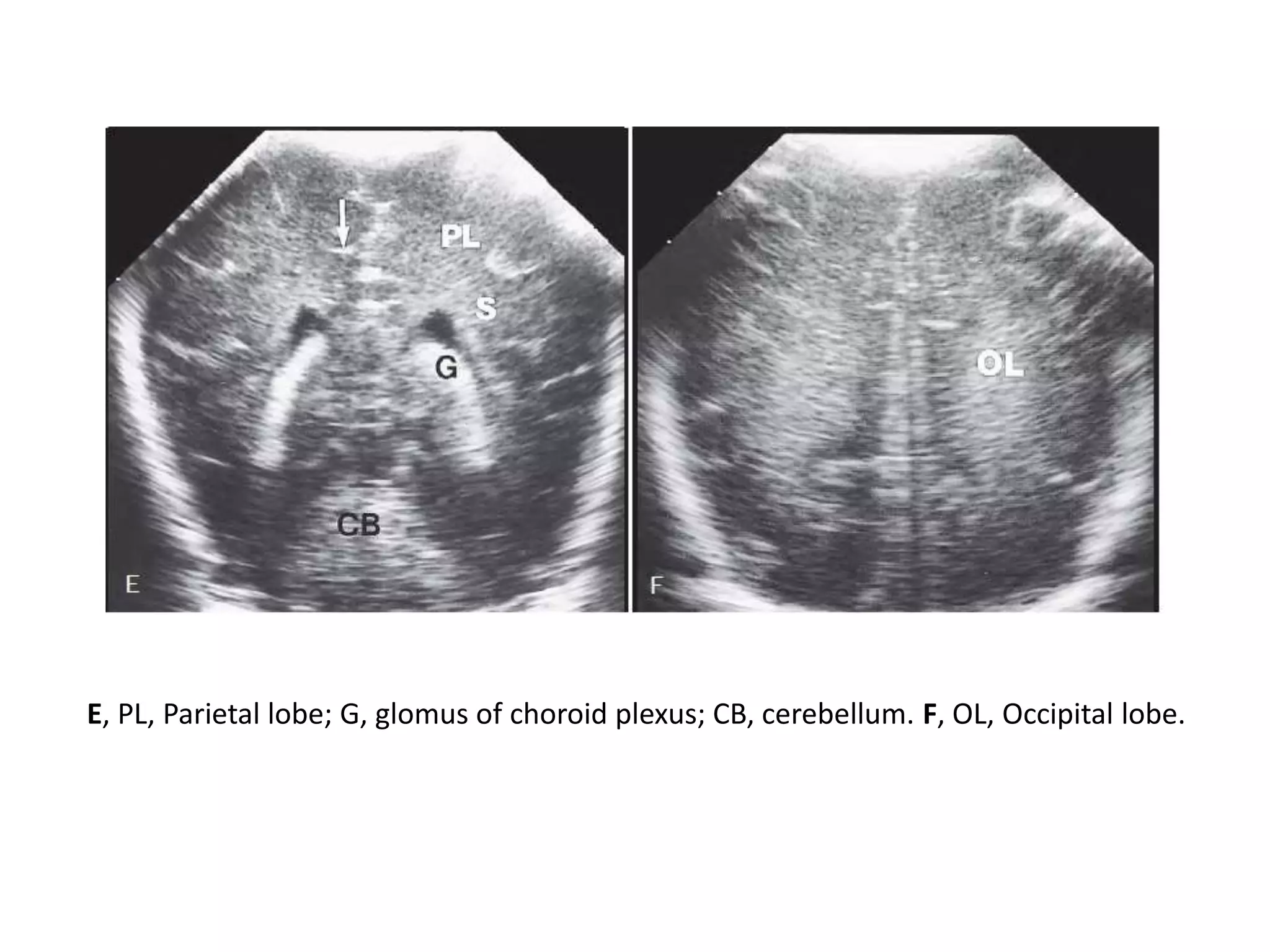
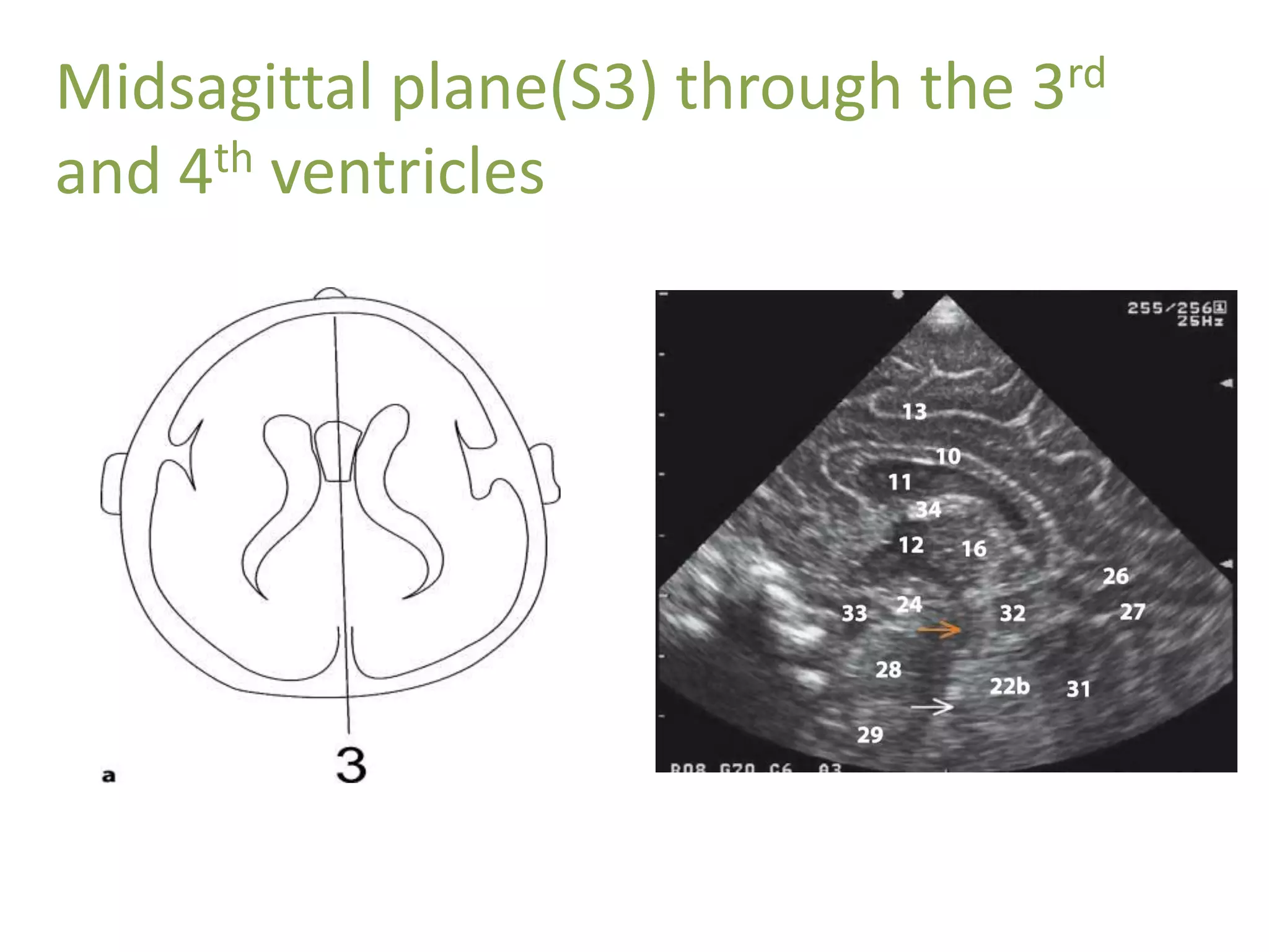
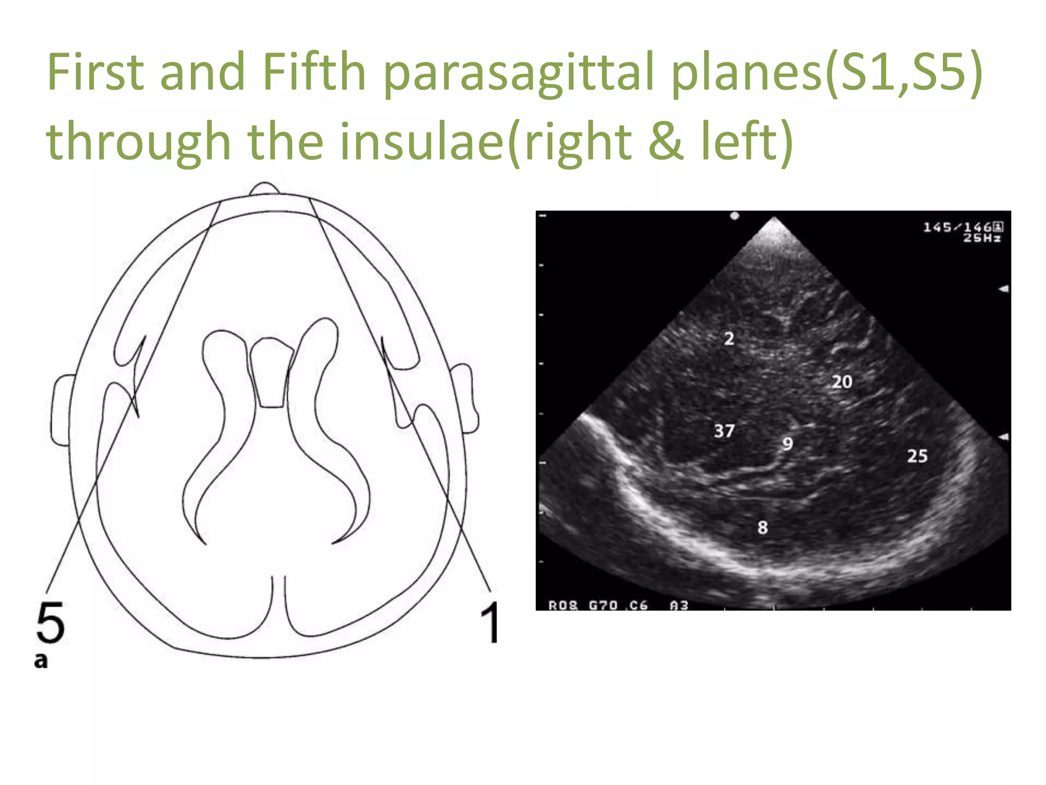
By dr Rabab Hashem, MRCPCH, pediatrician at El Nasr hospital Port said.
Cranial sonography is the most widely used neuroimaging procedure in premature infants. US helps in assessing the neurologic status of the child, since clinical examination and symptoms are often nonspecific. It gives information about immediate and long term prognosis.
This lecture prides 8 easy steps of scanning the neonatal brain through anterior fontanelle ,followed by examples of most commonly encountered abnormalities.
In this presentation we will discuss role of high resolution in characterizing normal variant and pathologies of spinal pathologies.
This is a pictoral review.
Description of various ultrasound features of benign and suspicious thyroid nodules with multiple ultrasound systems for risk stratification of malignancy.
Learn Chest X-Ray With Its Normal Positioning & Radio-AnatomyDr.Santosh Atreya
Learn Chest X-Ray With Its Normal Positioning & Radio-Anatomy..For some image description please go through the text book "David Sutton" because i have described these image during my presentation Verbally..There are many animations used inside this presentation so to see all the pictures which are placed layer by layer with the help of animations you simple need to download this presentation first.... Thanx.
Flu Vaccine Alert in Bangalore Karnatakaaddon Scans
As flu season approaches, health officials in Bangalore, Karnataka, are urging residents to get their flu vaccinations. The seasonal flu, while common, can lead to severe health complications, particularly for vulnerable populations such as young children, the elderly, and those with underlying health conditions.
Dr. Vidisha Kumari, a leading epidemiologist in Bangalore, emphasizes the importance of getting vaccinated. "The flu vaccine is our best defense against the influenza virus. It not only protects individuals but also helps prevent the spread of the virus in our communities," he says.
This year, the flu season is expected to coincide with a potential increase in other respiratory illnesses. The Karnataka Health Department has launched an awareness campaign highlighting the significance of flu vaccinations. They have set up multiple vaccination centers across Bangalore, making it convenient for residents to receive their shots.
To encourage widespread vaccination, the government is also collaborating with local schools, workplaces, and community centers to facilitate vaccination drives. Special attention is being given to ensuring that the vaccine is accessible to all, including marginalized communities who may have limited access to healthcare.
Residents are reminded that the flu vaccine is safe and effective. Common side effects are mild and may include soreness at the injection site, mild fever, or muscle aches. These side effects are generally short-lived and far less severe than the flu itself.
Healthcare providers are also stressing the importance of continuing COVID-19 precautions. Wearing masks, practicing good hand hygiene, and maintaining social distancing are still crucial, especially in crowded places.
Protect yourself and your loved ones by getting vaccinated. Together, we can help keep Bangalore healthy and safe this flu season. For more information on vaccination centers and schedules, residents can visit the Karnataka Health Department’s official website or follow their social media pages.
Stay informed, stay safe, and get your flu shot today!
Report Back from SGO 2024: What’s the Latest in Cervical Cancer?bkling
Are you curious about what’s new in cervical cancer research or unsure what the findings mean? Join Dr. Emily Ko, a gynecologic oncologist at Penn Medicine, to learn about the latest updates from the Society of Gynecologic Oncology (SGO) 2024 Annual Meeting on Women’s Cancer. Dr. Ko will discuss what the research presented at the conference means for you and answer your questions about the new developments.
Knee anatomy and clinical tests 2024.pdfvimalpl1234
This includes all relevant anatomy and clinical tests compiled from standard textbooks, Campbell,netter etc..It is comprehensive and best suited for orthopaedicians and orthopaedic residents.
New Drug Discovery and Development .....NEHA GUPTA
The "New Drug Discovery and Development" process involves the identification, design, testing, and manufacturing of novel pharmaceutical compounds with the aim of introducing new and improved treatments for various medical conditions. This comprehensive endeavor encompasses various stages, including target identification, preclinical studies, clinical trials, regulatory approval, and post-market surveillance. It involves multidisciplinary collaboration among scientists, researchers, clinicians, regulatory experts, and pharmaceutical companies to bring innovative therapies to market and address unmet medical needs.
NVBDCP.pptx Nation vector borne disease control programSapna Thakur
NVBDCP was launched in 2003-2004 . Vector-Borne Disease: Disease that results from an infection transmitted to humans and other animals by blood-feeding arthropods, such as mosquitoes, ticks, and fleas. Examples of vector-borne diseases include Dengue fever, West Nile Virus, Lyme disease, and malaria.
263778731218 Abortion Clinic /Pills In Harare ,sisternakatoto
263778731218 Abortion Clinic /Pills In Harare ,ABORTION WOMEN’S CLINIC +27730423979 IN women clinic we believe that every woman should be able to make choices in her pregnancy. Our job is to provide compassionate care, safety,affordable and confidential services. That’s why we have won the trust from all generations of women all over the world. we use non surgical method(Abortion pills) to terminate…Dr.LISA +27730423979women Clinic is committed to providing the highest quality of obstetrical and gynecological care to women of all ages. Our dedicated staff aim to treat each patient and her health concerns with compassion and respect.Our dedicated group ABORTION WOMEN’S CLINIC +27730423979 IN women clinic we believe that every woman should be able to make choices in her pregnancy. Our job is to provide compassionate care, safety,affordable and confidential services. That’s why we have won the trust from all generations of women all over the world. we use non surgical method(Abortion pills) to terminate…Dr.LISA +27730423979women Clinic is committed to providing the highest quality of obstetrical and gynecological care to women of all ages. Our dedicated staff aim to treat each patient and her health concerns with compassion and respect.Our dedicated group of receptionists, nurses, and physicians have worked together as a teamof receptionists, nurses, and physicians have worked together as a team wwww.lisywomensclinic.co.za/
Lung Cancer: Artificial Intelligence, Synergetics, Complex System Analysis, S...Oleg Kshivets
RESULTS: Overall life span (LS) was 2252.1±1742.5 days and cumulative 5-year survival (5YS) reached 73.2%, 10 years – 64.8%, 20 years – 42.5%. 513 LCP lived more than 5 years (LS=3124.6±1525.6 days), 148 LCP – more than 10 years (LS=5054.4±1504.1 days).199 LCP died because of LC (LS=562.7±374.5 days). 5YS of LCP after bi/lobectomies was significantly superior in comparison with LCP after pneumonectomies (78.1% vs.63.7%, P=0.00001 by log-rank test). AT significantly improved 5YS (66.3% vs. 34.8%) (P=0.00000 by log-rank test) only for LCP with N1-2. Cox modeling displayed that 5YS of LCP significantly depended on: phase transition (PT) early-invasive LC in terms of synergetics, PT N0—N12, cell ratio factors (ratio between cancer cells- CC and blood cells subpopulations), G1-3, histology, glucose, AT, blood cell circuit, prothrombin index, heparin tolerance, recalcification time (P=0.000-0.038). Neural networks, genetic algorithm selection and bootstrap simulation revealed relationships between 5YS and PT early-invasive LC (rank=1), PT N0—N12 (rank=2), thrombocytes/CC (3), erythrocytes/CC (4), eosinophils/CC (5), healthy cells/CC (6), lymphocytes/CC (7), segmented neutrophils/CC (8), stick neutrophils/CC (9), monocytes/CC (10); leucocytes/CC (11). Correct prediction of 5YS was 100% by neural networks computing (area under ROC curve=1.0; error=0.0).
CONCLUSIONS: 5YS of LCP after radical procedures significantly depended on: 1) PT early-invasive cancer; 2) PT N0--N12; 3) cell ratio factors; 4) blood cell circuit; 5) biochemical factors; 6) hemostasis system; 7) AT; 8) LC characteristics; 9) LC cell dynamics; 10) surgery type: lobectomy/pneumonectomy; 11) anthropometric data. Optimal diagnosis and treatment strategies for LC are: 1) screening and early detection of LC; 2) availability of experienced thoracic surgeons because of complexity of radical procedures; 3) aggressive en block surgery and adequate lymph node dissection for completeness; 4) precise prediction; 5) adjuvant chemoimmunoradiotherapy for LCP with unfavorable prognosis.
Couples presenting to the infertility clinic- Do they really have infertility...Sujoy Dasgupta
Dr Sujoy Dasgupta presented the study on "Couples presenting to the infertility clinic- Do they really have infertility? – The unexplored stories of non-consummation" in the 13th Congress of the Asia Pacific Initiative on Reproduction (ASPIRE 2024) at Manila on 24 May, 2024.
Acute scrotum is a general term referring to an emergency condition affecting the contents or the wall of the scrotum.
There are a number of conditions that present acutely, predominantly with pain and/or swelling
A careful and detailed history and examination, and in some cases, investigations allow differentiation between these diagnoses. A prompt diagnosis is essential as the patient may require urgent surgical intervention
Testicular torsion refers to twisting of the spermatic cord, causing ischaemia of the testicle.
Testicular torsion results from inadequate fixation of the testis to the tunica vaginalis producing ischemia from reduced arterial inflow and venous outflow obstruction.
The prevalence of testicular torsion in adult patients hospitalized with acute scrotal pain is approximately 25 to 50 percent
Prix Galien International 2024 Forum ProgramLevi Shapiro
June 20, 2024, Prix Galien International and Jerusalem Ethics Forum in ROME. Detailed agenda including panels:
- ADVANCES IN CARDIOLOGY: A NEW PARADIGM IS COMING
- WOMEN’S HEALTH: FERTILITY PRESERVATION
- WHAT’S NEW IN THE TREATMENT OF INFECTIOUS,
ONCOLOGICAL AND INFLAMMATORY SKIN DISEASES?
- ARTIFICIAL INTELLIGENCE AND ETHICS
- GENE THERAPY
- BEYOND BORDERS: GLOBAL INITIATIVES FOR DEMOCRATIZING LIFE SCIENCE TECHNOLOGIES AND PROMOTING ACCESS TO HEALTHCARE
- ETHICAL CHALLENGES IN LIFE SCIENCES
- Prix Galien International Awards Ceremony
Ozempic: Preoperative Management of Patients on GLP-1 Receptor Agonists Saeid Safari
Preoperative Management of Patients on GLP-1 Receptor Agonists like Ozempic and Semiglutide
ASA GUIDELINE
NYSORA Guideline
2 Case Reports of Gastric Ultrasound
2. • Neonatal sonography of the brain is an essential
part of newborn care, particularly in high-risk and
unstable premature infants.
• Current ultrasound (US) technology allows for
rapid evaluation of infants in the intensive care
nursery with virtually no risk.
• Color and spectral Doppler ultrasound of cranial
blood flow may prove valuable, particularly for
cystic lesions when the differential diagnosis
includes a vascular lesion, or for possible
subdural hematomas, and to separate normal
vascular structures from clot.
3. Advantage of cranial US (CUS)
• Bedside- compatible
• Reliable
• Early imaging
• Serial imaging:
Brain maturation
Evolution of lesions
• Inexpensive
• Suitable for screening
• no ionizing radiation
• no sedation.
4. EQUIPMENT
• Transducers : 5–10 MHz
• Appropriately sized
• Standard examination: use 7.5–8
MHz
• Small infant and/or superficial
structures: use additional higher
frequency (10 MHz)
• Large infant, thick hair, and/or
deep structures: use additional
lower frequency (5 MHz)
5. THE ACOUSTIC WINDOWS
Anterior Fontanel
The Standard view
window
Posterior Fontanel
Supplementary
view window
Mastoid Fontanel
Supplementary
view window
Temporal
Supplementary
view window
6. SONOGRAPHIC TECHNIQUE
• Every effort should be made to maintain normal
body temperature in premature infants when
performing ultrasound
• Handwashing and cleansing of the transducer
between patients are of paramount importance
to avoid the spread of infection in the intensive
care nursery.
• Standard brain scanning includes
sagittal and coronal view from anterior
fontanelle
axial view from posterior and mastoid fontanel
7. Coronal brain ultrasound planes through anterior fontanelle. A through F correspond to front to back. CC,
Cerebral cortex; BV, body of lateral ventricle; FH, frontal horn; OH, occipital horn; CN, caudate nucleus;
M, massa intermedia; PR, pineal recess; 3, third ventricle; TH, temporal horn; SR, supraoptic recess; IR,
infundibular recess; CP, choroid plexus; 4, fourth ventricle; CB, cerebellum.
Coronal view
•Coronal images are obtained by
placing the scan head of the transducer
transversely across the anterior
fontanelle
•The plane of the ultrasound beam
should then sweep in an anterior-to-
posterior direction, completely
through the brain.
•At least six standard coronal images
should be obtained during this anterior
to posterior sweep.
11. First coronal plane (C1) at the level of
frontal lobes
1. Interhemispheric
fissure
2. Frontal lobe
3. Skull
4. Orbit
12. Second coronal plane (C2) at the level of
frontal horns of the lateral ventricles
13. Second coronal plane (C2) at the level of
frontal horns of the lateral ventricles
2.Frontal lobe
5.Frontal horn of
lateral ventricle
6.Caudate nucleus
7.Basal ganglia
8.Temporal lobe
9.Sylvian fissure
17. Fourth coronal plane (C4) at the level of
the body of the lateral ventricle
1.Interhemispheric
fissure
8.Temporal lobe
9.Sylvian fissure
14.Body of lateral
ventricle
15.Choroid plexus
16.Thalamus
17.Hippocampal fissure
18.Aqueduct of Sylvius
19.Brain stem
20.Parietal lobe
18. Fifth coronal plane (C5) at the level of
the trigone of the lateral ventricle
19. Fifth coronal plane (C5) at the level of
the trigone of the lateral ventricle
8.Temporal lobe
10.Corpus callosum
15.Choroid plexus
20.Parietal lobe
21.Trigone of lateral
ventricle
22.Cerebellum(a:
hemispheres; b:
vermis)
23.Tentorium
24.Mesencephalon
21. Sixth coronal plane (C6) through the
parieto occipital lobes
20.Parietal lobe
25.Occipital lobe
26.Parieto-
occipital fissure
27.Calcarine
fissure
22. Coronal brain ultrasound images: normal full-term infant. Anterior to posterior corresponds to sections A to
F - A, FL, Frontal lobes; black arrow, interhemispheric fissure. B, P, Putamen; C, caudate nucleus; f,
frontal horns of lateral ventricles; TL, temporal lobe; arrowhead, corpus callosum; closed arrow, sylvian
fissure; open arrow, bifurcation of internal carotid artery. (On images A and B black arrow represents
interhemispheric fissure.) C, B, Brainstem; 3, location of third ventricle (third and fourth ventricles are
difficult to see in normal patients on coronal cuts). D, S, Centrum semiovale; b, body of lateral ventricle; c,
choroid plexus; T, thalamus; V, vermis of cerebellum; curved arrow, tentorium cerebelli; straight white
arrow, cingulate sulcus.
23. E, PL, Parietal lobe; G, glomus of choroid plexus; CB, cerebellum. F, OL, Occipital lobe.
24. Sagittal Imaging
•The sagittal images are obtained by
placing the transducer longitudinally
across the anterior fontanelle and
angling it to each side.
•The midline is first identified through
the interhemispheric fissure by
recognition of the curving line of the
corpus callosum above the cystic
cavum septi pellucidi and cavum
vergae, the third and fourth ventricles,
and the highly echogenic cerebellar
vermis.
36. •The transducer is placed over
posterior fontanelle and angled
slightly off midline with the
anterior portion of the probe
directed slightly medially, to
demonstrate the lateral
ventricular trigone with its
occipital horn in the near field.
•Angling the transducer into
the left and right parasagittal
planes will display each
occipital horn
Posterior fontanelle scan
39. Mastoid fontanelle images at fourth ventricle level in posterior fossa. A, Normal cerebellar
hemispheres (C), cerebellar vermis (V), fourth ventricle (4), and cisterna magna (CM).
•The ultrasound transducer is
placed about 1 cm behind the
helix of the ear and 1 cm above
the tragus.
•The mastoid fontanelle allows
assessment of the brainstem
and posterior fossa, which are
not well demonstrated in the
standard planes through the
anterior fontanelle.
Mastoid Fontanelle scan
40. Normal variants
• Frontal horn cyst.
• Cavum septum pellucidum and
cavum verge
• Cavum velum interpositum
• Calcar avis
• Mega cisterna magna
41. Frontal horn cysts, coronal and sagittal sonogram shows uncommon normal
cystic variants, also called frontal horn coarctation or connatal cysts. Frontal
horn cysts appear to be septations in the ventricles (arrow) but are thought to
be caused by folding of the frontal horn on itself.
Frontal horn cysts
•coarctation of the frontal horn
or connatal cysts
•exactly parallel (not above or
below) and adjacent to the frontal
horns.
•caused by folding of the frontal
horn on itself, resulting in kinking
(seen as a septation).
•typically bilateral and have
septations between the cyst and
the frontal horns
42. • continuous cystic midline structure in the septum
pellucidum during fetal life. The septum contains
the cavum septi pellucidi anterior to the foramen
of Monro and the cavum vergae posteriorly.
• Both parts are normally present early in
gestation, but they close from back to front,
starting at approximately 6 months’ gestation.
• By full term, closure has occurred posteriorly in
97% of infants so that there is only a cavum septi
pellucidi at birth.
Cavum Septi Pellucidum and Cavum Vergae
43. Normal premature infant brain. Sagittal. A, 25 weeks. No sulci; corpus callosum lies above
completely cystic cavum septi pellucidi and cavum vergae. Arrow, Occipitoparietal fissure. B, 27
weeks. Cingulate sulcus (arrow) is just developed, and cavum septi pellucidi (C) and cavum
vergae (V) are readily visible.
44. C, 30 weeks. Cingulate sulcus (short arrow) has a few branches. Long arrow, Cavum velum
interpositum.
•represents a potential space
above the choroid in the roof of
the third ventricle and below the
columns of the fornices.
•may appear as an anechoic,
inverted, helmetlike space
just inferior and posterior to the
splenium in the pineal region
Cavum velum interpositum
45. B, Calcar avisv(CA) can be averaged into the occipital horn and imitate
hemorrhage. Note continuity with the adjacent brain identifies this structure.
Calcar avis
•On posterior fontanelle views, a
normal gyrus, the calcar avis,
frequently protrudes into the
medial aspect of the lateral
ventricle at the junction of the
trigone and occipital horn.
•It can be recognized due to a
central echogenic sulcus (calcarine
fissure), its continuity with the
adjacent brain, and normal
vascularity on color Doppler
ultrasound.
46. Mega cisterna magna. A and B, Sagittal and coronal sonograms show an enlarged cisterna
magna (C) behind the vermis on A and below the tentorium on B, with no communication to
the fourth ventricle and no hydrocephalus.
Mega cisterna magna
47. Ventricular measurement
• Measurement of the ventricular system should be done in an easy
reproducable sonographic plane.
• Coronal section is used through the lateral ventricles slightly posterior to
the foramen of Monro.
• Two methods
1)Levine index- Up to 40 weeks of gestational age
2)Ventricular index-After 40 weeks
Levine index
•the absolute distance between the
falx and the lateral wall of the
anterior horn in the coronal plane at
the level of the third ventricle.
•This is performed for the left and
right side.
•measurements can be compared to
the reference curve and are quite
useful for further follow-up.
48. Ventricular index
•the ratio of the distance
between the lateral sides
of the ventricles and the
biparietal diameter.
51. DISORDERS OF NEURAL TUBE CLOSURE
Chiari Malformations
Chiari I malformation
• simply the downward displacement of the cerebellar
tonsils, without displacement of the fourth ventricle or
medulla.
Chiari II malformation
• the most common and of greatest clinical importance
because of its almost universal association with
myelomeningocele.
Chiari III malformation
• a high cervical encephalomeningocele in which the
medulla, fourth ventricle, and virtually the entire
cerebellum reside.
52.
53. Chiari II malformation. A, Lateral parasagittal sonogram, and B, MR image, show pointing of
frontal horn(curved arrow) and colpocephaly; O, occipital horn; T, trigone of lateral ventricle.
54. Chiari II malformation. C, Midline sagittal sonogram, and D, midline sagittal MR image, show
enlargement of massa intermedia (M) and tonsillar herniation through foramen magnum
(arrow).
55. Agenesis of Corpus Callosum
•depending on the timing of the intrauterine insult,
development of corpus callosum may be partially or
completely arrested . If partial, the genu is usually
present, and the dorsal splenium or the anterior
rostrum is absent.
•associated anomalies include
Chiari II and Dandy-Walker malformations,
holoprosencephaly, encephaloceles, lipomas,
arachnoid cysts, migrational abnormalities, and
Aicardi syndrome
56.
57. Agenesis of the corpus callosum, isolated anomaly. A, Tiny frontal horns are widely
separated. B, Starburst sulci radiate above the third ventricle. C and D, Occipital horns are
larger than frontal horns and widely separated.
58. Agenesis of the corpus callosum.
A and B, Sagittal and coronal sonograms show marked tapering of the frontal
horns with larger occipital horns (colpocephaly).
59. Dandy-Walker Malformation
• Characterised by cystic dilatation of the fourth
ventricle and an enlarged posterior fossa with
upward displacement of lateral sinuses,
tentorium and torcula herophili associated with
varying degree of vermian aplasia or hypoplasia.
• Due to failure of development of anterior
medullary velum( embryonic roof of 4th
ventricular) atresia of fourth ventricular outlet
foramina, and delayed opening of foramen
magendie.
60.
61. Dandy-Walker malformation: classic isolated malformation. A and B, Coronal and
sagittal sonograms. C, Dandy-Walker posterior fossa cyst; 3, third ventricle; 4, fourth
ventricle; L and LV, dilated lateral ventricles
62. Dandy-Walker malformation: classic isolated malformation C, Coronal ultrasound shows Dandy-Walker
malformation after ventriculoperitoneal shunt. Lateral and third ventricles have decompressed, but Dandy
Walker cyst (D) remains dilated. Frequently, posterior fossa cysts require additional shunts for
decompression.
63. Dandy Walker Variant
Dandy-Walker variant. Axial posterior fossa ultrasound shows a wide continuity between the
fourth ventricle and the cisterna magna, where the cerebellar vermis is hypoplastic and
separated behind the fourth ventricle.
•There is mild vermian hypoplasia with
a variable sized cystic space caused by
open communication of posteroinferior
4th ventricle and cisterna magna
through enlarged vallecula.
•4th ventricle is often slightly to
moderately enlarged but posterior
fossa is typically normal size.
•Brain stem is usually normal.
•Although hydrocephalus may be
present, 3rd and lateral ventricles are
usually normal.
64. DISORDERS OF DIVERTICULATION AND CLEAVAGE
Holoprosencephaly
• results from a failure of diverticulation when the primitive
prosencephalon does not divide into the telencephalon
and the diencephalon between the fourth and eighth
weeks of gestation.
• represents a spectrum of malformations that form a
continuum from most severe, with no separation of the
telencephalon into hemispheres (alobar
holoprosencephaly), to least severe, with partial
separation of the dorsal aspects of the brain (lobar
holoprosencephaly)
• The septum pellucidum is absent in all forms of
holoprosencephaly
65. Alobar
Holoprosencephaly
-the most severe form of
disorder.
Infants with this defect usually
die within the first months of
life or are stillborn.
-Facial features may include
cebocephaly , cyclopia, and
ethmocephaly (cyclopia or
hypotelorism) with a midline
proboscis above the eyes.
66. Alobar holoprosencephaly. A, Coronal sonogram shows single central ventricle (V) and
fused thalami (T). No falx or interhemispheric fissure is present. B, MR image, and C, pathology
specimen, show single central ventricle and fused thalami. D, Autopsy specimen shows
cebocephaly (severe hypotelorism and malformed nose).
67. Septo-optic Dysplasia
Septo-optic dysplasia. A and
B, Coronal and
sagittal sonograms; C,
pathology specimen.
Although the cavum septum
pellucidum is absent, the
corpus callosum is present.
-mildest form of lobar
holoprosencephaly
-absence of the septum
pellucidum and optic
nerve hypoplasia
-About two thirds of these
infants have hypothalamic-
pituitary dysfunction.
-may have visual
symptoms and growth
restriction.
68. DISORDERS OF SULCATION AND
CELLULAR MIGRATION
Schizencephaly
• Split brain
• a gray matter lined CSF filled cleft that extends from the
ependymal surface of the brain through the white matter
to the pia
• caused by a primary neuronal migration malformation in
utero
• Two types-
closed lip – cleft walls are in apposition
open lip- cleft walls are separated.
• Cleft may be unilateral or bilateral, symmetrical or
asymmetric
69. Schizencephaly. A, Coronal sonogram, and B, coronal MR image, of open-lip schizencephaly
show bilateral clefts (c) with wide openings to the ventricular (v) system. C, Sonogram, and D,
CT image, show closed-lip schizencephaly with calcification from in utero infection.
70. DESTRUCTIVE LESIONS
Porencephalic Cyst
• After 26 weeks’ gestation, a porencephalic cyst will develop
in an area of normally developed brain that has been
damaged and heals with scarring because of a lining of
gliotic white matter.
• By definition, porencephalic cysts always connect with the
ventricular system but do not extend to the surface cortex.
• usually occur after birth secondary to intraparenchymal
hemorrhage (IPH), infection (focal vasculitis, abscess), or
trauma.
71. Hydranencephaly
• believed to be caused by bilateral occlusion of
the internal carotid arteries during fetal
development, but it may result from any of
several intracranial destructive processes.
• It is the severest form of porencephaly in that
there is almost total destruction of the
cerebral cortex.
• These infants may appear surprisingly normal
at birth but are developmentally delayed from
an early age and frequently die within the first
year of life.
72. • USG findings:
-calvarium filled with CSF
-Structures that receive their blood supply
from the posterior cerebral artery and vertebral
artery, such as the thalamus, cerebellum,
brainstem, and posterior choroid plexus are
spared
• Blood flow will not be appreciated by color or
spectral Doppler in the carotid arteries.
• The presence of the falx helps differentiate this
lesion from alobar holoprosencephaly, in which
the falx does not form.
73. Hydranencephaly in two full-term
newborns. A, Coronal anterior; B, midline
sagittal; and C, coronal posterior
sonograms. Note that only anechoic
cerebrospinal fluid is seen above
thalamus (T), which at first looks like
holoprosencephaly, but the definitive
diagnosis is made because there is a falx
seen in the midline on all three views.
Note the echogenic falx on the midline
view.
74. Cystic Encephalomalacia
• Encephalomalacia is an area of focal brain damage that
pathologically has astrocytic proliferation and glial septations.
• In diffuse brain damage, there may be large areas of cystic
encephalomalacia
Cystic encephalomalacia. A , coronal ; B, sagittal , Severe hypoxic ischemic
encephalopathy has resulted in diffuse infarction, particularly severe in the
cerebral cortex, leading to multiple cystic areas in necrotic brain.
75. HYDROCEPHALUS
• Hydrocephalus occurs in 5 to 25 per 10,000
births and results from an imbalance between
CSF production and its drainage by the
arachnoid villi.
• Neonatal hydrocephalus is easily recognized
by routine coronal and sagittal imaging.
• Sonography is also helpful in following
ventricular decompression in patients shunted
for hydrocephalus.
76.
77. Aqueductal stenosis. A Coronal and midline sagittal ultrasound images; B, Sagittal T1-
weighted MR scan. Marked lateral and third (arrow) ventricular dilation demonstrated, but
fourth ventricle is normal (best seen anterior to the cerebellar vermis on sagittal MRI).
A B
78. Germinal Matrix Hemorrhage
• GMH is a common event occurring primarily in
premature infants less than 32 weeks’ gestational age.
• Infants at greatest risk are those at gestational ages of
less than 30 weeks, with birth weight less than 1500 g,
or both.
• GMH originates predominantly as hemorrhage in the
germinal matrix below the subependymal layer and
may be contained by the ependyma or may rupture
into the ventricular system or less often into the
adjacent parenchyma.
• Sonography is the most effective method for detecting
this hemorrhage in the newborn period and for follow-
up in the subsequent weeks.
80. Sequelae of subependymal
hemorrhage
(SEH). SEH may resolve, leaving a
normal scan; may resolve, leaving a
small subependymal cyst; or may
progress, rupturing into the ventricle,
causing intraventricular hemorrhage
(IVH), or extending into the
parenchyma, causing
intraparenchymal hemorrhage (IPH).
Hydrocephalus (HC) and porencephaly
(PC) are common sequelae of SEH.
81. Sagittal and coronal US of subependymal hemorrhage located
in the groove between the thalamus and the nucleus caudatue.
Grade I
82.
83. Coronal and Sagittal US Both lateral ventricles are filled with blood,
but there is no ventricular dilatation.
Grade II
84.
85. Coronal and sagittal sonograms show aging clot has become less echogenic
than choroid, lateral ventricle is dilated
Grade III
86.
87. Coronal US, a large subependymal bleeding but also a large area with increased
echogenicity in the brain parenchyma lateral to the ventricle.
Grade IV
88.
89. Peri Ventricular Leukomalacia (PVL)
• PVL is also known as Hypoxic-Ischemic Encephalopathy (HIE)
of the preterm.
• It is a white matter disease that affects the periventricular
zones.
• In prematures this white matter zone is a watershed zone
between deep and superficial vessels.
• PVL presents as areas of increased periventricular
echogenicity.
• Normally the echogenicity of the periventricular white matter
should be less than the echogenicity of the choroid plexus.
• PVL occurs most commonly in premature infants born at less
than 33 weeks gestation (38% PVL) and less than 1500 g birth
weight (45% PVL).
90. • Detection of PVL is important because a significant
percentage of surviving premature infants with PVL develop
cerebral palsy, intellectual impairment or visual disturbances.
• More than 50% of infants with PVL or grade III hemorrhage
develop cerebral palsy.
• Grading
91.
92. Transverse and sagittal image of a child with
PVL grade 2.
-small periventricular cysts.
Sagittal image of a child with PVL grade 1
-Increased periventricular echogenicity
93. Sagittal image demonstarting extensive
PVL grade 3
-areas of increased periventricular
echogenicity, that develop into extensive
periventricular cysts in the occipital and
fronto-parietal region.
Coronal and transverse images demonstrating
PVL grade 4
-areas of increased periventricular echogenicity
in the deep white matter developing into
extensive subcortical cysts.
94. Post traumatic injury
Subdural and Epidural Hematomas
• these hematomas present as unilateral or
bilateral hypoechoic fluid collections
surrounding the brain parenchyma.
• Small amounts of fluid may be difficult to
detect secondary to the nearfield artifact
inherent in every transducer. In such case a
high-frequency transducer (10-12 MHz) is
used or lower-frequency transducer,
interposing an acoustic gel pad between the
transducer and the fontanelle.
96. Galenic Venous Malformations
• represent dilation of the vein of Galen caused by a vascular
malformation that is fed by large arteries off the anterior or
posterior cerebral artery circulation.
• Infants with large shunts usually present in the first month of
life with congestive heart failure. In later childhood, smaller
shunts present with seizure, cranial bruit, hydrocephalus,
and cardiomegaly.
• Sonographically, a galenic venous malformation appears as
an anechoic cystic mass between the lateral ventricles ,
posterior to the foramen of Monro, superior to the third
ventricle, and primarily in the midline.
• Differentiated from other cystic masses by identification of a
large feeding vessel on Doppler USG.
Acoustic windows: anterior, posterior, and mastoid fontanelles
ethmocephaly - dimentary displaced nose Cebocephaly-defective nose with severe hypotelorism
Vein of Galen malformation. A , Coronal sonograms show enlarged vein of Galen (V) and straight sinus. Echogenic arterial feeding vessels below and anterior to dilated vein of Galen (curved arrow). B, Color Doppler and C, duplex Doppler ultrasound scans show turbulent flow, which clearly defines this cystic-appearing mass as vascular.