SlideShare a Scribd company logo
1 of 66
RADIOIMAGING OF MEDIASTINAL MASSES
DR NIRANJAN PATIL
MD(RADIO-DIAGNOSIS)
DY PATIL MEDICAL COLLEGE
HOSPITAL KOLHAPUR
MEDIASTINAL BOUNDARIES
ITMIG Definition of Mediastinal Compartments
HILUM OVERLAY SIGN
• If cause of opacity arises from hilum the silhouette of normal
pulmonary vessel is obliterated.causes of opacites include middle
med tumours.
• If edge of vessel are appreciated then cause of opacity is not in
contact with hilum therefore it may be anterior or posterior to it.
HILUM CONVERGENCE SIGN
CERVICO-THORACIC SIGN
THORACO-ABDOMINAL SIGN
ANTEROR JUNCTION LINE
POSTERIOR JUNCTION LINE
• This Line is also form by apposition of visceral and parital pleura of
posteromedial portion of lungs posterior to esophagus and anterior to third
through fifth vertebrae.
• posterior junction line demonstrates more cranial extension compare to
anterior junction line,it can be seen above clavicles.
RIGHT PARATRACHEAL STRIPE
• When the visceral and parietal pleura of the right upper lobe come in contact
with the right lateral border of the trachea and the intervening mediastinal fat, air
within the right lung and trachea outlines these entities to form the right
paratracheal stripe
• It begins superiorly at the level of the clavicles and extends inferiorly to the right
tracheobronchial angle at the level of the azygos arch.
LEFT PARATRACHEAL STRIPE
• It is formed by contact between the left upper lobe and either the
mediastinal fat adjacent to the left tracheal wall or the left tracheal wall
itself. Air within the trachea outlines the intervening soft tissue
• the left paratracheal stripe is seen less frequently it may be obscured by
contact between the left lung and either the proximal left common carotid
artery anteriorly or the left subclavian artery posteriorly.
Left Paraspinal Line
The left paraspinal line is formed by tangential contact of the left lung and pleura with the posterior mediastinal
fat, left paraspinal muscles, and adjacent soft tissues. The left paraspinal line extends vertically from the aortic
arch to the diaphragm and typically lies medial to the lateral wall of the descending thoracic aorta.
• The right paraspinal line is formed by the right lung and pleura coming in
tangential contact with the posterior mediastinal soft tissues.The right
paraspinal line is not a true mediastinal line.
Right Paraspinal Line
Azygoesophageal Recess
• The azygoesophageal recess represents a space lying lateral or posterior to
the esophagus and anterior to the spine, extending from the level of the
anterior turn of the azygos vein to the level of the aortic hiatus (T12)
inferiorly.
Aortopulmonary Window
• . It is bounded superiorly by the inferior wall of the aortic arch and inferiorly
by the superior wall of the left pulmonary artery. The posterior wall of the
ascending aorta forms the anterior boundary of the AP.
Lesions Fluid Fat Vascular
Anterior Thymoma
Lymphoma
Germ-cell tumor
Goiter
Thymic cyst
Thymoma
Pericardial cyst
Germ-cell tumor
Lymphoma
Duplication cyst
Necrotic nodes
Pericardial recess
Germ-cell tumor
Thymolipoma
Fat pad
Morgagni hernia
Thyroid
Cardiac coronary
aneurysm
Ascending aortic
aneurysm
Middle Lymph nodes
Duplication cyst
Arch anomaly
Esophageal mass
Duplication cyst
Necrotic nodes
Pericardial recess
Lipoma
Oesophageal
fibrovascular polyp
Arch anomaly
Azygos vein
Vascular node
Posterior Neurogenic
Bone and marrow
Neuroenteric cyst
Schwannoma
Meningocele
Extramedullary
hematopoiesis
Descending aorta
More than One
Lesion
Infection
Hemorrhage
Lung Cancer
Lymphangioma
Mediastinitis
Liposarcoma Hemangioma
April 27, 2022 26
ANTERIOR MEDIASTINAL MASS
• Silhouette with right cardiac border,ascending aorta
• Obliteration of Anterior junction line
• Hilum overlay Sign positive
• Obliteration of CP angle & Retrosternal space
• Mass effect on trachea
THYMOMAS
• It is the most common primary neoplasm of Anterior Mediastinum.
• Most thymomas arise near junction of heart and great vessel .
• Plain Radiographs
• Thymomas appear sharply marginated and smooth or lobulated in contour and
usually project one side of mediastinum.
• dense calcification may be seen throughout the mass or its periphery.
• Computed Tomography(CT)
• Almost all thymomas occur in pre-vascular space and displace great vessels.
• Thymomas usually appears homogenous in attenuation with or without
contrast. Some large thymomas appear cystic or contain areas of necrosis.
THYMIC CYST
• Thymic cysts are cysts that occur within or arise from the thymus.
• Thymic cysts can fluctuate both in size and attenuation over time on CT and MRI.
• Plain radiograph
• May not even be visible (especially small lesions). If visible thymic cysts often
indistinguishable from other non-lobulated thymic/anterior mediastinal lesions on
radiographs.
• CT
• Typically manifests as unilocular or multilocular cystic masses with well-defined
walls. Lesions can be lobulated and may have soft-tissue attenuation components.
• Some thymic cysts may have increased CT attenuation if haemorrhage or infection
occurs as a complication
Germ Cell Tumors(GCT)
• GCT accounts for 10% of primary mediastinal masses arises from primitive germ cells.
• They are most common in anterior mediastinum.
• GCT INCLUDES-
• Benign and Malignant teratoma
• Seminoma
• Embryonal Carcinoma
• Endodermal sinus tumour
• choriocarcinoma
Teratomas
• Mature teratomas constitute 60%-80% mediastinal GCT.
• Plain Radiographs:
• Usually Anterior med. mass projecting to one side of mediastinum.
• Teratoma appears smooth,rounded or lobulated mass.
• Computed Tomography(CT)
• Usually appear in prevascular space.
• CT often shows combination of fluid-filled cysts,fat,soft tissue and areas of calcification.
• teratomas well encapsulated and well demarcated.
SEMINOMA
Seminoma occurs entirely in men mean age is 29yrs.It is most common malignant mediastinal
Germ cell tumor.
PLAIN RADIOGRAPHS:
Large lobulated anterior mediastinal mass projecting to one or both sides often obscuring heart
borders.
CT:
Seminomas appear large,smooth,lobulated homogeneous soft tissue masses although small areas
of low attenuation Can be seen
THYROID ENLARGEMENT
• Thyroid enlargement commonly associated with extension into Anterior mediastinum.
• Multinodular Goitre most common condition in which med thyroid abnormality detected.
• Plain Radiographs:
• sharply marginated,superior med mass causing tracheal narrowing or displacement of
trachea contralateral side.
• Mass appears poorly marginated above level of clavicles.
• Computed Tomography(CT)
• Thyroid tissue is high attenuation relative to soft tissue because of its high iodine content.
• they usually dont project below arch of arota(differentiating them from thymomas
&teratomas)
HODGKINS LYMPHOMA
• Peak incidence third & eighth decades.
• Thoracic involvement in 85% of cases at diagnosis.
• HL most often involves superior mediastinal lymph nodes that is
Prevascular,paratracheal and aortopulmonary LN.
• HL associated with EBV infection in half of cases.it may occur in association
with AIDS.
NON HODGKIN LYMPHOMA
• It is more common then HL. Mean age is 55yr more common than HD in children.
• Thoracic involvement is only 40-50%.
• Superior mediastinal gp LN involved in nearly all patients.Involvement of single gp
of LN group is common.
• Posterior group of lymph node is more commonly involve then HL.
PERICARDIAL CYST
• Pericardial cysts are uncommon benign congenital anomalies of the anterior and
middle mediastinum.
• They are most commonly found on the right side, in particular the right anterior
cardiophrenic angle, but can be found almost anywhere adjacent to the heart.
• Plain radiograph
• Typically seen as a mass-like density at the cardiophrenic sulcus. They can be of different
shapes and are not always round. May change in shape and size with inspiration and position.
• CT
• Usually appears as a well-defined, non-enhancing, fluid-attenuation, rounded mass next to
the pericardium.
MIDDLE MEDIASTINUM
• Widening of paratracheal stripe
• Mass effect on trachea
• Convex border of AP window
• Distorsion of Azygoesophageal recess on right
Bronhiogenic cyst
• Bronchiogenic cyst can be present any part of mediastinum but most commonly present in
middle mediastinum.
• Most occur in conatct with tracheobronchial tree and within 5cm of carina.
• Plain Radiographs:
• Cyst appear smooth,sharply marginated,round,or eliptical masses.
• they may be large and can displace trachea or bronchii.
• subcarinal cyst may result in convexity in superior part of azygoesophageal recess.
• Computed Tomography(CT):
• cyst appear similar as on chest radiographs.
• Because of variable composition of fluid within cyst there attenuation is highly variable.
• Half of cyst are of water attenuation the other half higher then muscle.
POSTERIOR MEDIASTINAL MASS
• Distorsion of paraspinal lines.
• poterior ribs or vertebral erosion/remodelling.
• cervicothoracic sign
• obliteration of posterior junction line and descending aorta
• distorsion of azygoesophageal lines
NEUROGENIC TUMORS
• Neurogenic tumors is more common in children comapare to adults.
• 75% posterior mediastinal masses are neurogenic tumors.
• Types of neurogenic tumors arise from:
1. peripheral nerves(schwanoma,neurofibroma)
2. sympathetic ganglia(ganglioneuroma,neuroblastoma)
SCHWANNOMAS
• Schwannomas, less commonly called neurinomas or neurilemmomas, are benign tumors of Schwann
cell origin and are the most common tumor of peripheral nerves, including cranial nerves.
• Radiographic features
• General imaging features of schwannomas include:
• well-circumscribed masses which displace adjacent structures without direct invasion
• cystic and fatty degeneration is common
• the larger a schwannoma, the more likely it is to show heterogeneity because of cystic degeneration or haemorrhage
• CT
• low to intermediate attenuation
• intense contrast enhancement
• small tumors typically demonstrate homogeneous enhancement
• larger tumors may show heterogeneous enhancement
• adjacent bone remodelling with smooth corticated edges
NEUROBLASTOMAS
• Neuroblastomas are tumours of neuroblastic origin. Although they may occur anywhere
along the sympathetic chain.
• They represent the most common extracranial solid childhood malignancy.
Plain radiograph
• Appearances are non-specific, typically demonstrating an intrathoracic soft-tissue mass or an
intra-abdominal mass displacing adjacent organs.
• may cause remodelling of ribs, vertebral bodies or pedicle thinning. evidence of calcification
on the plain film.
CT
• On CT, the tumour typically is heterogeneous with calcifications seen in 80-90% of cases.
• Areas of necrosis are of low attenuation.
Mediastinal Hemangioma
• Med. hemangioma is most common in young patient.
• Hemangioma most commonly arise from anterior mediastinum compare to
posterior.
• ON CT:
• Tumors often hetrogenous in attenuation and fat is ocasionally present.
• Enhancement may be dense,multifocal,diffuse and cenral or peripheral.
• punctuate calcification maybe seen.
• Dilated mediastinal vessel may be associated with mass.
THANK YOU

More Related Content

What's hot

Imaging of the large bowel
Imaging of the large bowelImaging of the large bowel
Imaging of the large bowelArchana Koshy
 
Radiology day 3 mediastinal anatomy
Radiology day 3   mediastinal anatomyRadiology day 3   mediastinal anatomy
Radiology day 3 mediastinal anatomyVibhay Pareek
 
Radiological Evaluation of CNS Tumors
Radiological Evaluation of CNS TumorsRadiological Evaluation of CNS Tumors
Radiological Evaluation of CNS TumorsSubhash Thakur
 
Diagnostic Imaging of Mediastinal Masses
Diagnostic Imaging of Mediastinal MassesDiagnostic Imaging of Mediastinal Masses
Diagnostic Imaging of Mediastinal MassesMohamed M.A. Zaitoun
 
The mediastinum BY Dr Nikhil Bansal
The mediastinum BY Dr Nikhil BansalThe mediastinum BY Dr Nikhil Bansal
The mediastinum BY Dr Nikhil BansalNikhil Bansal
 
Radiological Anatomy of pharynx and esophagus abdul final
Radiological Anatomy of pharynx and esophagus abdul finalRadiological Anatomy of pharynx and esophagus abdul final
Radiological Anatomy of pharynx and esophagus abdul finalabduljelil nejmu
 
Retroperitoneal masses
 Retroperitoneal masses Retroperitoneal masses
Retroperitoneal massesVamshi Medico
 
Presentation2.pptx, radiological imaging of the rectal diseases.
Presentation2.pptx, radiological imaging of the rectal diseases.Presentation2.pptx, radiological imaging of the rectal diseases.
Presentation2.pptx, radiological imaging of the rectal diseases.Abdellah Nazeer
 
Breast imaging modalities
Breast  imaging modalitiesBreast  imaging modalities
Breast imaging modalitiesMadhu Reddy
 
Radiological imaging of mediastinal masses
Radiological imaging of mediastinal massesRadiological imaging of mediastinal masses
Radiological imaging of mediastinal massesPankaj Kaira
 
Presentation1.pptx, radiological imaging of scrotal diseases.
Presentation1.pptx, radiological imaging of scrotal diseases.Presentation1.pptx, radiological imaging of scrotal diseases.
Presentation1.pptx, radiological imaging of scrotal diseases.Abdellah Nazeer
 
Radiology day 1 mammography
Radiology day 1 mammographyRadiology day 1 mammography
Radiology day 1 mammographyVibhay Pareek
 
Mediastnum ppt
Mediastnum pptMediastnum ppt
Mediastnum pptjyomita
 
KEYS OF RADIOLOGY SPOTTERS GIT
KEYS OF RADIOLOGY SPOTTERS GITKEYS OF RADIOLOGY SPOTTERS GIT
KEYS OF RADIOLOGY SPOTTERS GITAnish Choudhary
 

What's hot (20)

Imaging of the large bowel
Imaging of the large bowelImaging of the large bowel
Imaging of the large bowel
 
Radiology day 3 mediastinal anatomy
Radiology day 3   mediastinal anatomyRadiology day 3   mediastinal anatomy
Radiology day 3 mediastinal anatomy
 
Radiological Evaluation of CNS Tumors
Radiological Evaluation of CNS TumorsRadiological Evaluation of CNS Tumors
Radiological Evaluation of CNS Tumors
 
Lines & mediastinal stripes 01
Lines & mediastinal stripes 01Lines & mediastinal stripes 01
Lines & mediastinal stripes 01
 
BI-RADS (MAMMOGRAPHY)
BI-RADS (MAMMOGRAPHY)BI-RADS (MAMMOGRAPHY)
BI-RADS (MAMMOGRAPHY)
 
Ca Rectum Imaging
Ca Rectum ImagingCa Rectum Imaging
Ca Rectum Imaging
 
Diagnostic Imaging of Mediastinal Masses
Diagnostic Imaging of Mediastinal MassesDiagnostic Imaging of Mediastinal Masses
Diagnostic Imaging of Mediastinal Masses
 
Radiology Spotters
Radiology Spotters Radiology Spotters
Radiology Spotters
 
The mediastinum BY Dr Nikhil Bansal
The mediastinum BY Dr Nikhil BansalThe mediastinum BY Dr Nikhil Bansal
The mediastinum BY Dr Nikhil Bansal
 
Adrenal imaging
Adrenal imagingAdrenal imaging
Adrenal imaging
 
Radiological Anatomy of pharynx and esophagus abdul final
Radiological Anatomy of pharynx and esophagus abdul finalRadiological Anatomy of pharynx and esophagus abdul final
Radiological Anatomy of pharynx and esophagus abdul final
 
Retroperitoneal masses
 Retroperitoneal masses Retroperitoneal masses
Retroperitoneal masses
 
Presentation2.pptx, radiological imaging of the rectal diseases.
Presentation2.pptx, radiological imaging of the rectal diseases.Presentation2.pptx, radiological imaging of the rectal diseases.
Presentation2.pptx, radiological imaging of the rectal diseases.
 
Breast imaging modalities
Breast  imaging modalitiesBreast  imaging modalities
Breast imaging modalities
 
Radiological imaging of mediastinal masses
Radiological imaging of mediastinal massesRadiological imaging of mediastinal masses
Radiological imaging of mediastinal masses
 
Presentation1.pptx, radiological imaging of scrotal diseases.
Presentation1.pptx, radiological imaging of scrotal diseases.Presentation1.pptx, radiological imaging of scrotal diseases.
Presentation1.pptx, radiological imaging of scrotal diseases.
 
Radiology day 1 mammography
Radiology day 1 mammographyRadiology day 1 mammography
Radiology day 1 mammography
 
Mediastnum ppt
Mediastnum pptMediastnum ppt
Mediastnum ppt
 
Radiological anatomy of the brain
Radiological anatomy of the brainRadiological anatomy of the brain
Radiological anatomy of the brain
 
KEYS OF RADIOLOGY SPOTTERS GIT
KEYS OF RADIOLOGY SPOTTERS GITKEYS OF RADIOLOGY SPOTTERS GIT
KEYS OF RADIOLOGY SPOTTERS GIT
 

Similar to Radiographic Imaging of Mediastinal Masses

Radio anatomy and approach of mediastinalmasses.pptx
Radio anatomy and approach of mediastinalmasses.pptxRadio anatomy and approach of mediastinalmasses.pptx
Radio anatomy and approach of mediastinalmasses.pptxdrgowthamr
 
radio anatomy of meidastinum and approach to mediastinal masses.pptx
radio anatomy of meidastinum and approach to mediastinal masses.pptxradio anatomy of meidastinum and approach to mediastinal masses.pptx
radio anatomy of meidastinum and approach to mediastinal masses.pptxsurekhanitave5
 
Tracheal pathologies
Tracheal pathologiesTracheal pathologies
Tracheal pathologiesJino Justin
 
Imaging in mediastinal masses by Dr. Milan Silwal
Imaging in mediastinal masses by Dr. Milan SilwalImaging in mediastinal masses by Dr. Milan Silwal
Imaging in mediastinal masses by Dr. Milan SilwalMilan Silwal
 
Pediatric chest (part 2)
Pediatric chest (part 2)Pediatric chest (part 2)
Pediatric chest (part 2)Dr. Mohit Goel
 
Paediatric chest by Dr. Kamal
Paediatric chest  by Dr. KamalPaediatric chest  by Dr. Kamal
Paediatric chest by Dr. KamalMilan Silwal
 
Middle mediastinal masses
Middle mediastinal massesMiddle mediastinal masses
Middle mediastinal massesairwave12
 
Radiological mapping of mediastinum.pptx
Radiological mapping of mediastinum.pptxRadiological mapping of mediastinum.pptx
Radiological mapping of mediastinum.pptxrambhoopal1
 
Anaesthesia for mediastinal masses
Anaesthesia for mediastinal massesAnaesthesia for mediastinal masses
Anaesthesia for mediastinal massesZIKRULLAH MALLICK
 
thorax med.pptx
thorax med.pptxthorax med.pptx
thorax med.pptxDRVISHAL12
 
19-10-09-mediastinum.ppt
19-10-09-mediastinum.ppt19-10-09-mediastinum.ppt
19-10-09-mediastinum.pptdrqazi7777
 
PHYSIOLOGICAL AND PATHOLOGICAL CALCIFICATION OF BRAIN
PHYSIOLOGICAL AND PATHOLOGICAL  CALCIFICATION OF BRAINPHYSIOLOGICAL AND PATHOLOGICAL  CALCIFICATION OF BRAIN
PHYSIOLOGICAL AND PATHOLOGICAL CALCIFICATION OF BRAINDr I Gurubharath .
 
Mediastinal syndrome
Mediastinal  syndromeMediastinal  syndrome
Mediastinal syndromeTai Alakawy
 
Mediastinum Tumors PPT
Mediastinum Tumors PPTMediastinum Tumors PPT
Mediastinum Tumors PPTTANER YEKE
 
Radiology respiratory new.ppt
Radiology respiratory new.pptRadiology respiratory new.ppt
Radiology respiratory new.pptDarshuBoricha
 

Similar to Radiographic Imaging of Mediastinal Masses (20)

Radio anatomy and approach of mediastinalmasses.pptx
Radio anatomy and approach of mediastinalmasses.pptxRadio anatomy and approach of mediastinalmasses.pptx
Radio anatomy and approach of mediastinalmasses.pptx
 
radio anatomy of meidastinum and approach to mediastinal masses.pptx
radio anatomy of meidastinum and approach to mediastinal masses.pptxradio anatomy of meidastinum and approach to mediastinal masses.pptx
radio anatomy of meidastinum and approach to mediastinal masses.pptx
 
Tracheal pathologies
Tracheal pathologiesTracheal pathologies
Tracheal pathologies
 
Imaging in mediastinal masses by Dr. Milan Silwal
Imaging in mediastinal masses by Dr. Milan SilwalImaging in mediastinal masses by Dr. Milan Silwal
Imaging in mediastinal masses by Dr. Milan Silwal
 
Pediatric chest (part 2)
Pediatric chest (part 2)Pediatric chest (part 2)
Pediatric chest (part 2)
 
Pediatric chest part 2
Pediatric chest part 2Pediatric chest part 2
Pediatric chest part 2
 
Paediatric chest by Dr. Kamal
Paediatric chest  by Dr. KamalPaediatric chest  by Dr. Kamal
Paediatric chest by Dr. Kamal
 
Middle mediastinal masses
Middle mediastinal massesMiddle mediastinal masses
Middle mediastinal masses
 
Radiological mapping of mediastinum.pptx
Radiological mapping of mediastinum.pptxRadiological mapping of mediastinum.pptx
Radiological mapping of mediastinum.pptx
 
Anaesthesia for mediastinal masses
Anaesthesia for mediastinal massesAnaesthesia for mediastinal masses
Anaesthesia for mediastinal masses
 
Medistinal mass seminar
Medistinal mass seminarMedistinal mass seminar
Medistinal mass seminar
 
thorax med.pptx
thorax med.pptxthorax med.pptx
thorax med.pptx
 
19-10-09-mediastinum.ppt
19-10-09-mediastinum.ppt19-10-09-mediastinum.ppt
19-10-09-mediastinum.ppt
 
Chest diseases
Chest diseasesChest diseases
Chest diseases
 
imaging.pptx
imaging.pptximaging.pptx
imaging.pptx
 
PHYSIOLOGICAL AND PATHOLOGICAL CALCIFICATION OF BRAIN
PHYSIOLOGICAL AND PATHOLOGICAL  CALCIFICATION OF BRAINPHYSIOLOGICAL AND PATHOLOGICAL  CALCIFICATION OF BRAIN
PHYSIOLOGICAL AND PATHOLOGICAL CALCIFICATION OF BRAIN
 
Mediastinal syndrome
Mediastinal  syndromeMediastinal  syndrome
Mediastinal syndrome
 
Mediastinum Tumors PPT
Mediastinum Tumors PPTMediastinum Tumors PPT
Mediastinum Tumors PPT
 
Radiology respiratory new.ppt
Radiology respiratory new.pptRadiology respiratory new.ppt
Radiology respiratory new.ppt
 
Ct Chest - Pleura
Ct Chest - PleuraCt Chest - Pleura
Ct Chest - Pleura
 

More from dypradio

Mammography -A ppt bt J K PATIL, Prof,dept of radiology
Mammography -A ppt bt J K PATIL, Prof,dept of radiologyMammography -A ppt bt J K PATIL, Prof,dept of radiology
Mammography -A ppt bt J K PATIL, Prof,dept of radiologydypradio
 
Ultrasound Features of Fetal Syndromes Part 1.pptx
Ultrasound Features of Fetal Syndromes Part 1.pptxUltrasound Features of Fetal Syndromes Part 1.pptx
Ultrasound Features of Fetal Syndromes Part 1.pptxdypradio
 
USG Fetal Gut .pptx
USG Fetal Gut .pptxUSG Fetal Gut .pptx
USG Fetal Gut .pptxdypradio
 
Anatomy and Anomalies of Aorta .pptx
Anatomy and Anomalies of Aorta .pptxAnatomy and Anomalies of Aorta .pptx
Anatomy and Anomalies of Aorta .pptxdypradio
 
Fetal cns ppt.pptx
Fetal cns ppt.pptxFetal cns ppt.pptx
Fetal cns ppt.pptxdypradio
 
CAROTID DOPPLER BY DR NITIN WADHWANI
CAROTID DOPPLER BY DR NITIN WADHWANICAROTID DOPPLER BY DR NITIN WADHWANI
CAROTID DOPPLER BY DR NITIN WADHWANIdypradio
 
ASPERGILLOSIS.pptx
ASPERGILLOSIS.pptxASPERGILLOSIS.pptx
ASPERGILLOSIS.pptxdypradio
 
evaluation of fetal anatomy in 1st trimester.pptx
evaluation of fetal anatomy in 1st trimester.pptxevaluation of fetal anatomy in 1st trimester.pptx
evaluation of fetal anatomy in 1st trimester.pptxdypradio
 
ULTRASOUND EVALUATION OF ANEUPLOIDY IN FIRST AND SECOND TRIMESTER
ULTRASOUND EVALUATION OF ANEUPLOIDY IN FIRST AND SECOND TRIMESTERULTRASOUND EVALUATION OF ANEUPLOIDY IN FIRST AND SECOND TRIMESTER
ULTRASOUND EVALUATION OF ANEUPLOIDY IN FIRST AND SECOND TRIMESTERdypradio
 
chromosomal anomalies.pptx
chromosomal anomalies.pptxchromosomal anomalies.pptx
chromosomal anomalies.pptxdypradio
 
scrotal doppler
scrotal doppler  scrotal doppler
scrotal doppler dypradio
 
mri diffusion .pptx
mri diffusion .pptxmri diffusion .pptx
mri diffusion .pptxdypradio
 
imaging of scrotum [Repaired] [Repaired].pptx
imaging of scrotum [Repaired] [Repaired].pptximaging of scrotum [Repaired] [Repaired].pptx
imaging of scrotum [Repaired] [Repaired].pptxdypradio
 
Radiological anatomy of brain.pptx
Radiological anatomy of brain.pptxRadiological anatomy of brain.pptx
Radiological anatomy of brain.pptxdypradio
 
Female infertility.pptx
Female infertility.pptxFemale infertility.pptx
Female infertility.pptxdypradio
 
radiation protection and personnel monitoring in radiology.pptx
radiation protection and personnel monitoring in radiology.pptxradiation protection and personnel monitoring in radiology.pptx
radiation protection and personnel monitoring in radiology.pptxdypradio
 
ANATOMY AND IMAGING OF LYMPHATIC SYSTEM
ANATOMY AND IMAGING OF LYMPHATIC SYSTEMANATOMY AND IMAGING OF LYMPHATIC SYSTEM
ANATOMY AND IMAGING OF LYMPHATIC SYSTEMdypradio
 
radiological procedures presentation.pptx
radiological procedures presentation.pptxradiological procedures presentation.pptx
radiological procedures presentation.pptxdypradio
 
CT ARTIFACTS.pptx
CT ARTIFACTS.pptxCT ARTIFACTS.pptx
CT ARTIFACTS.pptxdypradio
 
genitourinary trauma.pptx
genitourinary trauma.pptxgenitourinary trauma.pptx
genitourinary trauma.pptxdypradio
 

More from dypradio (20)

Mammography -A ppt bt J K PATIL, Prof,dept of radiology
Mammography -A ppt bt J K PATIL, Prof,dept of radiologyMammography -A ppt bt J K PATIL, Prof,dept of radiology
Mammography -A ppt bt J K PATIL, Prof,dept of radiology
 
Ultrasound Features of Fetal Syndromes Part 1.pptx
Ultrasound Features of Fetal Syndromes Part 1.pptxUltrasound Features of Fetal Syndromes Part 1.pptx
Ultrasound Features of Fetal Syndromes Part 1.pptx
 
USG Fetal Gut .pptx
USG Fetal Gut .pptxUSG Fetal Gut .pptx
USG Fetal Gut .pptx
 
Anatomy and Anomalies of Aorta .pptx
Anatomy and Anomalies of Aorta .pptxAnatomy and Anomalies of Aorta .pptx
Anatomy and Anomalies of Aorta .pptx
 
Fetal cns ppt.pptx
Fetal cns ppt.pptxFetal cns ppt.pptx
Fetal cns ppt.pptx
 
CAROTID DOPPLER BY DR NITIN WADHWANI
CAROTID DOPPLER BY DR NITIN WADHWANICAROTID DOPPLER BY DR NITIN WADHWANI
CAROTID DOPPLER BY DR NITIN WADHWANI
 
ASPERGILLOSIS.pptx
ASPERGILLOSIS.pptxASPERGILLOSIS.pptx
ASPERGILLOSIS.pptx
 
evaluation of fetal anatomy in 1st trimester.pptx
evaluation of fetal anatomy in 1st trimester.pptxevaluation of fetal anatomy in 1st trimester.pptx
evaluation of fetal anatomy in 1st trimester.pptx
 
ULTRASOUND EVALUATION OF ANEUPLOIDY IN FIRST AND SECOND TRIMESTER
ULTRASOUND EVALUATION OF ANEUPLOIDY IN FIRST AND SECOND TRIMESTERULTRASOUND EVALUATION OF ANEUPLOIDY IN FIRST AND SECOND TRIMESTER
ULTRASOUND EVALUATION OF ANEUPLOIDY IN FIRST AND SECOND TRIMESTER
 
chromosomal anomalies.pptx
chromosomal anomalies.pptxchromosomal anomalies.pptx
chromosomal anomalies.pptx
 
scrotal doppler
scrotal doppler  scrotal doppler
scrotal doppler
 
mri diffusion .pptx
mri diffusion .pptxmri diffusion .pptx
mri diffusion .pptx
 
imaging of scrotum [Repaired] [Repaired].pptx
imaging of scrotum [Repaired] [Repaired].pptximaging of scrotum [Repaired] [Repaired].pptx
imaging of scrotum [Repaired] [Repaired].pptx
 
Radiological anatomy of brain.pptx
Radiological anatomy of brain.pptxRadiological anatomy of brain.pptx
Radiological anatomy of brain.pptx
 
Female infertility.pptx
Female infertility.pptxFemale infertility.pptx
Female infertility.pptx
 
radiation protection and personnel monitoring in radiology.pptx
radiation protection and personnel monitoring in radiology.pptxradiation protection and personnel monitoring in radiology.pptx
radiation protection and personnel monitoring in radiology.pptx
 
ANATOMY AND IMAGING OF LYMPHATIC SYSTEM
ANATOMY AND IMAGING OF LYMPHATIC SYSTEMANATOMY AND IMAGING OF LYMPHATIC SYSTEM
ANATOMY AND IMAGING OF LYMPHATIC SYSTEM
 
radiological procedures presentation.pptx
radiological procedures presentation.pptxradiological procedures presentation.pptx
radiological procedures presentation.pptx
 
CT ARTIFACTS.pptx
CT ARTIFACTS.pptxCT ARTIFACTS.pptx
CT ARTIFACTS.pptx
 
genitourinary trauma.pptx
genitourinary trauma.pptxgenitourinary trauma.pptx
genitourinary trauma.pptx
 

Recently uploaded

Call Girls in Dwarka Mor Delhi Contact Us 9654467111
Call Girls in Dwarka Mor Delhi Contact Us 9654467111Call Girls in Dwarka Mor Delhi Contact Us 9654467111
Call Girls in Dwarka Mor Delhi Contact Us 9654467111Sapana Sha
 
SOCIAL AND HISTORICAL CONTEXT - LFTVD.pptx
SOCIAL AND HISTORICAL CONTEXT - LFTVD.pptxSOCIAL AND HISTORICAL CONTEXT - LFTVD.pptx
SOCIAL AND HISTORICAL CONTEXT - LFTVD.pptxiammrhaywood
 
mini mental status format.docx
mini    mental       status     format.docxmini    mental       status     format.docx
mini mental status format.docxPoojaSen20
 
Hybridoma Technology ( Production , Purification , and Application )
Hybridoma Technology  ( Production , Purification , and Application  ) Hybridoma Technology  ( Production , Purification , and Application  )
Hybridoma Technology ( Production , Purification , and Application ) Sakshi Ghasle
 
Employee wellbeing at the workplace.pptx
Employee wellbeing at the workplace.pptxEmployee wellbeing at the workplace.pptx
Employee wellbeing at the workplace.pptxNirmalaLoungPoorunde1
 
KSHARA STURA .pptx---KSHARA KARMA THERAPY (CAUSTIC THERAPY)————IMP.OF KSHARA ...
KSHARA STURA .pptx---KSHARA KARMA THERAPY (CAUSTIC THERAPY)————IMP.OF KSHARA ...KSHARA STURA .pptx---KSHARA KARMA THERAPY (CAUSTIC THERAPY)————IMP.OF KSHARA ...
KSHARA STURA .pptx---KSHARA KARMA THERAPY (CAUSTIC THERAPY)————IMP.OF KSHARA ...M56BOOKSTORE PRODUCT/SERVICE
 
Kisan Call Centre - To harness potential of ICT in Agriculture by answer farm...
Kisan Call Centre - To harness potential of ICT in Agriculture by answer farm...Kisan Call Centre - To harness potential of ICT in Agriculture by answer farm...
Kisan Call Centre - To harness potential of ICT in Agriculture by answer farm...Krashi Coaching
 
ECONOMIC CONTEXT - LONG FORM TV DRAMA - PPT
ECONOMIC CONTEXT - LONG FORM TV DRAMA - PPTECONOMIC CONTEXT - LONG FORM TV DRAMA - PPT
ECONOMIC CONTEXT - LONG FORM TV DRAMA - PPTiammrhaywood
 
Accessible design: Minimum effort, maximum impact
Accessible design: Minimum effort, maximum impactAccessible design: Minimum effort, maximum impact
Accessible design: Minimum effort, maximum impactdawncurless
 
Alper Gobel In Media Res Media Component
Alper Gobel In Media Res Media ComponentAlper Gobel In Media Res Media Component
Alper Gobel In Media Res Media ComponentInMediaRes1
 
Contemporary philippine arts from the regions_PPT_Module_12 [Autosaved] (1).pptx
Contemporary philippine arts from the regions_PPT_Module_12 [Autosaved] (1).pptxContemporary philippine arts from the regions_PPT_Module_12 [Autosaved] (1).pptx
Contemporary philippine arts from the regions_PPT_Module_12 [Autosaved] (1).pptxRoyAbrique
 
Interactive Powerpoint_How to Master effective communication
Interactive Powerpoint_How to Master effective communicationInteractive Powerpoint_How to Master effective communication
Interactive Powerpoint_How to Master effective communicationnomboosow
 
How to Configure Email Server in Odoo 17
How to Configure Email Server in Odoo 17How to Configure Email Server in Odoo 17
How to Configure Email Server in Odoo 17Celine George
 
Concept of Vouching. B.Com(Hons) /B.Compdf
Concept of Vouching. B.Com(Hons) /B.CompdfConcept of Vouching. B.Com(Hons) /B.Compdf
Concept of Vouching. B.Com(Hons) /B.CompdfUmakantAnnand
 
18-04-UA_REPORT_MEDIALITERAСY_INDEX-DM_23-1-final-eng.pdf
18-04-UA_REPORT_MEDIALITERAСY_INDEX-DM_23-1-final-eng.pdf18-04-UA_REPORT_MEDIALITERAСY_INDEX-DM_23-1-final-eng.pdf
18-04-UA_REPORT_MEDIALITERAСY_INDEX-DM_23-1-final-eng.pdfssuser54595a
 
Class 11 Legal Studies Ch-1 Concept of State .pdf
Class 11 Legal Studies Ch-1 Concept of State .pdfClass 11 Legal Studies Ch-1 Concept of State .pdf
Class 11 Legal Studies Ch-1 Concept of State .pdfakmcokerachita
 
Separation of Lanthanides/ Lanthanides and Actinides
Separation of Lanthanides/ Lanthanides and ActinidesSeparation of Lanthanides/ Lanthanides and Actinides
Separation of Lanthanides/ Lanthanides and ActinidesFatimaKhan178732
 
BASLIQ CURRENT LOOKBOOK LOOKBOOK(1) (1).pdf
BASLIQ CURRENT LOOKBOOK  LOOKBOOK(1) (1).pdfBASLIQ CURRENT LOOKBOOK  LOOKBOOK(1) (1).pdf
BASLIQ CURRENT LOOKBOOK LOOKBOOK(1) (1).pdfSoniaTolstoy
 

Recently uploaded (20)

Call Girls in Dwarka Mor Delhi Contact Us 9654467111
Call Girls in Dwarka Mor Delhi Contact Us 9654467111Call Girls in Dwarka Mor Delhi Contact Us 9654467111
Call Girls in Dwarka Mor Delhi Contact Us 9654467111
 
SOCIAL AND HISTORICAL CONTEXT - LFTVD.pptx
SOCIAL AND HISTORICAL CONTEXT - LFTVD.pptxSOCIAL AND HISTORICAL CONTEXT - LFTVD.pptx
SOCIAL AND HISTORICAL CONTEXT - LFTVD.pptx
 
mini mental status format.docx
mini    mental       status     format.docxmini    mental       status     format.docx
mini mental status format.docx
 
Hybridoma Technology ( Production , Purification , and Application )
Hybridoma Technology  ( Production , Purification , and Application  ) Hybridoma Technology  ( Production , Purification , and Application  )
Hybridoma Technology ( Production , Purification , and Application )
 
Staff of Color (SOC) Retention Efforts DDSD
Staff of Color (SOC) Retention Efforts DDSDStaff of Color (SOC) Retention Efforts DDSD
Staff of Color (SOC) Retention Efforts DDSD
 
Employee wellbeing at the workplace.pptx
Employee wellbeing at the workplace.pptxEmployee wellbeing at the workplace.pptx
Employee wellbeing at the workplace.pptx
 
TataKelola dan KamSiber Kecerdasan Buatan v022.pdf
TataKelola dan KamSiber Kecerdasan Buatan v022.pdfTataKelola dan KamSiber Kecerdasan Buatan v022.pdf
TataKelola dan KamSiber Kecerdasan Buatan v022.pdf
 
KSHARA STURA .pptx---KSHARA KARMA THERAPY (CAUSTIC THERAPY)————IMP.OF KSHARA ...
KSHARA STURA .pptx---KSHARA KARMA THERAPY (CAUSTIC THERAPY)————IMP.OF KSHARA ...KSHARA STURA .pptx---KSHARA KARMA THERAPY (CAUSTIC THERAPY)————IMP.OF KSHARA ...
KSHARA STURA .pptx---KSHARA KARMA THERAPY (CAUSTIC THERAPY)————IMP.OF KSHARA ...
 
Kisan Call Centre - To harness potential of ICT in Agriculture by answer farm...
Kisan Call Centre - To harness potential of ICT in Agriculture by answer farm...Kisan Call Centre - To harness potential of ICT in Agriculture by answer farm...
Kisan Call Centre - To harness potential of ICT in Agriculture by answer farm...
 
ECONOMIC CONTEXT - LONG FORM TV DRAMA - PPT
ECONOMIC CONTEXT - LONG FORM TV DRAMA - PPTECONOMIC CONTEXT - LONG FORM TV DRAMA - PPT
ECONOMIC CONTEXT - LONG FORM TV DRAMA - PPT
 
Accessible design: Minimum effort, maximum impact
Accessible design: Minimum effort, maximum impactAccessible design: Minimum effort, maximum impact
Accessible design: Minimum effort, maximum impact
 
Alper Gobel In Media Res Media Component
Alper Gobel In Media Res Media ComponentAlper Gobel In Media Res Media Component
Alper Gobel In Media Res Media Component
 
Contemporary philippine arts from the regions_PPT_Module_12 [Autosaved] (1).pptx
Contemporary philippine arts from the regions_PPT_Module_12 [Autosaved] (1).pptxContemporary philippine arts from the regions_PPT_Module_12 [Autosaved] (1).pptx
Contemporary philippine arts from the regions_PPT_Module_12 [Autosaved] (1).pptx
 
Interactive Powerpoint_How to Master effective communication
Interactive Powerpoint_How to Master effective communicationInteractive Powerpoint_How to Master effective communication
Interactive Powerpoint_How to Master effective communication
 
How to Configure Email Server in Odoo 17
How to Configure Email Server in Odoo 17How to Configure Email Server in Odoo 17
How to Configure Email Server in Odoo 17
 
Concept of Vouching. B.Com(Hons) /B.Compdf
Concept of Vouching. B.Com(Hons) /B.CompdfConcept of Vouching. B.Com(Hons) /B.Compdf
Concept of Vouching. B.Com(Hons) /B.Compdf
 
18-04-UA_REPORT_MEDIALITERAСY_INDEX-DM_23-1-final-eng.pdf
18-04-UA_REPORT_MEDIALITERAСY_INDEX-DM_23-1-final-eng.pdf18-04-UA_REPORT_MEDIALITERAСY_INDEX-DM_23-1-final-eng.pdf
18-04-UA_REPORT_MEDIALITERAСY_INDEX-DM_23-1-final-eng.pdf
 
Class 11 Legal Studies Ch-1 Concept of State .pdf
Class 11 Legal Studies Ch-1 Concept of State .pdfClass 11 Legal Studies Ch-1 Concept of State .pdf
Class 11 Legal Studies Ch-1 Concept of State .pdf
 
Separation of Lanthanides/ Lanthanides and Actinides
Separation of Lanthanides/ Lanthanides and ActinidesSeparation of Lanthanides/ Lanthanides and Actinides
Separation of Lanthanides/ Lanthanides and Actinides
 
BASLIQ CURRENT LOOKBOOK LOOKBOOK(1) (1).pdf
BASLIQ CURRENT LOOKBOOK  LOOKBOOK(1) (1).pdfBASLIQ CURRENT LOOKBOOK  LOOKBOOK(1) (1).pdf
BASLIQ CURRENT LOOKBOOK LOOKBOOK(1) (1).pdf
 

Radiographic Imaging of Mediastinal Masses

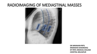
  • 1. RADIOIMAGING OF MEDIASTINAL MASSES DR NIRANJAN PATIL MD(RADIO-DIAGNOSIS) DY PATIL MEDICAL COLLEGE HOSPITAL KOLHAPUR
  • 3. ITMIG Definition of Mediastinal Compartments
  • 4.
  • 5.
  • 6.
  • 7.
  • 8.
  • 9. HILUM OVERLAY SIGN • If cause of opacity arises from hilum the silhouette of normal pulmonary vessel is obliterated.causes of opacites include middle med tumours. • If edge of vessel are appreciated then cause of opacity is not in contact with hilum therefore it may be anterior or posterior to it.
  • 10.
  • 15.
  • 16. POSTERIOR JUNCTION LINE • This Line is also form by apposition of visceral and parital pleura of posteromedial portion of lungs posterior to esophagus and anterior to third through fifth vertebrae. • posterior junction line demonstrates more cranial extension compare to anterior junction line,it can be seen above clavicles.
  • 17.
  • 18. RIGHT PARATRACHEAL STRIPE • When the visceral and parietal pleura of the right upper lobe come in contact with the right lateral border of the trachea and the intervening mediastinal fat, air within the right lung and trachea outlines these entities to form the right paratracheal stripe • It begins superiorly at the level of the clavicles and extends inferiorly to the right tracheobronchial angle at the level of the azygos arch.
  • 19.
  • 20. LEFT PARATRACHEAL STRIPE • It is formed by contact between the left upper lobe and either the mediastinal fat adjacent to the left tracheal wall or the left tracheal wall itself. Air within the trachea outlines the intervening soft tissue • the left paratracheal stripe is seen less frequently it may be obscured by contact between the left lung and either the proximal left common carotid artery anteriorly or the left subclavian artery posteriorly.
  • 21.
  • 22. Left Paraspinal Line The left paraspinal line is formed by tangential contact of the left lung and pleura with the posterior mediastinal fat, left paraspinal muscles, and adjacent soft tissues. The left paraspinal line extends vertically from the aortic arch to the diaphragm and typically lies medial to the lateral wall of the descending thoracic aorta.
  • 23. • The right paraspinal line is formed by the right lung and pleura coming in tangential contact with the posterior mediastinal soft tissues.The right paraspinal line is not a true mediastinal line. Right Paraspinal Line
  • 24. Azygoesophageal Recess • The azygoesophageal recess represents a space lying lateral or posterior to the esophagus and anterior to the spine, extending from the level of the anterior turn of the azygos vein to the level of the aortic hiatus (T12) inferiorly.
  • 25. Aortopulmonary Window • . It is bounded superiorly by the inferior wall of the aortic arch and inferiorly by the superior wall of the left pulmonary artery. The posterior wall of the ascending aorta forms the anterior boundary of the AP.
  • 26. Lesions Fluid Fat Vascular Anterior Thymoma Lymphoma Germ-cell tumor Goiter Thymic cyst Thymoma Pericardial cyst Germ-cell tumor Lymphoma Duplication cyst Necrotic nodes Pericardial recess Germ-cell tumor Thymolipoma Fat pad Morgagni hernia Thyroid Cardiac coronary aneurysm Ascending aortic aneurysm Middle Lymph nodes Duplication cyst Arch anomaly Esophageal mass Duplication cyst Necrotic nodes Pericardial recess Lipoma Oesophageal fibrovascular polyp Arch anomaly Azygos vein Vascular node Posterior Neurogenic Bone and marrow Neuroenteric cyst Schwannoma Meningocele Extramedullary hematopoiesis Descending aorta More than One Lesion Infection Hemorrhage Lung Cancer Lymphangioma Mediastinitis Liposarcoma Hemangioma April 27, 2022 26
  • 27. ANTERIOR MEDIASTINAL MASS • Silhouette with right cardiac border,ascending aorta • Obliteration of Anterior junction line • Hilum overlay Sign positive • Obliteration of CP angle & Retrosternal space • Mass effect on trachea
  • 28. THYMOMAS • It is the most common primary neoplasm of Anterior Mediastinum. • Most thymomas arise near junction of heart and great vessel . • Plain Radiographs • Thymomas appear sharply marginated and smooth or lobulated in contour and usually project one side of mediastinum. • dense calcification may be seen throughout the mass or its periphery. • Computed Tomography(CT) • Almost all thymomas occur in pre-vascular space and displace great vessels. • Thymomas usually appears homogenous in attenuation with or without contrast. Some large thymomas appear cystic or contain areas of necrosis.
  • 29.
  • 30.
  • 31. THYMIC CYST • Thymic cysts are cysts that occur within or arise from the thymus. • Thymic cysts can fluctuate both in size and attenuation over time on CT and MRI. • Plain radiograph • May not even be visible (especially small lesions). If visible thymic cysts often indistinguishable from other non-lobulated thymic/anterior mediastinal lesions on radiographs. • CT • Typically manifests as unilocular or multilocular cystic masses with well-defined walls. Lesions can be lobulated and may have soft-tissue attenuation components. • Some thymic cysts may have increased CT attenuation if haemorrhage or infection occurs as a complication
  • 32.
  • 33. Germ Cell Tumors(GCT) • GCT accounts for 10% of primary mediastinal masses arises from primitive germ cells. • They are most common in anterior mediastinum. • GCT INCLUDES- • Benign and Malignant teratoma • Seminoma • Embryonal Carcinoma • Endodermal sinus tumour • choriocarcinoma
  • 34. Teratomas • Mature teratomas constitute 60%-80% mediastinal GCT. • Plain Radiographs: • Usually Anterior med. mass projecting to one side of mediastinum. • Teratoma appears smooth,rounded or lobulated mass. • Computed Tomography(CT) • Usually appear in prevascular space. • CT often shows combination of fluid-filled cysts,fat,soft tissue and areas of calcification. • teratomas well encapsulated and well demarcated.
  • 35.
  • 36.
  • 37. SEMINOMA Seminoma occurs entirely in men mean age is 29yrs.It is most common malignant mediastinal Germ cell tumor. PLAIN RADIOGRAPHS: Large lobulated anterior mediastinal mass projecting to one or both sides often obscuring heart borders. CT: Seminomas appear large,smooth,lobulated homogeneous soft tissue masses although small areas of low attenuation Can be seen
  • 38.
  • 39.
  • 40. THYROID ENLARGEMENT • Thyroid enlargement commonly associated with extension into Anterior mediastinum. • Multinodular Goitre most common condition in which med thyroid abnormality detected. • Plain Radiographs: • sharply marginated,superior med mass causing tracheal narrowing or displacement of trachea contralateral side. • Mass appears poorly marginated above level of clavicles. • Computed Tomography(CT) • Thyroid tissue is high attenuation relative to soft tissue because of its high iodine content. • they usually dont project below arch of arota(differentiating them from thymomas &teratomas)
  • 41.
  • 42. HODGKINS LYMPHOMA • Peak incidence third & eighth decades. • Thoracic involvement in 85% of cases at diagnosis. • HL most often involves superior mediastinal lymph nodes that is Prevascular,paratracheal and aortopulmonary LN. • HL associated with EBV infection in half of cases.it may occur in association with AIDS.
  • 43.
  • 44.
  • 45.
  • 46. NON HODGKIN LYMPHOMA • It is more common then HL. Mean age is 55yr more common than HD in children. • Thoracic involvement is only 40-50%. • Superior mediastinal gp LN involved in nearly all patients.Involvement of single gp of LN group is common. • Posterior group of lymph node is more commonly involve then HL.
  • 47.
  • 48. PERICARDIAL CYST • Pericardial cysts are uncommon benign congenital anomalies of the anterior and middle mediastinum. • They are most commonly found on the right side, in particular the right anterior cardiophrenic angle, but can be found almost anywhere adjacent to the heart. • Plain radiograph • Typically seen as a mass-like density at the cardiophrenic sulcus. They can be of different shapes and are not always round. May change in shape and size with inspiration and position. • CT • Usually appears as a well-defined, non-enhancing, fluid-attenuation, rounded mass next to the pericardium.
  • 49.
  • 50.
  • 51. MIDDLE MEDIASTINUM • Widening of paratracheal stripe • Mass effect on trachea • Convex border of AP window • Distorsion of Azygoesophageal recess on right
  • 52. Bronhiogenic cyst • Bronchiogenic cyst can be present any part of mediastinum but most commonly present in middle mediastinum. • Most occur in conatct with tracheobronchial tree and within 5cm of carina. • Plain Radiographs: • Cyst appear smooth,sharply marginated,round,or eliptical masses. • they may be large and can displace trachea or bronchii. • subcarinal cyst may result in convexity in superior part of azygoesophageal recess. • Computed Tomography(CT): • cyst appear similar as on chest radiographs. • Because of variable composition of fluid within cyst there attenuation is highly variable. • Half of cyst are of water attenuation the other half higher then muscle.
  • 53.
  • 54.
  • 55.
  • 56. POSTERIOR MEDIASTINAL MASS • Distorsion of paraspinal lines. • poterior ribs or vertebral erosion/remodelling. • cervicothoracic sign • obliteration of posterior junction line and descending aorta • distorsion of azygoesophageal lines
  • 57. NEUROGENIC TUMORS • Neurogenic tumors is more common in children comapare to adults. • 75% posterior mediastinal masses are neurogenic tumors. • Types of neurogenic tumors arise from: 1. peripheral nerves(schwanoma,neurofibroma) 2. sympathetic ganglia(ganglioneuroma,neuroblastoma)
  • 58. SCHWANNOMAS • Schwannomas, less commonly called neurinomas or neurilemmomas, are benign tumors of Schwann cell origin and are the most common tumor of peripheral nerves, including cranial nerves. • Radiographic features • General imaging features of schwannomas include: • well-circumscribed masses which displace adjacent structures without direct invasion • cystic and fatty degeneration is common • the larger a schwannoma, the more likely it is to show heterogeneity because of cystic degeneration or haemorrhage • CT • low to intermediate attenuation • intense contrast enhancement • small tumors typically demonstrate homogeneous enhancement • larger tumors may show heterogeneous enhancement • adjacent bone remodelling with smooth corticated edges
  • 59.
  • 60.
  • 61. NEUROBLASTOMAS • Neuroblastomas are tumours of neuroblastic origin. Although they may occur anywhere along the sympathetic chain. • They represent the most common extracranial solid childhood malignancy. Plain radiograph • Appearances are non-specific, typically demonstrating an intrathoracic soft-tissue mass or an intra-abdominal mass displacing adjacent organs. • may cause remodelling of ribs, vertebral bodies or pedicle thinning. evidence of calcification on the plain film. CT • On CT, the tumour typically is heterogeneous with calcifications seen in 80-90% of cases. • Areas of necrosis are of low attenuation.
  • 62.
  • 63.
  • 64. Mediastinal Hemangioma • Med. hemangioma is most common in young patient. • Hemangioma most commonly arise from anterior mediastinum compare to posterior. • ON CT: • Tumors often hetrogenous in attenuation and fat is ocasionally present. • Enhancement may be dense,multifocal,diffuse and cenral or peripheral. • punctuate calcification maybe seen. • Dilated mediastinal vessel may be associated with mass.
  • 65.

Editor's Notes

  1. prevascular compartment (purple) wraps around the heart and pericardium, which are located in the visceral compartment (blue). Yellow = paravertebral compartment, green line = visceral-paravertebral compartment boundary line. INTERNATIONAL THYMIC MAIGNANCY INTRESITING GP definition of mediastinal compartments. Sagittal CT images at the levels of the aortic arch The most common abnormalities encountered in the prevascular compartment include thymic lesions ( thymoma, thymic carcinoma, and neuroendocrine neoplasms); germ cell neoplasms ; lymphoma; and intrathoracic goiter. he most common abnormalities in the visceral compartment include lymphadenopathy (related to lymphoma or metastatic disease), duplication cysts, BRONCHIOGENIC CYST tracheal lesions, and esophageal neoplasms. most abnormalities in this region are neurogenic neoplasms that arise from the dorsal root ganglia/neurons adjacent to the intervertebral foramina. Other potential lesions in this compartment are of infectious
  2. (b), left pulmonary artery (c), and left atrium
  3. LEFT - LUNG ABUTS THE MEDIASTINAL SRF AND CREATES ACUTE ANGLE WITH LUNG RIGHT-MED MASS SIT UNDER THE SRF OF MEDSTINUM CREATES OBTUSE ANGLES WITH LUNG
  4. This Sign is useful differntiating whether an opacity on fronal chest radiograph in the region of lung hilum is loacted within the hilum versus anterior or posterior.
  5. Chest xray pa shows we can easily appreciate pulmonary vessel within the mass so this mass may be in middle med
  6. A-IF VESSEL CONVERGE MEDIAL TO HILAR SHADOW THEN ENLAGEMENT DT MASS B I VESSEL CONVERGE N MERGE DIRECTLY ON HILAR SHADOW THEN ENLARGEMENT IS D/T ENLARGED VESSEL.
  7. 1 SHOWS INFERIOR MARGIN OF MASS IS CONVERGING TOWARDS SPINE.THIS SIGNIFIES MASS IS ALMOST INTRATHORACIC 2 SHOWS INFE MARGIN OF MED MASS DIVERGES AWAY FROM MIDLINE THIS SIGNIFIES LARGE COMPENENT OF MASS LIES WITHIN ABDOMEN.
  8. CT scan demonstrates a normal anterior junction line (arrow) obliteration or abnormal convexity of the line suggests underlying anterior mediastinal disease such as thyroid masses, lymphadenopathy, neoplasms, thymic masses, or lipomatosis (,4). Volume loss and hyperinflation of the surrounding lung can also displace the line (,Fig 3,).
  9. and frontal chest radiograph demonstrate a normal posterior junction line CT scan demonstrates a normal posterior junction line (arrow) Abnormal bulging or convexity of the posterior junction line suggests a posterior mediastinal abnormality such as esophageal masses, lymphadenopathy, aortic disease, or neurogenic tumors.
  10. It has a maximum normal thickness of 4 mm. ARCH OF AZYGOUS VEIN T4-T5
  11. frontal chest radiograph demonstrate a normal right paratracheal stripe ( arrows in). cT scan shows that the right paratracheal stripe (arrow) is formed by air within the right upper lobe and trachea outlining the right lateral tracheal wall, right upper lobe pleura, and intervening soft tissues. A wide variety of disease entities can cause widening or abnormal contour of the right paratracheal stripe, such as paratracheal lymphadenopathy, thyroid or parathyroid neoplasms (,Fig 8,), and tracheal carcinoma or stenosis. Pleural disease such as effusion or thickening is among the most common causes for widening of the right paratracheal stripe (,1,,3,,4).
  12. The stripe extends superiorly from the aortic arch to join with the reflection from the left subclavian artery
  13. Normal lt paratracheal stripe seen.
  14. the left paraspinal line is seen more frequently than the right paraspinal line due to the presence of the descending thoracic aorta on the left, which promotes the tangential contact of the left lung necessary to produce the lung-mediastinum interface an abnormal contour of the left paraspinal line; however, tortuosity of the descending thoracic aorta may also displace it. Abnormal contour or displacement may also suggest additional posterior mediastinal abnormalities such as a mediastinal hematoma, a mass, extramedullary hematopoiesis, or esophageal varices (
  15. The right paraspinal line appears straight and typically extends from the 8th through the 12th thoracic vertebral levels CT ARROW SHOWS RT N LT PARASPINAL LINES The right paraspinal line may be displaced laterally by osteophytes or prominent mediastinal fat. However, abnormal contour or displacement may also suggest a posterior mediastinal abnormality such as a mediastinal hematoma
  16. CT scan shows that the azygoesophageal recess (arrow) is an interface formed by the right lower lobe outlining the mediastinum adjacent to the esophagus and azygos vein. XRAY demonstrate a normal azygoesophageal recess with mild leftward convexity superiorly and a straight edge in middle one third lower one third straight line. Abnormal contour and convexity may be due to lymphadenopathy, hiatal hernias,bronchopulmonary-foregut malformations, esophageal neoplasms, pleural abnormalities, and cardiomegaly with left atrial enlargement
  17. XRAY normal AP window as a shallow concave interface (*) between the aorta and the pulmonary artery.normal aortic-pulmonary stripe (arrows) and its relation to the AP window. AP STRIPE REPRESENTS MEDIASTINAL STRIPES reflection or interface formed by the pleura of theanterior left lung coming in contact with and tan-gentially reflecting over the mediastinal fat an-terolateral to the left pulmonary artery and aortic arch. CT scan shows the normal AP window (*). The concave interface seen in actually represents the lateral border (arrow) of the AP window formed by the left lung and pleura contacting the aortic arch and extending to the pulmonary artery.
  18. WE CAN LOACTE MASS ANATOMICAL LOCATION ON THE BASIS OF XRAY we can differentiate lesioN on CT N MRI IN CT WE HAVE DIFFERENT HU UNITS LIKE FOR FAT -50 TO -100 VASCULAR/BLOOD 40 FLUID -10 TO 20
  19. Most common age is 50-60yrs rare before 20yrs age a/w MG
  20. PA SHOWS LARGE THYMOMAS (T ARROE) OBSCURING RT HEaRT BORDER EXTENDING INTO RT CARDIOPHERENIC BORDER LAT SHOWS THYMOMAS(t) IS VISILE OVERLYING HEART SHADOW (ARROW)
  21. CT SHOWS CYSTIC FLUID ATTENUATION MASS IN RT CP ANGLE MASS SHOW DENSE CALCIFICATION INCLUDING CALCIFICATION OF CAPSULE
  22. axial contrast-enhanced CT image below the level of the carina shows a well-defined low-attenuation lesion (arrow) in the thymic bed of the prevascular mediastinum. The internal Hounsfield unit value was 15. MR imaging is the most useful imaging modality for distinguishing cystic from solid lesions, discerning cystic and/or necrotic components within solid masses, and identifying septa and/or soft tissue in cystic lesions.
  23. MOST COMMON IN 2-4 DECADE OF LIFE.
  24. most commonly occur in children and young 4types-mature,cystic,immature malignant
  25. CHEST PA SHOWS SMOOTH SHARPLY MARGINATED LEFT MED MASS IT OBSCURES LEFT HEART BORDER INDICATING ANTERIOR LOACTIONS. LAT SHOWS INC DENSITY OF RETROSTERNAL CSPACE(ARROW) BUT DISCRETE mASS NOT VISBLE
  26. TERATOMAS APPEARS LARGE MASS COMPRESSING RT CARDIAC STR IT CONTAINS CALCIFICATION AREAS OF FAT(ARROW)AND CYSTIC OR SOFT TISSUE REGIONS. The presence of visible regions of intralesional fat measuring between −40 and −120 HU at multidetector CT within a heterogeneous prevascular mediastinal mass is highly suggestive of a mature teratoma. These benign lesions characteristically demonstrate varying amounts of fat, fluid, calcification, and soft tissue
  27. PA CHEST RADIO SHOWS LARGE LOBULATED MED MASS (ARROW)OBSCURING RT HEART BORDER RT PLEURAL EFFUSION IS ALSO VISIBLE. LAT XRAY SHOWS LARGE ANTR MED MASS SEEN.
  28. BOTH CT SCAN IMAGE SHOWS LARGE RELATIVELY HOMOGENOUS MASS IN PREVASCULAR SPACE DISPLACING MEDIASTINAL STR TO LEFT.PLEURAL EFFUSION IS ALSO NOTED.
  29. with Hounsfield unit values of 70–85 due to the presence of iodine),
  30. ct shows enhancing mass is visble in right paratracheal med XRAY shows displacement of trachea towards contralateral side sharp margin of mass is seen pumonary vessel can be appreciated
  31. PLEURAL EFUSION DT LYMPHATIC OBSTRUCTION Pericardium invasion dt direct ivasion by tumor CHEST WALL INVASION TUMOR INVOLVE RIBS STERNUM RESULT IN LYTIC BONE LESION
  32. PA RADIOGRAPHS SHOWS B/L LOBULATED MEDSTINAL MASS LAT VIEW SHOWS MASS IN ANTERIOR MED
  33. CT SHOWS PREVASCULAR ANTERIOR MED LYMPHNODE ENLARGEMENT (ARROW) PRETRACHEAAL LYMPHNODE ENLARGEMENT (*) LN ENLARGE TYPICAL FOR HODGKIN LYMPHOMA BUT FOR CONFIRM DIAGNIOSIS WE NEED CT GUIDED BIOPSY
  34. XRAY PA SHOWS LEFT SIDE MED MASS WITH OBSCURING LEFT HEART BORDER.ELEVATION OF LEFT SIDE HEMI DIAPHRAGM MAY REFLECT DIAPHRAGMATIC PARALYSIS RELATED TO COMPRESSION OF LEFT PHERENIC NERVE. CT SHOWS THAT LYMPHADENOPATHY IS LIMITED TO PREVASCULAR COMPARTMENT(arrow).INVOLVEMENT OF SINGLE LN is common in NHL.
  35. XRAY PA SHOWS MASS IN RT CARDIOPHERENIC SULCUS WITH SHARP BORDERS OBSCURING RT HEART BORDERS.SIMILARLY WE CAN SEE ON LATERAL CHEST XRAY MASS IN ANTERIOR MEDIASTIUM
  36. axial contrast-enhanced multidetector CT image at the level of the ventricles shows a well-circumscribed, homogeneous, fluid attenuation lesion (M) with imperceptible walls in the right cardiophrenic angle. These findings are pathognomonic for a pericardial cyst.
  37. PA SHOWS SUBACRINAL MASS WITH INCREASE OPACITY(*) SPLAYING CARINA ABNORMAL CONVEXITY OF UPPER AND MIDDLE THIRD OF AZYGOESOPHAGEAL RECESS CT CONFIRMS SUBCARINAL MASS (ARROW)
  38. bronchogenic cysts appear at a subcarinal location (most commonly) oesophageal cysts appear more tubular and in close relation to the oesophagus ON CT Thin-walled bronchogenic cyst relatively thick-walled oesophageal cyst (due to the presence of smooth muscle)
  39. axial contrast-enhanced CT image at the level of the aortic arch shows a well-circumscribed, homogeneous, fluid attenuation mass (M) adjacent to and intimately associated with the upper thoracic esophagus. The location of the abnormality enables definitive diagnosis of an esophageal duplication cyst with imaging alone.
  40. PA WELL DEFINED MASS SHARP BORDERS CERVICOTHORACIC SIGN POSITIVE LARERAL VIEW MASS LOCATED POSTERIORLY
  41. A-WELL DEFINED HOMOGENOUS MASS VISIBLE ON CT B- CT SHOWS PARAVERTEBRAL MASS IN HETROGENOUS PERIPHERAL ENAHNCEMENT MASS EXTENDINTO NEURAL FORAMEN (ARROES)WHICH IS ENLARGED COMAPRE TO RIGHT
  42. mediastinal neuroblastoma- xray pa and lat shows large superior posterior med mass
  43. CT IMAGE SHOWS LARGE PARAVERTEBRAL POSTERIOR MEDIASTINAL MASSES CONTAING SMALL AREAS OF CALCIFICATION T2W MRI SHOWS MASS IN POSTERIOR MED TUMOR EXTEND INTO SPINAL CANAL THROUGH NEURAL FORAMEN(WHITE ARROW) DISPLACING SPINAL CORD IN RIGHT (BLACK ARROW)
  44. Med HEMANGIOMA CAN ARISE FROM ANY PART OF MEDIASTINUM
  45. mediastinal hemangioma A CT SHOWS LARGE MASS IN SUBCARINAL SPACE CONATAIN PUNCTUATE CALCIFICATION ALSO KNOWN AS PHLEBOLITHS B)AT HIGHER LEVEL SVC (large arrow) and AZYGOUS VEIN(SMALL ARROE) DILATED. DILATED VEINS ARE VISBLE IN MEDIASTINUM POSTERIOR TO SVC.