This document discusses pleural effusions, including their causes, characteristics, diagnosis and evaluation. Key points:
- Pleural effusions can be transudative or exudative based on their mechanism of formation and fluid chemistry. Common causes include heart failure, pneumonia, malignancy and pulmonary embolism.
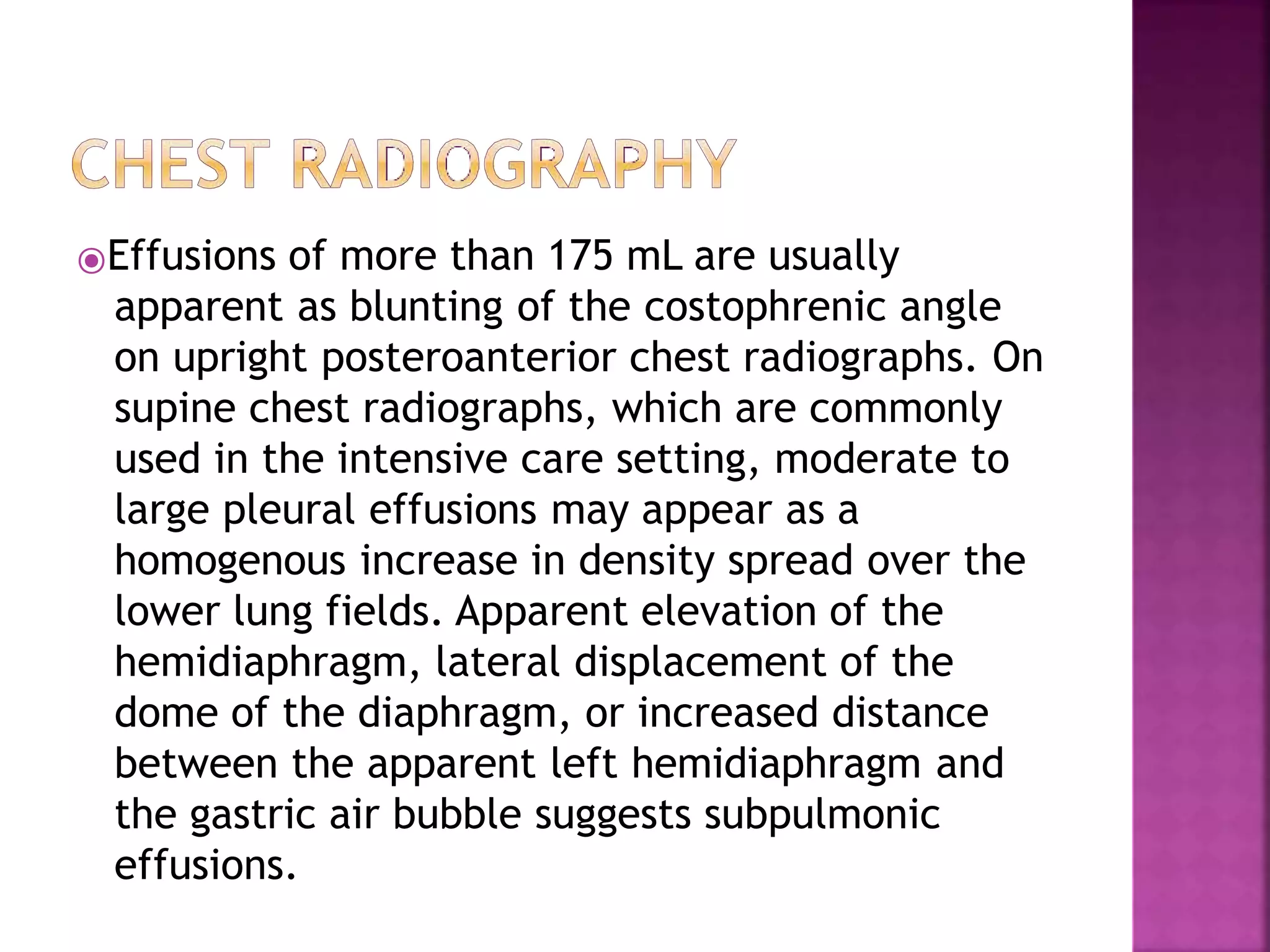
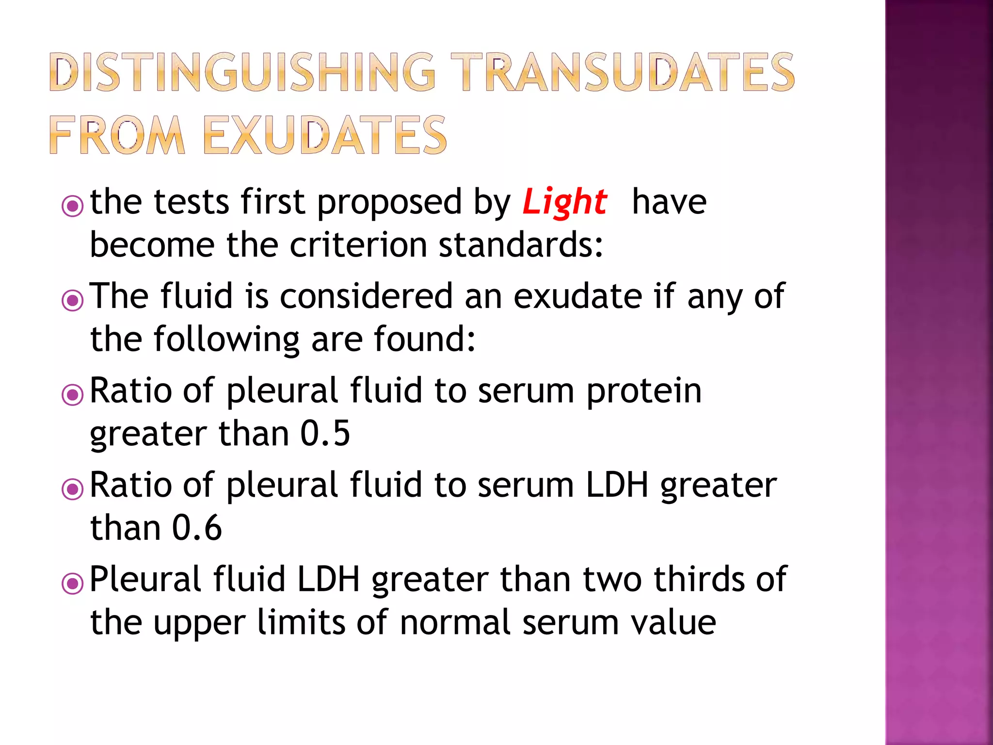
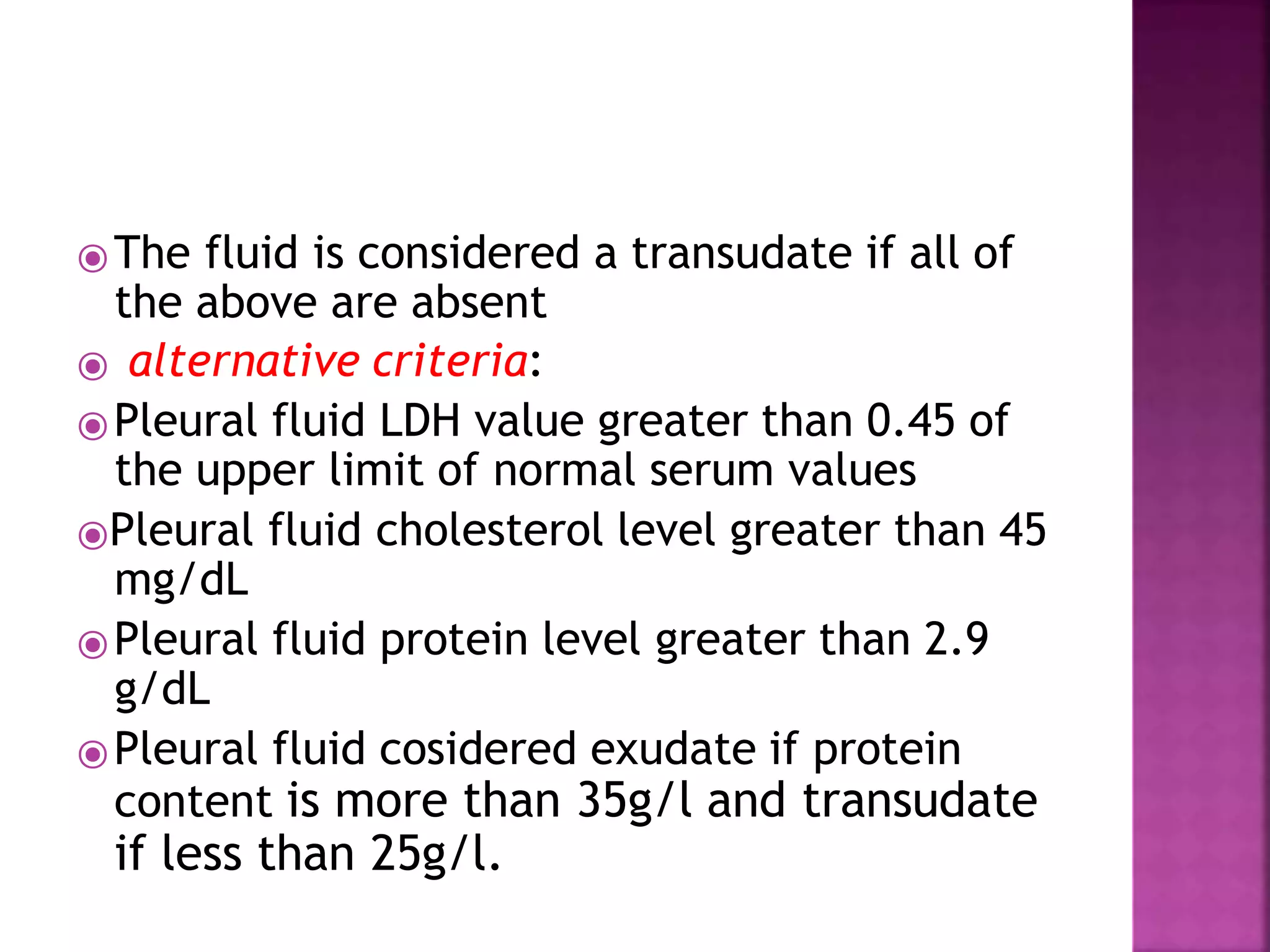
- Diagnosis involves chest imaging, diagnostic thoracentesis and fluid analysis to determine if the fluid is an exudate or transudate based on pleural fluid to serum ratios of protein and LDH. Additional fluid tests provide clues to specific causes.
- Pleural fluid characteristics like glucose, pH and cell differentials provide diagnostic information and indicate need for drainage in some cases like parapneumonic effusions