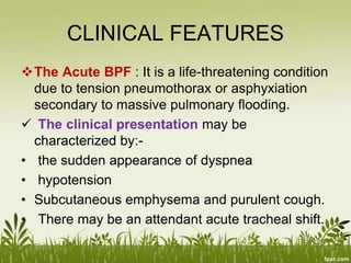
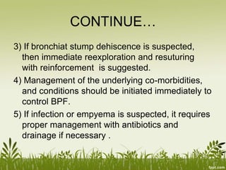
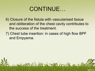
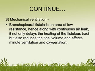
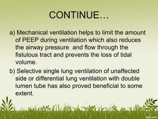
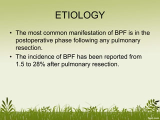
INTRODUCTION
• Bronchopleural fistula is defined as a
communication involving both the pleural space
and the bronchial tree.
ETIOLOGY
• The most common manifestation of BPF is in the
postoperative phase following any pulmonary
resection.
• The incidence of BPF has been reported from
1.5 to 28% after pulmonary resection.
CONTINUE…
 This most common manifestation is followed by:-
• lung necrosis
• persistent spontaneous pneumothorax
• chemotherapy or radiotherapy, (as a treatment
for lung cancer)
• tuberculosis
RISK
• Patients at risk for the development of BPF
include patients with:-
 ARDS
 chest trauma
 invasive chest procedures
 debilitated patients
 patients with pneumonia
CONTINUE…
 lung abscess
 COPD
 bullous lung disease
 spontaneous pneumothorax
 parenchymal abnormalities
CLASSIFICATION
Postoperatively a BPF may be classified as:-
• Acute
• Sub acute
• Chronic
 The acute form of BPF is typically related to
stump dehiscence and necessitates early re-
operation.
CONTINUE…
 The sub-acute and chronic forms are
commonly associated with infection and
immunocompromised or debilitated patients with
various comorbidities.
CONTINUE…
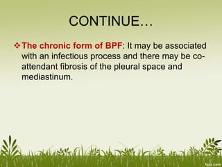
• Varoli et al7 classified fistulas according to
the time of onset after the operation:
1) early [1 to 7 days]
2) intermediate [8 to 30days]
3) late fistulas [more than 30 days].
CLINICAL FEATURES
The Acute BPF : It is a life-threatening condition
due to tension pneumothorax or asphyxiation
secondary to massive pulmonary flooding.
 The clinical presentation may be
characterized by:-
• the sudden appearance of dyspnea
• hypotension
• Subcutaneous emphysema and purulent cough.
• There may be an attendant acute tracheal shift.
CONTINUE…
• An acute presentation generally requires a
visit to the operating suite and should be
treated as quickly as possible.
CONTINUE…
The Sub-acute presentation: It is rather
insidious and is characterized by:-
• a general wasting
• malaise
• fever
• minimally productive cough.
CONTINUE…
The chronic form of BPF: It may be associated
with an infectious process and there may be co-
attendant fibrosis of the pleural space and
mediastinum.
DIAGNOSIS
• methylene blue test
• Bronchography
• CT-scan to diagnose the etiology
of bronchopleural fistula.
INVESTIGATIONS
• Chest x-ray
• Bronchoscopy
• Biopsy
• Routine examination:-
 Blood grouping and crossmatching
 Serology
 CBC
CONTINUE…
 sugar
 urea
 creatinine
 serum
CONTINUE…
 amylase
 Electrolytes
• Urine RE/ME
• ECG
COMPLICATIONS
• Tension pneumothorax
• Infection
• Medistenitis
• Inflammation
• Subcutaneous emphysema
TREATMENT
• Surgical and Medical treatment are the two
options as per the clinical condition of the
patient.
1) The control of any life threatening complication
like tension pneumothorax by emergent
drainage is required.
2) In some life threatening conditions, airway
control and postural drainage by placing the
affected lung down is required.
CONTINUE…
3) If bronchiat stump dehiscence is suspected,
then immediate reexploration and resuturing
with reinforcement is suggested.
4) Management of the underlying co-morbidities,
and conditions should be initiated immediately to
control BPF.
5) If infection or empyema is suspected, it requires
proper management with antibiotics and
drainage if necessary .
CONTINUE…
6) Closure of the fistula with vascularised tissue
and obliteration of the chest cavity contributes to
the success of the treatment.
7) Chest tube insertion: in cases of high flow BPF
and Empyema.
CONTINUE…
8) Mechanical ventilation:-
• Bronchopleural fistula is an area of low
resistance, hence along with continuous air leak,
it not only delays the healing of the fistulous tract
but also reduces the tidal volume and affects
minute ventilation and oxygenation.
CONTINUE…
a) Mechanical ventilation helps to limit the amount
of PEEP during ventilation which also reduces
the airway pressure and flow through the
fistulous tract and prevents the loss of tidal
volume.
b) Selective single lung ventilation of unaffected
side or differential lung ventilation with double
lumen tube has also proved beneficial to some
extent.
CONTINUE…
c) High frequency ventilation is used only with
proximal BPF and when the lung parenchyma is
normal.
CONTINUE…
9) Surgery
10) Bronchoscopy
Fig:Management of bronchopleural
fistula.
REFERENCE
• Das.S, Manual on clinical surgery including
special investigations and differential diagnosis,
4th edition, 13, Old Mayors’ Court, Culcutta.
Page no 305
• Hauser.kasper, Longo Braunwald, Jameson
Fauci, Harrison’s, Principles of Internal
Medicine, 16th edition, volume 1, McGraw-Hill,
Medical Publishing Division, New York. Page no
1497-1507
•
•
CONTINUE…
• Arnold Hodder, Williams Normans, Bailey and
Love’s Short practice of surgery, 25th edition.
Page no 891.
• Retrieved from www.google/bronchopleural
fistula .com

Bronchopleuralfistula

  • 2.
    INTRODUCTION • Bronchopleural fistulais defined as a communication involving both the pleural space and the bronchial tree.
  • 3.
    ETIOLOGY • The mostcommon manifestation of BPF is in the postoperative phase following any pulmonary resection. • The incidence of BPF has been reported from 1.5 to 28% after pulmonary resection.
  • 4.
    CONTINUE…  This mostcommon manifestation is followed by:- • lung necrosis • persistent spontaneous pneumothorax • chemotherapy or radiotherapy, (as a treatment for lung cancer) • tuberculosis
  • 5.
    RISK • Patients atrisk for the development of BPF include patients with:-  ARDS  chest trauma  invasive chest procedures  debilitated patients  patients with pneumonia
  • 6.
    CONTINUE…  lung abscess COPD  bullous lung disease  spontaneous pneumothorax  parenchymal abnormalities
  • 7.
    CLASSIFICATION Postoperatively a BPFmay be classified as:- • Acute • Sub acute • Chronic  The acute form of BPF is typically related to stump dehiscence and necessitates early re- operation.
  • 8.
    CONTINUE…  The sub-acuteand chronic forms are commonly associated with infection and immunocompromised or debilitated patients with various comorbidities.
  • 9.
    CONTINUE… • Varoli etal7 classified fistulas according to the time of onset after the operation: 1) early [1 to 7 days] 2) intermediate [8 to 30days] 3) late fistulas [more than 30 days].
  • 10.
    CLINICAL FEATURES The AcuteBPF : It is a life-threatening condition due to tension pneumothorax or asphyxiation secondary to massive pulmonary flooding.  The clinical presentation may be characterized by:- • the sudden appearance of dyspnea • hypotension • Subcutaneous emphysema and purulent cough. • There may be an attendant acute tracheal shift.
  • 11.
    CONTINUE… • An acutepresentation generally requires a visit to the operating suite and should be treated as quickly as possible.
  • 12.
    CONTINUE… The Sub-acute presentation:It is rather insidious and is characterized by:- • a general wasting • malaise • fever • minimally productive cough.
  • 13.
    CONTINUE… The chronic formof BPF: It may be associated with an infectious process and there may be co- attendant fibrosis of the pleural space and mediastinum.
  • 14.
    DIAGNOSIS • methylene bluetest • Bronchography • CT-scan to diagnose the etiology of bronchopleural fistula.
  • 15.
    INVESTIGATIONS • Chest x-ray •Bronchoscopy • Biopsy • Routine examination:-  Blood grouping and crossmatching  Serology  CBC
  • 16.
  • 17.
  • 18.
    COMPLICATIONS • Tension pneumothorax •Infection • Medistenitis • Inflammation • Subcutaneous emphysema
  • 19.
    TREATMENT • Surgical andMedical treatment are the two options as per the clinical condition of the patient. 1) The control of any life threatening complication like tension pneumothorax by emergent drainage is required. 2) In some life threatening conditions, airway control and postural drainage by placing the affected lung down is required.
  • 20.
    CONTINUE… 3) If bronchiatstump dehiscence is suspected, then immediate reexploration and resuturing with reinforcement is suggested. 4) Management of the underlying co-morbidities, and conditions should be initiated immediately to control BPF. 5) If infection or empyema is suspected, it requires proper management with antibiotics and drainage if necessary .
  • 21.
    CONTINUE… 6) Closure ofthe fistula with vascularised tissue and obliteration of the chest cavity contributes to the success of the treatment. 7) Chest tube insertion: in cases of high flow BPF and Empyema.
  • 22.
    CONTINUE… 8) Mechanical ventilation:- •Bronchopleural fistula is an area of low resistance, hence along with continuous air leak, it not only delays the healing of the fistulous tract but also reduces the tidal volume and affects minute ventilation and oxygenation.
  • 23.
    CONTINUE… a) Mechanical ventilationhelps to limit the amount of PEEP during ventilation which also reduces the airway pressure and flow through the fistulous tract and prevents the loss of tidal volume. b) Selective single lung ventilation of unaffected side or differential lung ventilation with double lumen tube has also proved beneficial to some extent.
  • 24.
    CONTINUE… c) High frequencyventilation is used only with proximal BPF and when the lung parenchyma is normal.
  • 25.
  • 26.
  • 27.
    REFERENCE • Das.S, Manualon clinical surgery including special investigations and differential diagnosis, 4th edition, 13, Old Mayors’ Court, Culcutta. Page no 305 • Hauser.kasper, Longo Braunwald, Jameson Fauci, Harrison’s, Principles of Internal Medicine, 16th edition, volume 1, McGraw-Hill, Medical Publishing Division, New York. Page no 1497-1507 • •
  • 28.
    CONTINUE… • Arnold Hodder,Williams Normans, Bailey and Love’s Short practice of surgery, 25th edition. Page no 891. • Retrieved from www.google/bronchopleural fistula .com