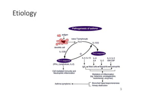
Status Asthmaticus
Fakhir Raza
Background
• Acute Exacerbation
• Unresponsive
• Mild to Severe form
• Trend is towards less number of admissions in intensive care1
• Han P, Cole RP. Evolving differences in the presentation of severe asthma requiring intensive care unit
admission. Respiration. Sep-Oct 2004;71(5):458-62.
Treatment goals
• Reverse airway obstruction
• Correct Hypoxemia
• Prevent or treat complications like pneumothorax and respiratory
arrest
Etiology
Etiology
• Acute Bronchospastic component marked by smooth muscle
bronchoconstriction.
• Later inflammatory airway swelling and edema
Early bronchospastic response
• Exposure to allergen
• Mast cell degranulation
• Release of histamine, PGD2, LT-C4
• airway smooth muscle contraction, increased capillary permeability,
mucus secretion, and activation of neuronal reflexes
• Bronchoconstriction typically responds to bronchodilator therapy like beta 2
agonist
Later inflammatory response
• Inflammatory mediators prime endothelium and epithelium of
bronchial mucosa.
• Inflammatory cells like eosinophils, neutrophils and basophils attach
to primed endothelium and epithelium and later enter into the
tissues
• Eosinophils release ECP and MBP which induce desquamation of
airway epithelium and expose nerve endings
• It leads to further hyper responsiveness.
Later inflammatory response
• Airway resistance and obstruction
• caused by Bronchospasm, mucus plugging, and edema in the
peripheral
• Air trapping
• results in lung hyperinflation, ventilation/perfusion (V/Q)
mismatch, and increased dead space ventilation.
Later inflammatory response
• Increase in pleural and intra alveolar pressure and distended alveoli
leads to VQ mismatch, hypoxemia and increase in minute ventilation.
Complications
• Slow compartments vs fast compartments
• Respiratory alkalosis vs hypercarbia
• Cardiac arrest
• Respiratory failure or arrest
• Hypoxemia with hypoxic ischemic central nervous system (CNS) injury
• Pneumothorax or pneumomediastinum
• Toxicity from medications
Risk factors
• Genetic
• GERD
• Viral infections
• Air pollutants
• Medications
• Cold exposure
• Exercise
Prognosis
• Generally good except when combined with heart failure or COPD
• Poor prognostic factors include delay in starting treatment especially
steroids
History
• Severe dyspnea or hours or days.
• Previous intubation and ventilation
Risk factors for developing status asthmaticus
• Increased use of home bronchodilators without improvement or
effect
• Previous intensive care unit (ICU) admissions, with or without
intubation
• Asthma exacerbation despite recent or current use of corticosteroids
• Frequent emergency department visits and/or hospitalization
• Less than 10% improvement in peak expiratory flow rate (PEFR)
• History of syncope or seizures during acute exacerbation
• Oxygen saturation below 92% despite supplemental oxygen
Asthma with No Wheezing
• Silent chest
• Severe obstruction
• fatigue
Physical Examination
• Tachypnea
• Wheezing in early stages
• Initially expiratory
• Later in both phases, may have absent breath sound in advance stage
• Use of accessory muscles
• Inability to speak more than 1 to 2 words
• Decreased oxygen saturation
• Tachycardia and Hypertension
• Signs of complication, tension pneumothorax, pneumomediastinum
• Peak expiratory flow meter measurement
Assessment of severity of asthma
exacerbation
• Moderate asthma exacerbation:
• Increasing symptoms.
• PEFR >50-75% best or predicted.
• No features of acute severe asthma.
• Acute severe asthma - any one of:
• PEFR 33-50% best or predicted.
• Respiratory rate ≥25 breaths/minute.
• Heart rate ≥110 beats/minute.
• Inability to complete sentences in one breath.
• Life-threatening asthma - any one of the following in a patient with severe
asthma:
• Clinical signs: altered conscious level, exhaustion, arrhythmia, hypotension, cyanosis, silent
chest, poor respiratory effort.
• Measurements: PEFR <33% best or predicted, SpO2 <92%, PaO2 <8 kPa, 'normal' PaCO2 (4.6-
6.0 kPa).
Differential diagnosis
• In children
• Viral infections, bronchiolitis
• Foreign body
• Congestive heart failure
• Extrinsic compression, lymph node, tumor, blood vessel
• Tracheomalacia, primary or secondary
• Inhalational injury
• Other diagnosis, like cystic fibrosis, bronchiectasis etc
Workup
• Blood test
• CBC, ABG, Electrolytes, RBS, Theophillne level
• Chest X-ray
• To rule out pneumothorax, pneumomediastinum, heart failure, pneumonia
Complete blood count
• CBC with differential to evaluate for pneumonia, ABPA, Churg-Strauss
vasculitis
• It could vary because of treatment as well with or without
neutrophilia
• Serum lactate level
Arterial blood gases
• If peak expiratory flow rate is less than 30% of predicted or patient
best
• Signs of fatigue or progressive airflow obstruction
• Stages of progression
4 stages of blood gas progression with status
asthmaticus
PaCO2 PaO2
Stage 1 Decrease Normal
Stage 2 Decrease Decreased
Stage 3 NORMAL Decreased
Stage 4 High Decreased
Electrolytes and glucose
• Hypokalemia as a result of medications
• Hyperglycemia and in infants hypoglycemia
Need for hospitalization
• If after treatment PEF and FEV1 is between 50% to 70%
• If less than 50% then intensive care admission is indicated
National Heart, Lung, and Blood Institute. Managing exacerbations of asthma. In: National
Asthma Education and Prevention Program (NAEPP). Expert panel report 3: guidelines for
the diagnosis and management of asthma. National Guideline Clearinghouse
Response to treatment
• Response to treatment is assessed by Pulse oximetry and spirometry
Impulse Oscillometry Testing
• Almost independent of patient cooperation
• Valid for all ages from 4 years and older children, adult and geriatric
patients.
• Quite breathing i.e Tidal volume breathing for 30 seconds
• It measures impedance at different frequencies indicative of central
and peripheral airway resistance.
• Bronchodilator therapy often does not reach the peripheral airways.
IOS can provide objective response to drug therapy even when FEV1
can't.
Impulse Oscillometry Testing
Histologic finding
• Autopsy of patients dying in few hours showed Neutrophil infiltration
• Those who die in days showed Eosinophilic infiltration.
• Extensive mucus production and severe bronchial smooth muscle
hypertrophy
Treatment
• Mainstay of treatment of status asthmaticus are beta 2 agonist, systemic
steroids and theophyllines.
• Pregnant and non pregnant are treated in the same manner
• Fluid replacement, hypokalemia and hypophosphatemia are important to
treat.
• Routine use of antibiotics is discouraged
• Oxygen monitoring and therapy
• Maintain SatO2 above 92% except in pregnant and cardiac patients where maintain
above 95%.
• Endotracheal intubation, ventilation and chest tube placement as needed.
• ECMO when needed.
Beta2 Agonists
• Albuterol neubulizer continuously 10 – 15 mg/hour or q5 to 20 min
• Albuterol MDI 4 puff with chamber 15 to 30 minute interval
• Endotracheal epinephrine has no role.
• Intravenous beta2 agonist when inhalation is not possible
• Epinephrine 0.3 to 0.5mg subcutaneously (caution in CHF and history
of arrhythmias)
Anticholinergics
• Ipratropium bromide every 4 to 6 hours
• Synergistic effect with beta2 agonist.
• Does not cross blood brain barrier like atropine
Glucocorticoids
• Most important treatment in status asthmaticus
• decrease mucus production
• Improve oxygenation
• Reduce beta-agonist or theophylline requirements
• Decrease bronchial hypersensitivity
• Help to regenerate the bronchial epithelial cells.
• Oral and IV have same onset of action
• No role of nebulized steroids
• Name any ten Adverse effects of steroids
Bronchodilators
• Methylxanthines theophylline, aminophylline
• bronchodilatation, increased diaphragmatic function, and central
stimulation of breathing
• Narrow therapeutic index, needs monitoring
• Smokers and patients on phenytoin need higher doses
• Side effects, nausea, vomiting, palpitation
• 6mg/kg loading followed by 1mg/kg/hour
Bronchodilators
• Magnesium Sulfate
• relax smooth muscle and hence cause bronchodilation
• Usually 1 gm to 2.5gm is administered as a single dose.
• No studies on repeated doses
• More effective in children. 40mg/kg over 20 minutes
Sedatives
• Usually reserved for intubated patients
• In very agitated patients on high bronchodilator therapy a dose of
lorazepam 0.5mg to 1mg intravenous
Therapies for severe and resistant status
despite mechanical ventilation
• Ketamine
• Inhaled anesthetic agents
• NMBA
• Other treatments in case reports and personal experiences
Extracorporeal life support
• high risk of developing refractory status asthmaticus.
• Patients with a history of multiple incubations
• Respiratory failure requiring intubation within 6 hours of admission
• Hemodynamic instability
• Neurologic impairment at the time of admission
• Duration of respiratory failure greater than 12 hours despite maximal medical
therapy.
• Practiced in limited centers of the world
• references
1. Mikkelsen ME, Pugh ME, Hansen-Flaschen JH, Woo YJ, Sager JS. Emergency extracorporeal life support for asphyxic status
asthmaticus. Respir Care. Nov 2007;52(11):1525-9
2. Coleman NE, Dalton HJ. Extracorporeal life support for status asthmaticus: the breath of life that's often forgotten. Crit Care.
2009;13(2):136
3. Hebbar KB, Petrillo-Albarano T,Coto-Puckett W,Heard M, Rycus PT, Fortenberry JD. Experience with use of extracorporeal life
support for severe refractory status asthmaticus in children. Crit Care. 2009;13(2):R29
Non invasive ventilation
• Limited to weaning from ventilation
• Not effective in most of the acute cases unlike acute exacerbation of
COPD
Mechanical ventilation
• Indications --- already discussed
• Considerations
• Low volume, lower rate, I:E 1:3-4, addition of PEEP to prevent airway collapse
during expiration (cautiously)
• Heavy sedation
• Steroids and NMBA can cause prolong paralysis
• Monitor flow volume loop, exhaled tidal volume, autoPEEP
• Decreased cardiac output due to decreased preload, diastolic hypotension
• Fluid and judicious use of noradrenaline / phenylephrine
• Arterial line for repeated blood gases
• Replace electrolytes
Heliox
• Mixture of Helium and Oxygen
• Effective when percentage of Helium is at least 60%, so limiting its use
when FiO2 requirement is high
• It has more laminar flow and less turbulence in small airways so the
Oxygen reach to lower airways besides nebulized aerosols.
• No effect on caliber of bronchi.
A word about transfer, prevention and long
term care
• Features of stability
• Monitorting FEV1 and IOS
Comments and suggestion
Thank you and JazakAllah
• I am thankful to Medscape and internet to prepare this presentation
beside my teachers.

statusasthmaticus-140524030120-phpapp01.pptx

  • 1.
  • 2.
    Background • Acute Exacerbation •Unresponsive • Mild to Severe form • Trend is towards less number of admissions in intensive care1 • Han P, Cole RP. Evolving differences in the presentation of severe asthma requiring intensive care unit admission. Respiration. Sep-Oct 2004;71(5):458-62.
  • 3.
    Treatment goals • Reverseairway obstruction • Correct Hypoxemia • Prevent or treat complications like pneumothorax and respiratory arrest
  • 4.
  • 5.
    Etiology • Acute Bronchospasticcomponent marked by smooth muscle bronchoconstriction. • Later inflammatory airway swelling and edema
  • 6.
    Early bronchospastic response •Exposure to allergen • Mast cell degranulation • Release of histamine, PGD2, LT-C4 • airway smooth muscle contraction, increased capillary permeability, mucus secretion, and activation of neuronal reflexes • Bronchoconstriction typically responds to bronchodilator therapy like beta 2 agonist
  • 7.
    Later inflammatory response •Inflammatory mediators prime endothelium and epithelium of bronchial mucosa. • Inflammatory cells like eosinophils, neutrophils and basophils attach to primed endothelium and epithelium and later enter into the tissues • Eosinophils release ECP and MBP which induce desquamation of airway epithelium and expose nerve endings • It leads to further hyper responsiveness.
  • 8.
    Later inflammatory response •Airway resistance and obstruction • caused by Bronchospasm, mucus plugging, and edema in the peripheral • Air trapping • results in lung hyperinflation, ventilation/perfusion (V/Q) mismatch, and increased dead space ventilation.
  • 9.
    Later inflammatory response •Increase in pleural and intra alveolar pressure and distended alveoli leads to VQ mismatch, hypoxemia and increase in minute ventilation.
  • 10.
    Complications • Slow compartmentsvs fast compartments • Respiratory alkalosis vs hypercarbia • Cardiac arrest • Respiratory failure or arrest • Hypoxemia with hypoxic ischemic central nervous system (CNS) injury • Pneumothorax or pneumomediastinum • Toxicity from medications
  • 11.
    Risk factors • Genetic •GERD • Viral infections • Air pollutants • Medications • Cold exposure • Exercise
  • 12.
    Prognosis • Generally goodexcept when combined with heart failure or COPD • Poor prognostic factors include delay in starting treatment especially steroids
  • 13.
    History • Severe dyspneaor hours or days. • Previous intubation and ventilation
  • 14.
    Risk factors fordeveloping status asthmaticus • Increased use of home bronchodilators without improvement or effect • Previous intensive care unit (ICU) admissions, with or without intubation • Asthma exacerbation despite recent or current use of corticosteroids • Frequent emergency department visits and/or hospitalization • Less than 10% improvement in peak expiratory flow rate (PEFR) • History of syncope or seizures during acute exacerbation • Oxygen saturation below 92% despite supplemental oxygen
  • 15.
    Asthma with NoWheezing • Silent chest • Severe obstruction • fatigue
  • 16.
    Physical Examination • Tachypnea •Wheezing in early stages • Initially expiratory • Later in both phases, may have absent breath sound in advance stage • Use of accessory muscles • Inability to speak more than 1 to 2 words • Decreased oxygen saturation • Tachycardia and Hypertension • Signs of complication, tension pneumothorax, pneumomediastinum • Peak expiratory flow meter measurement
  • 17.
    Assessment of severityof asthma exacerbation • Moderate asthma exacerbation: • Increasing symptoms. • PEFR >50-75% best or predicted. • No features of acute severe asthma. • Acute severe asthma - any one of: • PEFR 33-50% best or predicted. • Respiratory rate ≥25 breaths/minute. • Heart rate ≥110 beats/minute. • Inability to complete sentences in one breath. • Life-threatening asthma - any one of the following in a patient with severe asthma: • Clinical signs: altered conscious level, exhaustion, arrhythmia, hypotension, cyanosis, silent chest, poor respiratory effort. • Measurements: PEFR <33% best or predicted, SpO2 <92%, PaO2 <8 kPa, 'normal' PaCO2 (4.6- 6.0 kPa).
  • 18.
    Differential diagnosis • Inchildren • Viral infections, bronchiolitis • Foreign body • Congestive heart failure • Extrinsic compression, lymph node, tumor, blood vessel • Tracheomalacia, primary or secondary • Inhalational injury • Other diagnosis, like cystic fibrosis, bronchiectasis etc
  • 19.
    Workup • Blood test •CBC, ABG, Electrolytes, RBS, Theophillne level • Chest X-ray • To rule out pneumothorax, pneumomediastinum, heart failure, pneumonia
  • 20.
    Complete blood count •CBC with differential to evaluate for pneumonia, ABPA, Churg-Strauss vasculitis • It could vary because of treatment as well with or without neutrophilia • Serum lactate level
  • 21.
    Arterial blood gases •If peak expiratory flow rate is less than 30% of predicted or patient best • Signs of fatigue or progressive airflow obstruction • Stages of progression
  • 22.
    4 stages ofblood gas progression with status asthmaticus PaCO2 PaO2 Stage 1 Decrease Normal Stage 2 Decrease Decreased Stage 3 NORMAL Decreased Stage 4 High Decreased
  • 23.
    Electrolytes and glucose •Hypokalemia as a result of medications • Hyperglycemia and in infants hypoglycemia
  • 24.
    Need for hospitalization •If after treatment PEF and FEV1 is between 50% to 70% • If less than 50% then intensive care admission is indicated National Heart, Lung, and Blood Institute. Managing exacerbations of asthma. In: National Asthma Education and Prevention Program (NAEPP). Expert panel report 3: guidelines for the diagnosis and management of asthma. National Guideline Clearinghouse
  • 25.
    Response to treatment •Response to treatment is assessed by Pulse oximetry and spirometry
  • 26.
    Impulse Oscillometry Testing •Almost independent of patient cooperation • Valid for all ages from 4 years and older children, adult and geriatric patients. • Quite breathing i.e Tidal volume breathing for 30 seconds • It measures impedance at different frequencies indicative of central and peripheral airway resistance. • Bronchodilator therapy often does not reach the peripheral airways. IOS can provide objective response to drug therapy even when FEV1 can't.
  • 27.
  • 28.
    Histologic finding • Autopsyof patients dying in few hours showed Neutrophil infiltration • Those who die in days showed Eosinophilic infiltration. • Extensive mucus production and severe bronchial smooth muscle hypertrophy
  • 29.
    Treatment • Mainstay oftreatment of status asthmaticus are beta 2 agonist, systemic steroids and theophyllines. • Pregnant and non pregnant are treated in the same manner • Fluid replacement, hypokalemia and hypophosphatemia are important to treat. • Routine use of antibiotics is discouraged • Oxygen monitoring and therapy • Maintain SatO2 above 92% except in pregnant and cardiac patients where maintain above 95%. • Endotracheal intubation, ventilation and chest tube placement as needed. • ECMO when needed.
  • 30.
    Beta2 Agonists • Albuterolneubulizer continuously 10 – 15 mg/hour or q5 to 20 min • Albuterol MDI 4 puff with chamber 15 to 30 minute interval • Endotracheal epinephrine has no role. • Intravenous beta2 agonist when inhalation is not possible • Epinephrine 0.3 to 0.5mg subcutaneously (caution in CHF and history of arrhythmias)
  • 31.
    Anticholinergics • Ipratropium bromideevery 4 to 6 hours • Synergistic effect with beta2 agonist. • Does not cross blood brain barrier like atropine
  • 32.
    Glucocorticoids • Most importanttreatment in status asthmaticus • decrease mucus production • Improve oxygenation • Reduce beta-agonist or theophylline requirements • Decrease bronchial hypersensitivity • Help to regenerate the bronchial epithelial cells. • Oral and IV have same onset of action • No role of nebulized steroids • Name any ten Adverse effects of steroids
  • 33.
    Bronchodilators • Methylxanthines theophylline,aminophylline • bronchodilatation, increased diaphragmatic function, and central stimulation of breathing • Narrow therapeutic index, needs monitoring • Smokers and patients on phenytoin need higher doses • Side effects, nausea, vomiting, palpitation • 6mg/kg loading followed by 1mg/kg/hour
  • 34.
    Bronchodilators • Magnesium Sulfate •relax smooth muscle and hence cause bronchodilation • Usually 1 gm to 2.5gm is administered as a single dose. • No studies on repeated doses • More effective in children. 40mg/kg over 20 minutes
  • 35.
    Sedatives • Usually reservedfor intubated patients • In very agitated patients on high bronchodilator therapy a dose of lorazepam 0.5mg to 1mg intravenous
  • 36.
    Therapies for severeand resistant status despite mechanical ventilation • Ketamine • Inhaled anesthetic agents • NMBA • Other treatments in case reports and personal experiences
  • 37.
    Extracorporeal life support •high risk of developing refractory status asthmaticus. • Patients with a history of multiple incubations • Respiratory failure requiring intubation within 6 hours of admission • Hemodynamic instability • Neurologic impairment at the time of admission • Duration of respiratory failure greater than 12 hours despite maximal medical therapy. • Practiced in limited centers of the world • references 1. Mikkelsen ME, Pugh ME, Hansen-Flaschen JH, Woo YJ, Sager JS. Emergency extracorporeal life support for asphyxic status asthmaticus. Respir Care. Nov 2007;52(11):1525-9 2. Coleman NE, Dalton HJ. Extracorporeal life support for status asthmaticus: the breath of life that's often forgotten. Crit Care. 2009;13(2):136 3. Hebbar KB, Petrillo-Albarano T,Coto-Puckett W,Heard M, Rycus PT, Fortenberry JD. Experience with use of extracorporeal life support for severe refractory status asthmaticus in children. Crit Care. 2009;13(2):R29
  • 38.
    Non invasive ventilation •Limited to weaning from ventilation • Not effective in most of the acute cases unlike acute exacerbation of COPD
  • 39.
    Mechanical ventilation • Indications--- already discussed • Considerations • Low volume, lower rate, I:E 1:3-4, addition of PEEP to prevent airway collapse during expiration (cautiously) • Heavy sedation • Steroids and NMBA can cause prolong paralysis • Monitor flow volume loop, exhaled tidal volume, autoPEEP • Decreased cardiac output due to decreased preload, diastolic hypotension • Fluid and judicious use of noradrenaline / phenylephrine • Arterial line for repeated blood gases • Replace electrolytes
  • 40.
    Heliox • Mixture ofHelium and Oxygen • Effective when percentage of Helium is at least 60%, so limiting its use when FiO2 requirement is high • It has more laminar flow and less turbulence in small airways so the Oxygen reach to lower airways besides nebulized aerosols. • No effect on caliber of bronchi.
  • 41.
    A word abouttransfer, prevention and long term care • Features of stability • Monitorting FEV1 and IOS
  • 42.
  • 43.
    Thank you andJazakAllah • I am thankful to Medscape and internet to prepare this presentation beside my teachers.