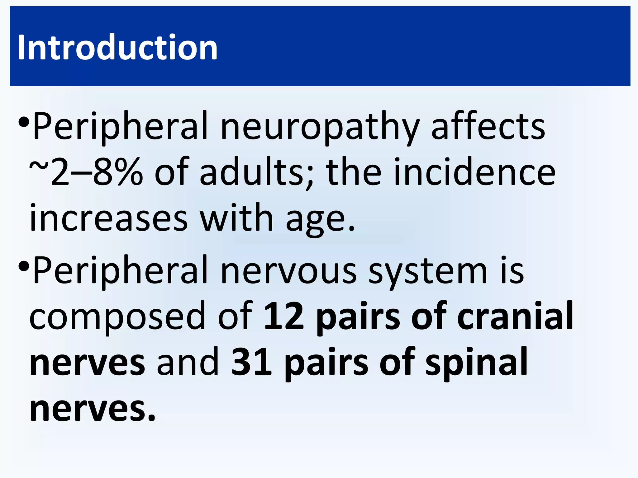
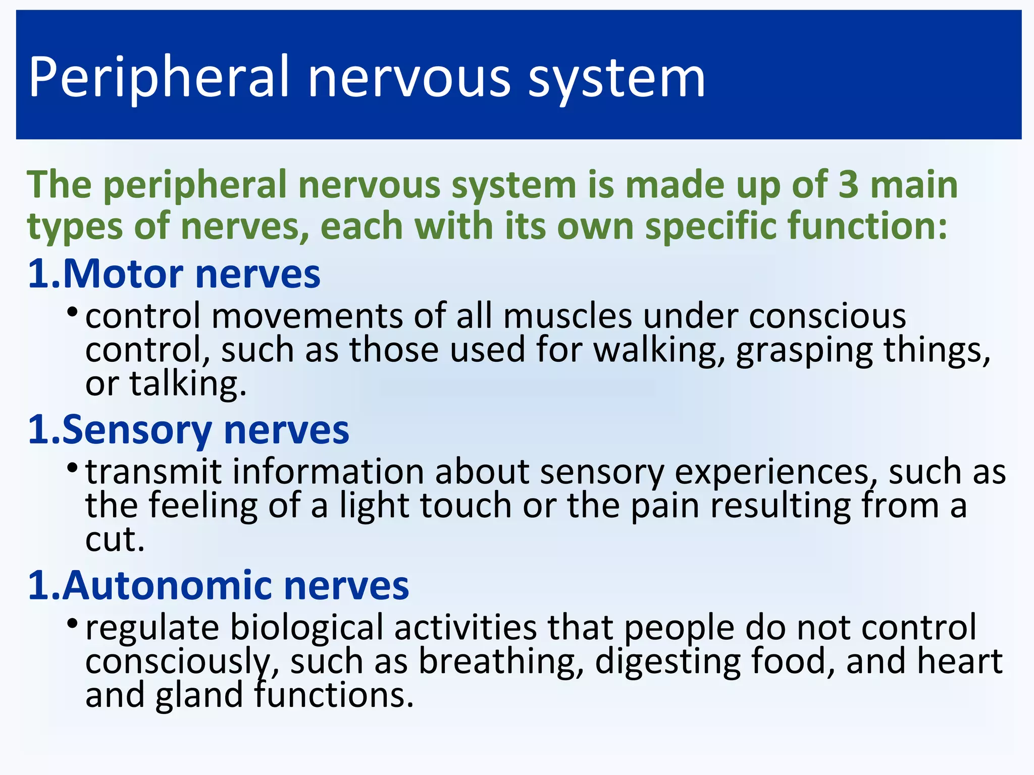
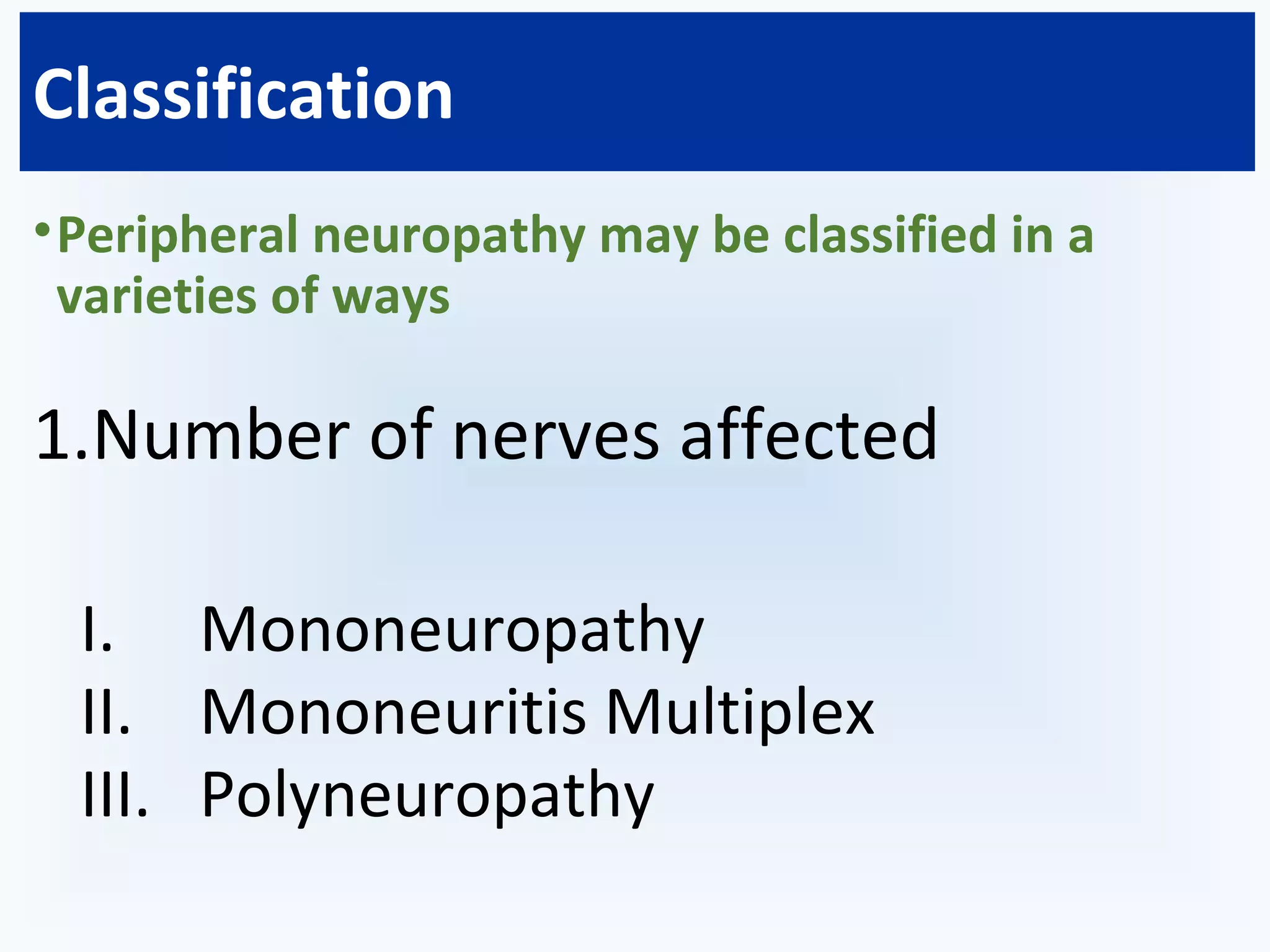
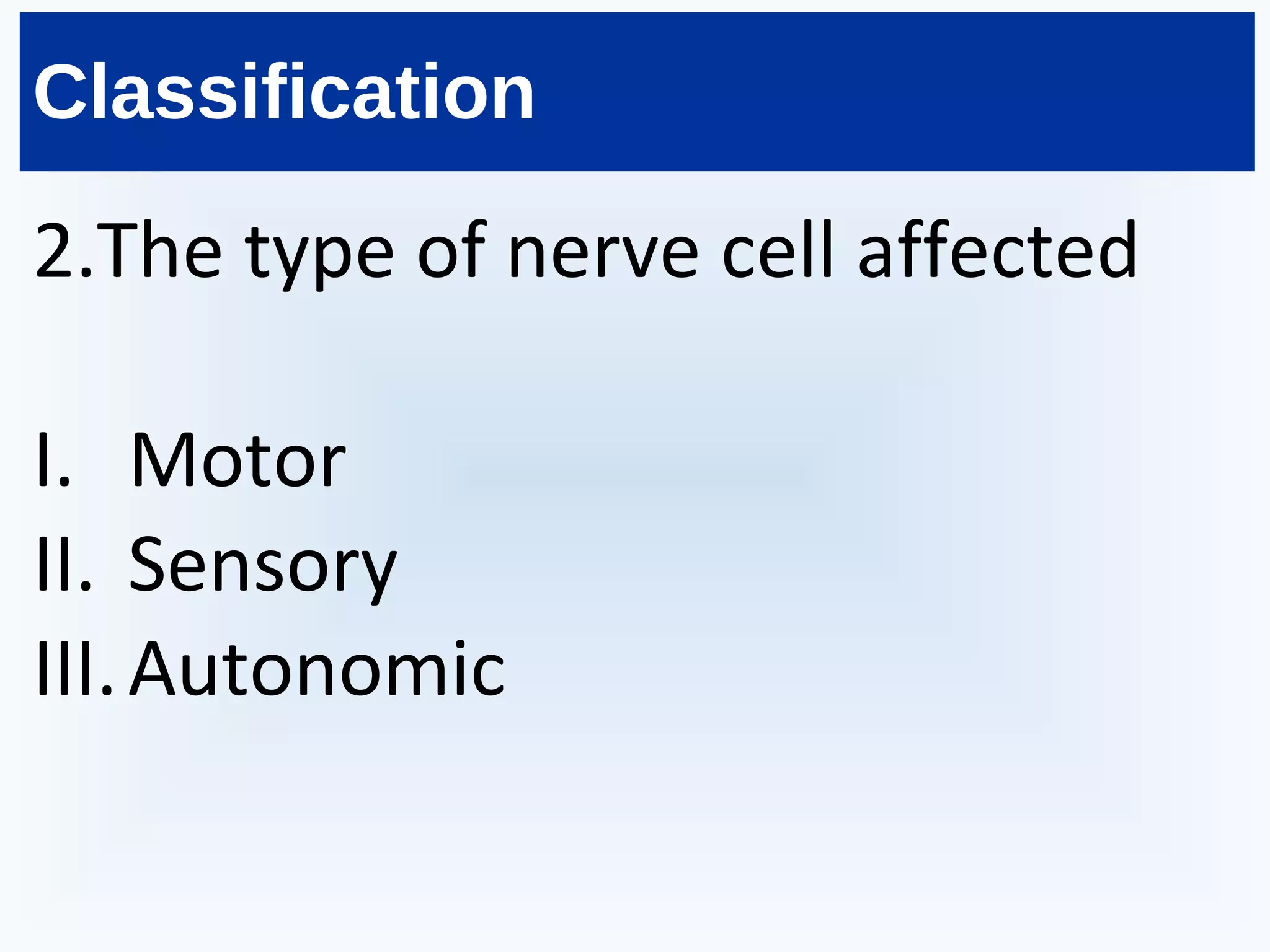
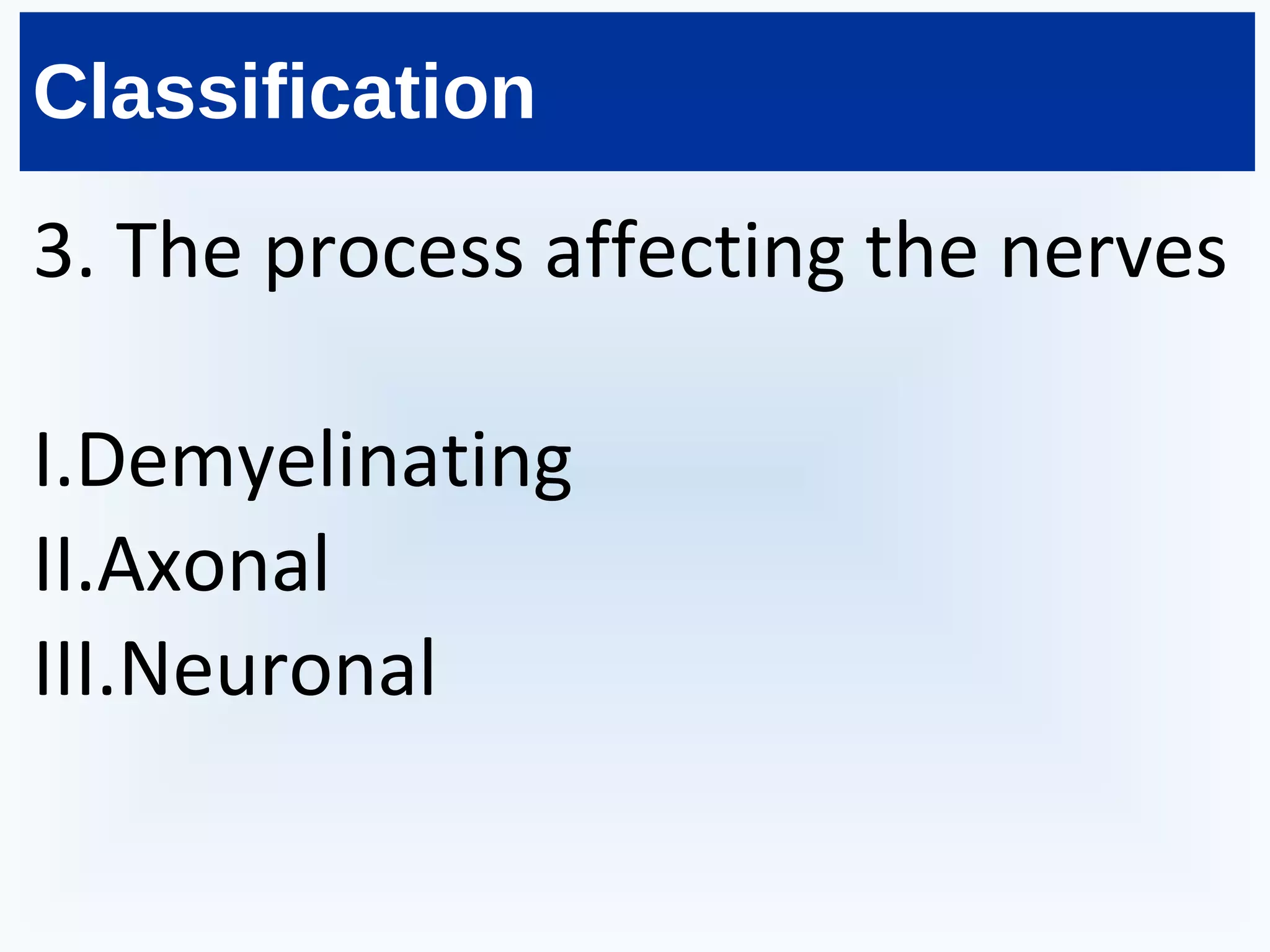
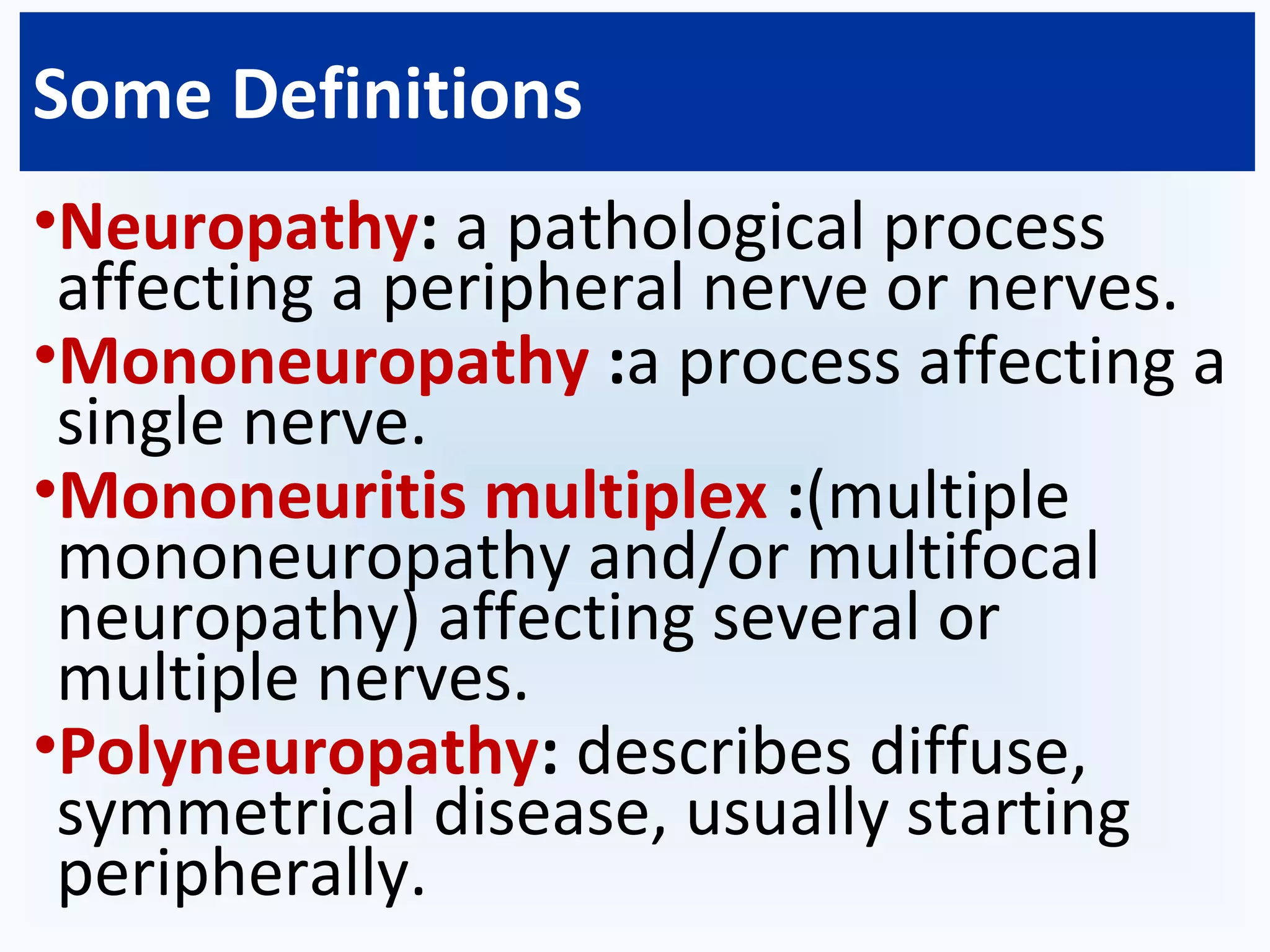
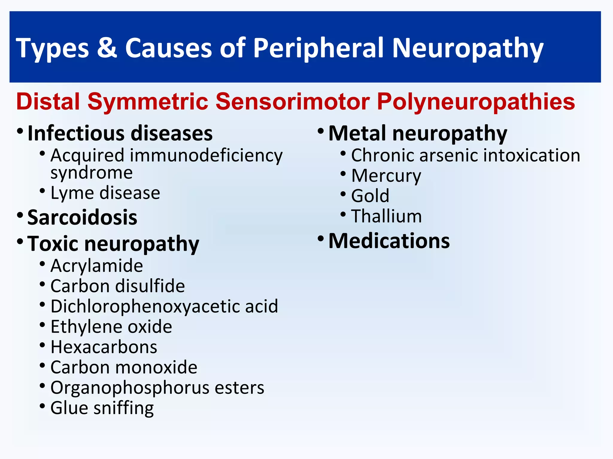
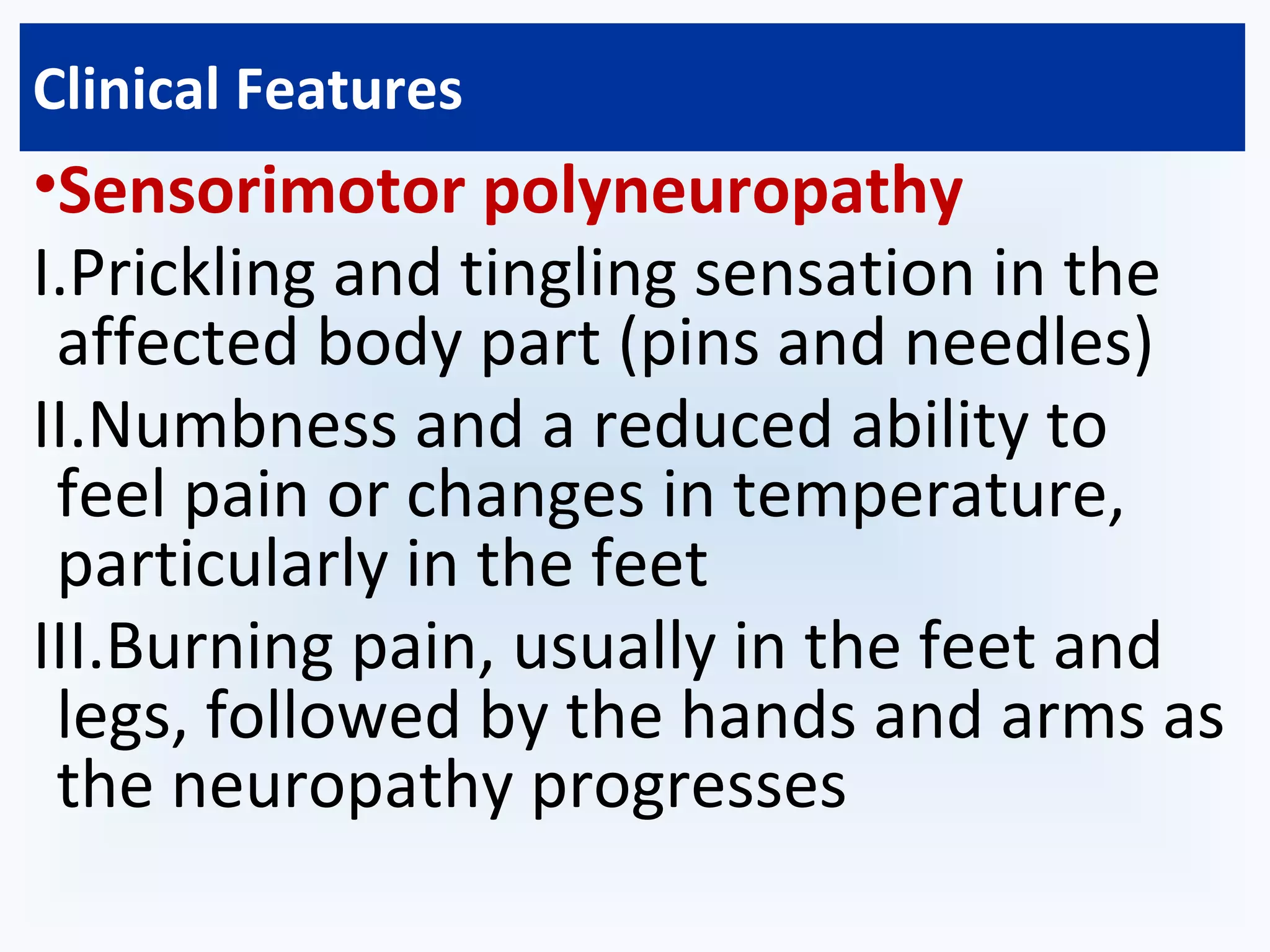
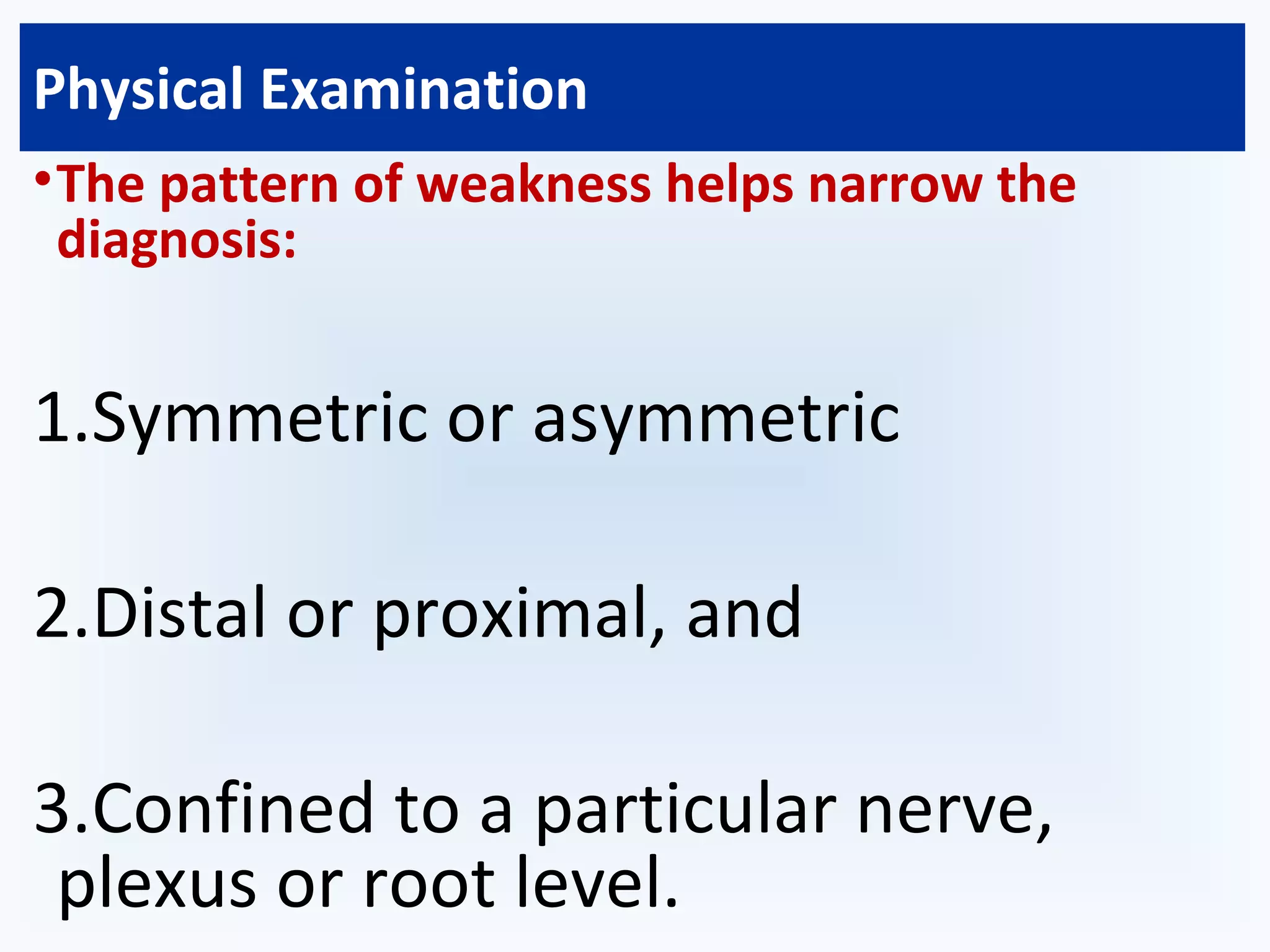
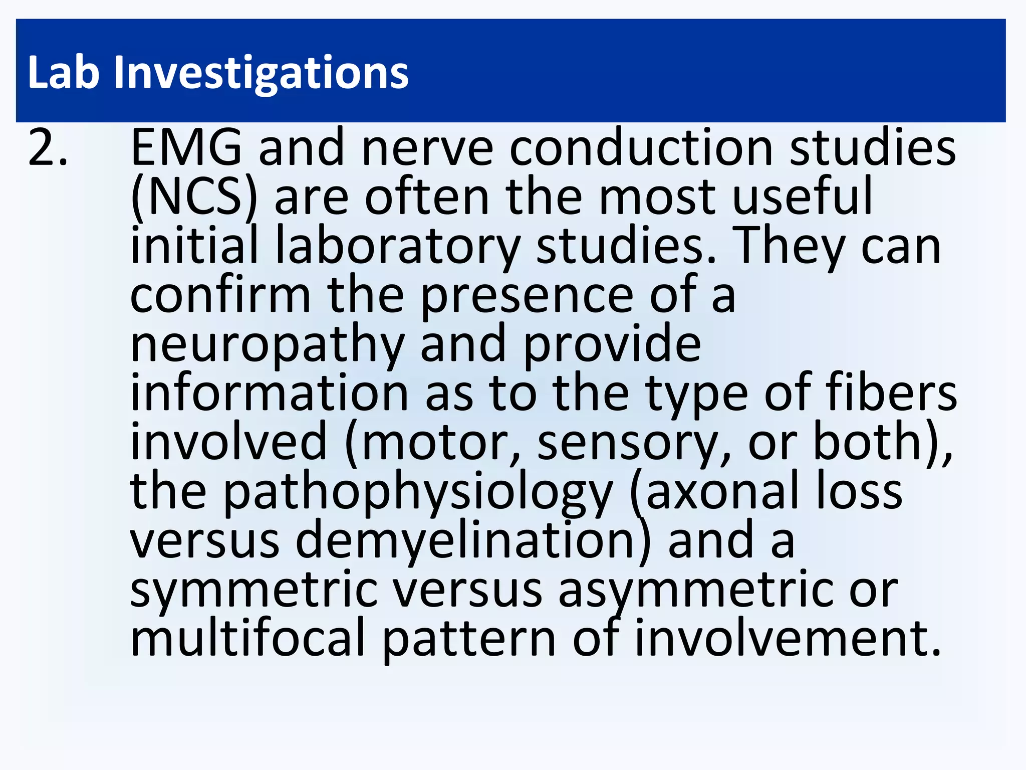
Peripheral neuritis, or peripheral neuropathy, is damage to nerves in the peripheral nervous system caused by diseases of the nerves or systemic illness. The peripheral nervous system consists of cranial and spinal nerves and contains motor, sensory, and autonomic nerves. Peripheral neuropathy can be classified based on the number, type, or process affecting nerves and may be caused by diseases like diabetes, infections, toxins, and medications. Physical exams can reveal motor and sensory signs while electrodiagnostic tests help evaluate the type and pattern of nerve involvement. Treatment involves managing underlying causes, relieving symptoms, and mechanical aids or surgery for specific nerve damage.
2. Definition:
Peripheral Neuritis or Peripheral
neuropathy is the term used for
damage to nerves of the
peripheral nervous system, which
may be caused either by diseases
of the nerve or from the side-
effects of systemic illness.
4. Peripheral nervous system
The peripheral nervous system is made up of 3 main
types of nerves, each with its own specific function:
1.Motor nerves
•control movements of all muscles under conscious
control, such as those used for walking, grasping things,
or talking.
1.Sensory nerves
•transmit information about sensory experiences, such as
the feeling of a light touch or the pain resulting from a
cut.
1.Autonomic nerves
•regulate biological activities that people do not control
consciously, such as breathing, digesting food, and heart
and gland functions.
5. Classification
•Peripheral neuropathy may be classified in a
varieties of ways
1.Number of nerves affected
I. Mononeuropathy
II. Mononeuritis Multiplex
III. Polyneuropathy
8. Some Definitions
•Neuropathy: a pathological process
affecting a peripheral nerve or nerves.
•Mononeuropathy :a process affecting a
single nerve.
•Mononeuritis multiplex :(multiple
mononeuropathy and/or multifocal
neuropathy) affecting several or
multiple nerves.
•Polyneuropathy: describes diffuse,
symmetrical disease, usually starting
peripherally.
11. Clinical Features
•Sensorimotor polyneuropathy
I.Prickling and tingling sensation in the
affected body part (pins and needles)
II.Numbness and a reduced ability to
feel pain or changes in temperature,
particularly in the feet
III.Burning pain, usually in the feet and
legs, followed by the hands and arms as
the neuropathy progresses
12. Clinical Features
•Sensorimotor polyneuropathy
IV.Sharp stabbing pain, which is often
worse at night (the feet and legs are
affected first, followed by the hands
and arms)
V.Muscle weakness
VI.Loss of co-ordination
VII.Muscle paralysis
VIII.Increased risk of developing
problems that affect the feet, such as
skin infections and foot ulcers
14. Signs
•Signs depend on the types, severity & nerve
cells involved.
I.Motor signs- lower motor neuron
lesion-wasting ,fasciculation and loss of
reflexes
II.Sensory signs-diminished or loss of
sensation of all modalities of sensation
15. Physical Examination
•A cranial nerve examination can
provide evidence of
mononeuropathies or proximal
involvement.
•Funduscopic examination may show
abnormalities such as optic pallor,
which can be present in
leukodystrophies and vitamin B12
deficiency.
16. Physical Examination
•Direct strength testing of muscles
enervated by cranial nerves V, VII, IX/X,
XI and XII is important, as mild bilateral
weakness can be missed by observation
only.
•The motor examination includes a
search for fasciculations or cramps, or
loss of muscle bulk.
•Tone is normal or reduced.
17. Physical Examination
•The pattern of weakness helps narrow the
diagnosis:
1.Symmetric or asymmetric
2.Distal or proximal, and
3.Confined to a particular nerve,
plexus or root level.
18. Physical Examination
•Deep tendon reflexes are reduced or
absent.
•A bilateral foot drop may result in a
steppage gait in which the patient must lift
the knees very high in order to clear the
toes.
•Proximal weakness results in an inability to
squat or to rise unassisted from a chair.
20. Lab Investigations
•The most common presentation is that of a distal
symmetric sensorimotor neuropathy.
1. Initial evaluation should include:
I. Fasting serum glucose
II. Glycosylated hemoglobin
III. Blood urea nitrogen
IV. Creatinine
V. Complete blood cell count
VI. Erythrocyte sedimentation rate
VII. Urinalysis
VIII.Vitamin B12
IX. Hyrotropin stimulating hormone levels.
21. Lab Investigations
2. EMG and nerve conduction studies
(NCS) are often the most useful
initial laboratory studies. They can
confirm the presence of a
neuropathy and provide
information as to the type of fibers
involved (motor, sensory, or both),
the pathophysiology (axonal loss
versus demyelination) and a
symmetric versus asymmetric or
multifocal pattern of involvement.
23. Lab Investigations
5. Other studies useful in specific clinical
contexts are:
•cytology (lymphoma)
•special studies such as Lyme polymerase chain
reaction and cytomegalovirus branched chain
DNA (polyradiculopathy or mononeuritis
multiplex in AIDS).
24. Treatment
•The goal of treatment is to manage
the underlying condition causing the
neuropathy and repair damage, as
well as provide symptom relief.
•Controlling a chronic condition may
not eliminate the neuropathy, but it
can play a key role in managing it.