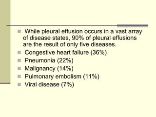
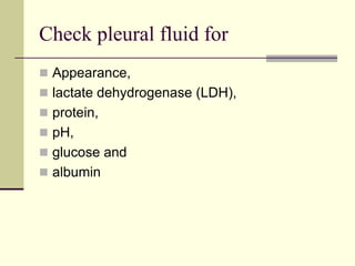
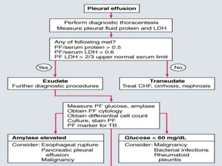
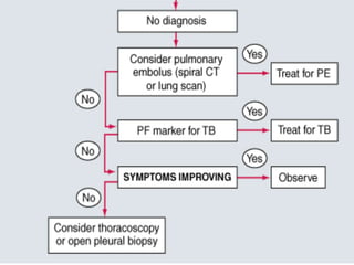
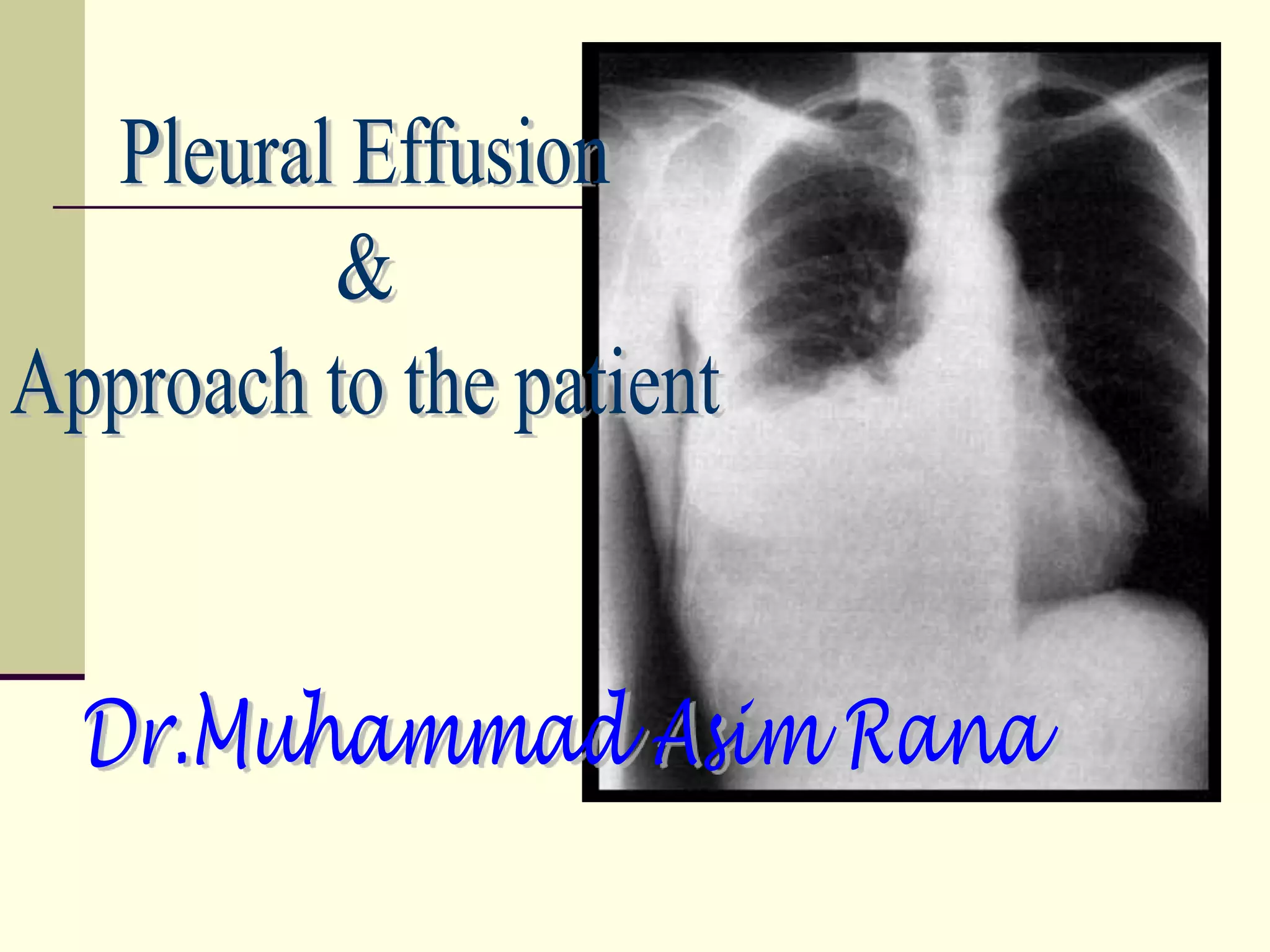
SlideShare a Scribd company logo
1 of 91
Download to read offline
Definition
 Pleural effusion is the accumulation of fluid in
the pleural space.
 The pleural space lies between the lung and
chest wall and normally contains a very thin
layer of fluid, which serves as a coupling
system.
 A pleural effusion is present when there is an
excess quantity of fluid in the pleural space.
Etiology
 Normally, fluid enters the pleural space from
the capillaries in the parietal pleura and is
removed via the lymphatics situated in the
parietal pleura.
 Fluid can also enter the pleural space from
the interstitial spaces of the lung via the
visceral pleura or from the peritoneal cavity
via small holes in the diaphragm.
 Pleural fluid accumulates when pleural fluid formation
exceeds pleural fluid absorption.
 The lymphatics have the capacity to absorb 20 times
more fluid than is normally formed. Accordingly, a
pleural effusion may develop when there is excess
pleural fluid formation (from the interstitial spaces of
the lung, the parietal pleura, or the peritoneal cavity)
or when there is decreased fluid removal by the
lymphatics.
Types of pleural effusion
Transudative pleural effusions
result from alteration of hydrostatic and
oncotic factors that increase the formation or
decrease the absorption of pleural fluid (e.g.,
increased mean capillary pressure [heart
failure] or decreased oncotic pressure
[cirrhosis or nephrotic syndrome]).
Exudative pleural effusions
occur when damage or disruption of the
normal pleural membranes or vasculature
(e.g., tumor involvement of the pleural space,
infection, inflammatory conditions, or trauma)
leads to increased capillary permeability or
decreased lymphatic drainage.
Diagnostic Approach
Clinical Presentation
 The underlying cause of the effusion usually
dictates the symptoms, although patients may
be asymptomatic.
 Pleural inflammation, abnormal pulmonary
mechanics, and worsened alveolar gas
exchange produce symptoms and signs of
disease.
symptoms and signs
 Inflammation of the parietal pleura leads to
pain in local (intercostal) involved areas or
referred (phrenic) distributions (shoulder).
 Dyspnea is frequent and may be present
and out of proportion to the size of the
effusion.
 Cough can occur.
Chest examination is notable for
 dullness to percussion, decreased or
absent tactile fremitus, and decreased
breath sounds.
 Tracheal shift to the contralateral side or
an ipsilateral pleural rub may be present.
 The clinical setting is crucial to establishing a proper
diagnosis. A definitive diagnosis based solely upon
pleural fluid analysis is possible in the minority of
pleural effusions.
 History or physical examination findings suggestive
of congestive heart failure, malignancy, pneumonia,
pulmonary embolism, myocardial infarction, surgery,
cirrhosis, or rheumatologic arthritis provide important
clues to the underlying diagnosis.
Laboratory and Imaging
Studies
Chest Roentginogram
 Pleural effusions are typically detected by
chest radiography as blunting of the
costophrenic angle or opacification of the
base of the hemithorax without loss of volume
of the hemithorax (which would suggest
atelectasis), and may be accompanied by air
bronchograms (which would suggest an
alveolar filling process such as pneumonia).
 Prior to invasive diagnostic or therapeutic
procedures, the patient should undergo
imaging to confirm the presence and size of
the effusion. Preferred modalities include:
Decubitus chest radiography
 Showing layering fluid will confirm the
presence of pleural effusion and
demonstrates that at least a portion of the
fluid is not loculated.
Thoracic ultrasonography
 Is one of the best modalities to assess for
pleural fluid loculations.
 Ultrasonography can also provide real-time
guidance for pleural procedures and can
reduce both the complication and failure rate
of thoracentesis.
Computed tomography of the chest
 With contrast helps differentiate pleural fluid
from lung masses and atelectatic lung, and
 helps define the extent of pleural thickening,
pleural nodularity, and other associated
findings.
Pleural fluid analysis
 Thoracentesis can be performed safely at the
bedside, in the absence of disorders of hemostasis,
on effusions that extend >10 mm from the inner chest
wall on a lateral decubitus film.
 Loculated effusions can be localized with
ultrasonography or CT scan.
 Proper technique and sonographic guidance
minimize the risk of pneumothorax and other
complications.
 The first step is to determine whether the
effusion is a transudate or an exudate.
 A transudative pleural effusion occurs when
systemic factors that influence the formation
and absorption of pleural fluid are altered.
The leading causes of transudative pleural
effusions are left ventricular failure and
cirrhosis.
 An exudative pleural effusion occurs when
local factors that influence the formation and
absorption of pleural fluid are altered. The
leading causes of exudative pleural effusions
are bacterial pneumonia, malignancy, viral
infection, and pulmonary embolism.
 The primary reason to make this
differentiation is that additional diagnostic
procedures are indicated with exudative
effusions to define the cause of the local
disease.
 While pleural effusion occurs in a vast array
of disease states, 90% of pleural effusions
are the result of only five diseases.
 Congestive heart failure (36%)
 Pneumonia (22%)
 Malignancy (14%)
 Pulmonary embolism (11%)
 Viral disease (7%)
Check pleural fluid for
 Appearance,
 lactate dehydrogenase (LDH),
 protein,
 pH,
 glucose and
 albumin
 Serum lactate dehydrogenase (LDH), protein,
pH, glucose and albumin should be
measured within hour of the thoracentesis to
allow appropriate comparison.
Pleural fluid appearance
 Most transudates are clear, straw colored,
nonviscid, and without odor
 Red-tinged pleural effusions indicate the
presence of blood.
 In exudative pleural effusions,
serosanguineous fluid is usually not helpful in
narrowing the diagnosis.
Bloody pleural fluid
 If the blood is due to thoracentesis, the
degree of discoloration should clear during
the aspiration.
 Bloody pleural fluid usually indicates the
presence of malignancy, pulmonary
embolism (PE), or trauma.
Hemothorax
 The presence of gross blood should lead to
the measurement of a pleural fluid
hematocrit.
 Hemothorax is defined as a pleural fluid to
blood hematocrit ratio of >0.5, and chest tube
drainage should be considered.
 Exudative pleural effusions meet at least one
of the Light's criteria , whereas transudative
pleural effusions meet none:
Light's criteria
 (a) a pleural fluid-to-serum protein ratio of
>0.5,
 (b) a pleural fluid-to-serum LDH ratio of >0.6,
 (c) a pleural fluid LDH of more than two-thirds
of the upper limit of normal for serum LDH
 The above criteria misidentify ~25% of
transudates as exudates.
 If one or more of the exudative criteria are
met and the patient is clinically thought to
have a condition producing a transudative
effusion, like in whom clinical suspicion for
heart, liver, or kidney disease is high what
should be done?
 The difference between the protein levels in the
serum and the pleural fluid should be measured.
 If this gradient is greater than 31 g/L (3.1 g/dL), the
exudative categorization by the above criteria can be
ignored because almost all such patients have a
transudative pleural effusion.
 In some texts a gradient of >1.2 g/dL suggests that
the pleural fluid is transudate.
If a patient has an exudative pleural
effusion
 Description of the fluid,
 Glucose level,
 Differential cell count,
 Microbiologic studies,
 Cytology.
 Cultures,
 Triglycerides,
 Amylase, and
 pH
WBC differential
 The WBC differential is often not diagnostic,
although neutrophilia is suggestive of
infection.
 Eosinophilia (>10% of total nucleated cell
count) is suggestive of air or blood in the
pleural space. If air or blood is not present in
the pleural space, consideration should be
given to fungal and parasitic infection, drug-
induced disease, PE, asbestos-related
disease, and Churg-Strauss syndrome.
 Lymphocytosis (>50% of the total nucleated
cell count) is suggestive of malignancy or
tuberculosis.
 Mesothelial cells argues against the
diagnosis of tuberculosis.
 Plasma cells suggest a diagnosis of multiple
myeloma.
 Exudative effusions with normal protein but
high LDH are likely to be parapneumonic or
secondary to malignancy.
 LDH is an indicator of the degree of pleural
inflammation.
Glucose concentration
 A glucose concentration of <60 mg/dL is
probably due to
 tuberculosis,
 malignancy,
 rheumatoid arthritis, or
 parapneumonic effusion.
 For parapneumonic pleural effusions with a
glucose of <60 mg/dL, tube thoracostomy
should be considered.
Pleural fluid with a low pH
 A pH of <7.3 is seen with
 empyema,
 tuberculosis,
 malignancy,
 collagen vascular disease, or
 esophageal rupture.
 For parapneumonic pleural effusions with a pH of
<7.20, tube thoracostomy should be considered.
 Pleural fluid for pH testing should be collected
anaerobically in a heparinized syringe and placed on
ice.
 Pleural fluid with a low pH usually has a low glucose
and a high LDH; otherwise, the low pH may be due to
poor sample collection technique.
Amylase
 An elevation of amylase suggests that the
patient has pancreatic disease, malignancy,
or esophageal rupture.
 Malignancy and esophageal rupture have
salivary amylase elevations and not
pancreatic amylase elevations.
Turbid or milky fluid
 should be centrifuged.
 If the supernatant clears, the cloudiness is likely due
to cells and debris.
 If the supernatant remains turbid, pleural lipids should
be measured. Elevation of triglycerides (>110 mg/dL)
suggests that a chylothorax is present, usually due to
disruption of the thoracic duct from trauma, surgery,
or malignancy (i.e., lymphoma).
Cytology
 Cytology is positive in approximately 60% of
malignant effusions.
 Priming the fluid collection bag with
unfractionated heparin (UFH; e.g., 1,000
International Units) may increase the yield.
 The volume of pleural fluid analyzed does not
impact the yield of cytologic diagnosis.
 Repeat thoracentesis increases the
diagnostic yield.
Surgical diagnostic
procedures
Closed pleural biopsy
 Closed pleural biopsy adds little to the diagnostic
yield of thoracentesis, except in the diagnosis of
tuberculosis.
 For tuberculous effusions, pleural fluid cultures alone
are positive in only 20% to 25% of cases. However,
the combination of pleural fluid studies and pleural
biopsy (demonstrating granulomas or organisms) is
90% sensitive in establishing tuberculosis as the
etiology of the effusion.
Diagnostic thoracoscopy
 Diagnostic thoracoscopy has largely
replaced closed pleural biopsy. Thoracoscopy
allows directed biopsies that increase the
diagnostic yield for malignancy while
maintaining the high diagnostic yield of
closed pleural biopsy for TB.
Indications for diagnostic
thoracoscopy
 Pleural effusion of unknown etiology
 Mesothelioma
 Lung cancer
 Tuberculosis
 Other benign pleural disorders
 Pulmonary parenchymal disease
Other diagnostic
procedures
 Other diagnostic procedures that are useful in
establishing the etiology of a pleural effusion
when the aforementioned tests are
nondiagnostic include:
 Evaluation of liver function
 Renal function,
 Cardiac echo,
 Biopsy of other abnormal sites (e.g., a
mediastinal or lung mass), and
 Evaluation of PE.
Differential Diagnoses of
Pleural Effusions
Transudative Pleural Effusions
1. Congestive heart failure
2. Cirrhosis
3. Pulmonary embolization
4. Nephrotic syndrome
5. Peritoneal dialysis
6. Superior vena cava obstruction
7. Myxedema
8. Urinothorax
Exudative Pleural Effusions
 1. Neoplastic diseases
a. Metastatic disease
b. Mesothelioma
 2. Infectious diseases
a. Bacterial infections
b. Tuberculosis
c. Fungal infections
d. Viral infections
e. Parasitic infections
 3. Pulmonary embolization
 4. Gastrointestinal disease
a. Esophageal perforation
b. Pancreatic disease
c. Intraabdominal abscesses
d. Diaphragmatic hernia
e. After abdominal surgery
f. Endoscopic variceal sclerotherapy
g. After liver transplant
 5. Collagen-vascular diseases
a. Rheumatoid pleuritis
b. Systemic lupus erythematosus
c. Drug-induced lupus
d. Immunoblastic lymphadenopathy
e. Sjögren's syndrome
f. Wegener's granulomatosis
g. Churg-Strauss syndrome
 6. Post-coronary artery bypass surgery
 7. Asbestos exposure
 8. Sarcoidosis
 9. Uremia
 10. Meigs' syndrome
 11. Yellow nail syndrome
 12. Drug-induced pleural disease
a. Nitrofurantoin
b. Dantrolene
c. Methysergide
d. Bromocriptine
e. Procarbazine
f. Amiodarone
 13. Trapped lung
 14. Radiation therapy
 15. Post-cardiac injury syndrome
 16. Hemothorax
 17. Iatrogenic injury
 18. Ovarian hyperstimulation syndrome
 19. Pericardial disease
 20. Chylothorax
"Classic" exudates that can be
transudates
Malignancy:
Due to early lymphatic obstruction, obstructive
atelectasis, or concomitant disease (CHF).
Pulmonary embolism:
23 percent incidence; due to atelectasis.
Sarcoidosis:
Stage II and III disease.
Hypothyroid pleural effusion:
From hypothyroid heart disease or hypothyroidism per
se.
Treatment
Transudates resolve with treatment of the
underlying heart, kidney, or liver disease.
Uncommonly, more aggressive approaches
including pleurodesis and shunts are
required.
 Parapneumonic effusions and empyema
should be managed with tube drainage when
indicated based on the size, gross
appearance, or biochemical analysis of the
pleural fluid or the presence of loculations
Multiple tubes are sometimes required to
adequately drain the pleural space.
Failure to adequately and quickly drain a
complicated parapneumonic effusion can
lead to organization of the pleural fluid and
formation of a thick pleural adhesions which
may necessitate surgical removal known as
decortication.
Indications for Tube
Thoracostomy in
Parapneumonic Effusions
Radiographic criteria
Pleural fluid loculations
Effusion filling more than half the hemithorax
Air fluid level
Microbiologic criteria
Pus in the pleural space
Positive stain for microorganisms
Positive pleural fluid cultures
Chemical criteria
Pleural fluid pH <7.2
Pleural fluid glucose <60 mg/dL
CONTRAINDICATIONS
 There are no absolute contraindications to
tube thoracostomy, particularly if the patient is
in respiratory distress.
 Anticoagulation or a bleeding diathesis is a
relative contraindication in a patient
undergoing elective chest tube placement for
pleurodesis. Blind insertion of a chest tube is
dangerous in a patient with adhesions from
infection, previous pleurodesis, or a lung
transplant; guidance by CT scan without
contrast is preferred in these patients.
Type of tube
 Silastic® tubes are preferred because older
rubber tubes have fewer drainage holes, are
not well visualized on chest radiographs, and
produce more pleural inflammation. Silastic
chest tubes contain a radiopaque strip with a
gap that serves to mark the most proximal
drainage hole.
Size of tube
 A chest tube's internal diameter and length
are the critical determinants of flow.
 Select the appropriate chest tube size to
account for the viscosity and accumulation
rate of the pleural material to be drained.
 As an example, drainage of viscous fluids
requires a larger bore chest tube than that
required for drainage of a similar volume of
air.
 Malignant effusion— A small-bore catheter (8
to 14 Fr) placed under ultrasound or CT
guidance is usually adequate to drain a
malignant pleural effusion and achieve
pleurodesis.
 Empyema — For a complicated
parapneumonic effusion or empyema that is
amenable to drainage with a single catheter.
Prefer initial image-guided placement of
small-bore catheters (10 to 14 Fr), with or
without intrapleural fibrinolytic agents.
 It is preferred to use the smaller tube size as
this is generally more comfortable for
patients, particularly if more than one tube is
needed. Alternatively, when the fluid appears
viscous, a larger bore tube (16-24 Fr) may be
used.
 Unsuccessful drainage with a small-bore
catheter either indicates the presence of
multiple loculations or very viscous material.
Multiple small-bore catheters may be used in
multiloculated effusions or large bore
catheters in case of very viscous material.
Failure to drain with a single small-bore tube
should also lead to thoracic surgery
consultation to avoid delays in case video
assisted thoracoscopy (VATS) becomes
necessary.
 Hemothorax — The goals of tube
thoracostomy in acute hemothorax are
drainage of fresh blood, quantification of the
rate of bleeding, evacuation of any coexisting
pneumothorax, and tamponade of the
bleeding site. Large bore catheters (32 to 40
Fr) are required to reliably achieve these
goals.
 Once a hemothorax is defibrinated in situ,
that is after the acute phase, success of
drainage is less dependent on the size of the
tube, than on the degree and mode of clot
formation. Large amounts of clotted blood
should be evacuated via video assisted
thoracoscopy.
 Occasionally, a hemothorax may result in a
sonographically complex septate pattern and
may be treated with small-bore catheters.
Treatment of hemothorax should be
individualized and done in consultation with
thoracic surgery.
Malignant pleural effusions
 Observation without invasive interventions
may be appropriate for some patients with
malignant pleural effusions.
 Therapeutic thoracentesis may improve
patient comfort and relieve dyspnea. The
rapid removal of more than 1 L of pleural fluid
may rarely result in re-expansion pulmonary
edema, especially if the lung is unable to re-
expand.
 Repeated thoracenteses are reasonable if
they achieve symptomatic relief and if fluid
reaccumulation is slow. Unfortunately, 95% of
malignant effusions will recur with a median
time to recurrence of less than a week. When
frequent or repeated thoracentesis is required
for effusions that reaccumulate, early
consideration should be given to tube
drainage with pleurodesis or placement of a
chronic indwelling pleural catheter.
Chemical pleurodesis
 Chemical pleurodesis is an effective therapy
for recurrent effusions. This treatment is
recommended in patients whose symptoms
are relieved with initial drainage but who have
rapid reaccumulation of fluid.
Talc pleurodesis
Effective and inexpensive.
Fever and hypoxia are common following
instillation of talc into the pleural space, and
respiratory failure has been described on
occasion.
Overall efficacy is similar for talc slurry
delivered via chest tube versus dry talc
insufflated during thoracoscopy.
Doxycycline or minocycline
 Doxycycline or minocycline can also be
instilled into the pleural space via a chest
tube.
 Pain is more prevalent and severe following
doxycycline and minocycline than following
talc.
Bleomycin
 Bleomycin appears to be less effective and
more expensive than other drugs.
 Systemic analgesics and the administration of
lidocaine in the sclerosing agent solution help
to decrease the appreciable discomfort
associated with the procedure.
 If the chest tube drainage remains high (>100
mL/d) more than 2 days after the initial
pleurodesis, a second dose of the sclerosing
agent can be administered.
Chronic indwelling pleural catheters
 Provide good control of effusion-related
symptoms via intermittent drainage.
 The Pleurx catheter is better at controlling
symptoms than doxycycline administered via a
chest tube. Furthermore, repeated drainage via
a Pleurx catheter leads to pleurodesis in roughly
50% of patients, allowing the catheter to be
removed.
Pleurectomy or pleural abrasion
 Requires thoracic surgery and should be
reserved for patients with a good prognosis who
have had ineffective pleurodesis or inadequate
response to chronic pleural drainage.
Chemotherapy and mediastinal
radiotherapy
 May control effusions in responsive tumors, such
as lymphoma or small-cell bronchogenic
carcinoma, although it has poor efficacy in
metastatic carcinoma.
Thank you very much

More Related Content

Similar to approachtopleuraleffusion-140205151929-phpapp01.pdf

L5 pleural effusion
L5 pleural effusionL5 pleural effusion
L5 pleural effusionbilal natiq
 
Diseases of the pleura and pleural space
Diseases of the pleura and pleural spaceDiseases of the pleura and pleural space
Diseases of the pleura and pleural spaceDr Vaziri
 
6.Pleural Effusions
6.Pleural Effusions6.Pleural Effusions
6.Pleural Effusionsghalan
 
20.5.pleural effusion &amp;empyema
20.5.pleural effusion &amp;empyema20.5.pleural effusion &amp;empyema
20.5.pleural effusion &amp;empyemapediatricsmgmcri
 
Pleural Effusion for Undergraduates
Pleural Effusion for UndergraduatesPleural Effusion for Undergraduates
Pleural Effusion for UndergraduatesSesha Sai
 
Pleural effusion Seminar Presentation
Pleural effusion Seminar PresentationPleural effusion Seminar Presentation
Pleural effusion Seminar Presentationpradeepmk8
 
Aproach To Diagnosis of Pleural Effusion
Aproach To Diagnosis of Pleural EffusionAproach To Diagnosis of Pleural Effusion
Aproach To Diagnosis of Pleural EffusionAmitKalne1
 
hydrothorax presentation.pptx
hydrothorax presentation.pptxhydrothorax presentation.pptx
hydrothorax presentation.pptxAshraf Shaik
 
Disorders of pleura
Disorders of pleuraDisorders of pleura
Disorders of pleuraFiroz Hakkim
 
4 pleural effusions
4 pleural effusions4 pleural effusions
4 pleural effusionsinternalmed
 
Diagnostic value of pleural effusion
Diagnostic value of pleural effusionDiagnostic value of pleural effusion
Diagnostic value of pleural effusionSarfraz Saleemi
 
Pleural effusion by nurse peter
Pleural effusion by nurse peterPleural effusion by nurse peter
Pleural effusion by nurse peterPeter Shirima
 
Malignant Pleural Effusion.pptx
Malignant Pleural Effusion.pptxMalignant Pleural Effusion.pptx
Malignant Pleural Effusion.pptxYY19921
 
Pleural effusion analysis
Pleural effusion  analysisPleural effusion  analysis
Pleural effusion analysiswalaaelfar
 

Similar to approachtopleuraleffusion-140205151929-phpapp01.pdf (20)

Approach to a case of pleural effusion
Approach to a case of pleural effusionApproach to a case of pleural effusion
Approach to a case of pleural effusion
 
L5 pleural effusion
L5 pleural effusionL5 pleural effusion
L5 pleural effusion
 
Diseases of the pleura and pleural space
Diseases of the pleura and pleural spaceDiseases of the pleura and pleural space
Diseases of the pleura and pleural space
 
Pleural effusion
Pleural effusionPleural effusion
Pleural effusion
 
6.Pleural Effusions
6.Pleural Effusions6.Pleural Effusions
6.Pleural Effusions
 
20.5.pleural effusion &amp;empyema
20.5.pleural effusion &amp;empyema20.5.pleural effusion &amp;empyema
20.5.pleural effusion &amp;empyema
 
Pleural effusion
Pleural effusionPleural effusion
Pleural effusion
 
Pleural Effusion for Undergraduates
Pleural Effusion for UndergraduatesPleural Effusion for Undergraduates
Pleural Effusion for Undergraduates
 
Pleural effusion Seminar Presentation
Pleural effusion Seminar PresentationPleural effusion Seminar Presentation
Pleural effusion Seminar Presentation
 
Aproach To Diagnosis of Pleural Effusion
Aproach To Diagnosis of Pleural EffusionAproach To Diagnosis of Pleural Effusion
Aproach To Diagnosis of Pleural Effusion
 
hydrothorax presentation.pptx
hydrothorax presentation.pptxhydrothorax presentation.pptx
hydrothorax presentation.pptx
 
Disorders of pleura
Disorders of pleuraDisorders of pleura
Disorders of pleura
 
4 pleural effusions
4 pleural effusions4 pleural effusions
4 pleural effusions
 
Pleural Effusions
Pleural  EffusionsPleural  Effusions
Pleural Effusions
 
Diagnostic value of pleural effusion
Diagnostic value of pleural effusionDiagnostic value of pleural effusion
Diagnostic value of pleural effusion
 
Pleuresy
PleuresyPleuresy
Pleuresy
 
Pleural effusion
Pleural effusionPleural effusion
Pleural effusion
 
Pleural effusion by nurse peter
Pleural effusion by nurse peterPleural effusion by nurse peter
Pleural effusion by nurse peter
 
Malignant Pleural Effusion.pptx
Malignant Pleural Effusion.pptxMalignant Pleural Effusion.pptx
Malignant Pleural Effusion.pptx
 
Pleural effusion analysis
Pleural effusion  analysisPleural effusion  analysis
Pleural effusion analysis
 

More from NellyPhiri5

Lecture 3 -Screening tests.ppt presentation
Lecture 3 -Screening tests.ppt presentationLecture 3 -Screening tests.ppt presentation
Lecture 3 -Screening tests.ppt presentationNellyPhiri5
 
Lecture 16 APH.pptx. power point lecture
Lecture 16 APH.pptx. power point lectureLecture 16 APH.pptx. power point lecture
Lecture 16 APH.pptx. power point lectureNellyPhiri5
 
BURNS.pptx lecture presentation surgery.
BURNS.pptx lecture presentation surgery.BURNS.pptx lecture presentation surgery.
BURNS.pptx lecture presentation surgery.NellyPhiri5
 
SUTURING AND SUTURE MATERIALS.lecture.pptx
SUTURING AND SUTURE MATERIALS.lecture.pptxSUTURING AND SUTURE MATERIALS.lecture.pptx
SUTURING AND SUTURE MATERIALS.lecture.pptxNellyPhiri5
 
TYPES OF INCISIONS. presentation lecture
TYPES OF INCISIONS. presentation lectureTYPES OF INCISIONS. presentation lecture
TYPES OF INCISIONS. presentation lectureNellyPhiri5
 
Spirochetes ppt microbiology and immunology
Spirochetes ppt  microbiology and immunologySpirochetes ppt  microbiology and immunology
Spirochetes ppt microbiology and immunologyNellyPhiri5
 
Contraception_Lecture_i mweene.pptx
Contraception_Lecture_i mweene.pptxContraception_Lecture_i mweene.pptx
Contraception_Lecture_i mweene.pptxNellyPhiri5
 
Lecture 19 (Tapeworms).pptx
Lecture 19 (Tapeworms).pptxLecture 19 (Tapeworms).pptx
Lecture 19 (Tapeworms).pptxNellyPhiri5
 
Lecture 3 (T. cruzi) .pptx
Lecture 3 (T. cruzi) .pptxLecture 3 (T. cruzi) .pptx
Lecture 3 (T. cruzi) .pptxNellyPhiri5
 
12.2NUCLEOTIDES & NUCLEIC ACIDS-1.pptx
12.2NUCLEOTIDES & NUCLEIC ACIDS-1.pptx12.2NUCLEOTIDES & NUCLEIC ACIDS-1.pptx
12.2NUCLEOTIDES & NUCLEIC ACIDS-1.pptxNellyPhiri5
 
shockbyara1-141208091205-conversion-gate02 (1).pdf
shockbyara1-141208091205-conversion-gate02 (1).pdfshockbyara1-141208091205-conversion-gate02 (1).pdf
shockbyara1-141208091205-conversion-gate02 (1).pdfNellyPhiri5
 
Lecture 12 (G+ & G- cocci ;STAPH& NEISSERIA).pptx
Lecture 12 (G+ & G- cocci ;STAPH& NEISSERIA).pptxLecture 12 (G+ & G- cocci ;STAPH& NEISSERIA).pptx
Lecture 12 (G+ & G- cocci ;STAPH& NEISSERIA).pptxNellyPhiri5
 
Lecture 2 - NATURAL HISTORY OF A DISEASE.pptx
Lecture 2 - NATURAL HISTORY OF A DISEASE.pptxLecture 2 - NATURAL HISTORY OF A DISEASE.pptx
Lecture 2 - NATURAL HISTORY OF A DISEASE.pptxNellyPhiri5
 
Legg-Calve-Perthes Disease.pptx
Legg-Calve-Perthes Disease.pptxLegg-Calve-Perthes Disease.pptx
Legg-Calve-Perthes Disease.pptxNellyPhiri5
 
FOURNIER’S GANGRENE.pptx
FOURNIER’S GANGRENE.pptxFOURNIER’S GANGRENE.pptx
FOURNIER’S GANGRENE.pptxNellyPhiri5
 
Lecture 18B (Viruses).pptx
Lecture 18B (Viruses).pptxLecture 18B (Viruses).pptx
Lecture 18B (Viruses).pptxNellyPhiri5
 
SURGICAL_WOUNDS,CLASSIFICATIONS,WOUND_HEALING_AND_COMPLICATIONS.ppt
SURGICAL_WOUNDS,CLASSIFICATIONS,WOUND_HEALING_AND_COMPLICATIONS.pptSURGICAL_WOUNDS,CLASSIFICATIONS,WOUND_HEALING_AND_COMPLICATIONS.ppt
SURGICAL_WOUNDS,CLASSIFICATIONS,WOUND_HEALING_AND_COMPLICATIONS.pptNellyPhiri5
 
OPEN FRACTURES.pptx
OPEN FRACTURES.pptxOPEN FRACTURES.pptx
OPEN FRACTURES.pptxNellyPhiri5
 

More from NellyPhiri5 (20)

Lecture 3 -Screening tests.ppt presentation
Lecture 3 -Screening tests.ppt presentationLecture 3 -Screening tests.ppt presentation
Lecture 3 -Screening tests.ppt presentation
 
Lecture 16 APH.pptx. power point lecture
Lecture 16 APH.pptx. power point lectureLecture 16 APH.pptx. power point lecture
Lecture 16 APH.pptx. power point lecture
 
BURNS.pptx lecture presentation surgery.
BURNS.pptx lecture presentation surgery.BURNS.pptx lecture presentation surgery.
BURNS.pptx lecture presentation surgery.
 
SUTURING AND SUTURE MATERIALS.lecture.pptx
SUTURING AND SUTURE MATERIALS.lecture.pptxSUTURING AND SUTURE MATERIALS.lecture.pptx
SUTURING AND SUTURE MATERIALS.lecture.pptx
 
TYPES OF INCISIONS. presentation lecture
TYPES OF INCISIONS. presentation lectureTYPES OF INCISIONS. presentation lecture
TYPES OF INCISIONS. presentation lecture
 
Spirochetes ppt microbiology and immunology
Spirochetes ppt  microbiology and immunologySpirochetes ppt  microbiology and immunology
Spirochetes ppt microbiology and immunology
 
Contraception_Lecture_i mweene.pptx
Contraception_Lecture_i mweene.pptxContraception_Lecture_i mweene.pptx
Contraception_Lecture_i mweene.pptx
 
Lecture 19 (Tapeworms).pptx
Lecture 19 (Tapeworms).pptxLecture 19 (Tapeworms).pptx
Lecture 19 (Tapeworms).pptx
 
Lecture 3 (T. cruzi) .pptx
Lecture 3 (T. cruzi) .pptxLecture 3 (T. cruzi) .pptx
Lecture 3 (T. cruzi) .pptx
 
12.2NUCLEOTIDES & NUCLEIC ACIDS-1.pptx
12.2NUCLEOTIDES & NUCLEIC ACIDS-1.pptx12.2NUCLEOTIDES & NUCLEIC ACIDS-1.pptx
12.2NUCLEOTIDES & NUCLEIC ACIDS-1.pptx
 
shockbyara1-141208091205-conversion-gate02 (1).pdf
shockbyara1-141208091205-conversion-gate02 (1).pdfshockbyara1-141208091205-conversion-gate02 (1).pdf
shockbyara1-141208091205-conversion-gate02 (1).pdf
 
Lecture 12 (G+ & G- cocci ;STAPH& NEISSERIA).pptx
Lecture 12 (G+ & G- cocci ;STAPH& NEISSERIA).pptxLecture 12 (G+ & G- cocci ;STAPH& NEISSERIA).pptx
Lecture 12 (G+ & G- cocci ;STAPH& NEISSERIA).pptx
 
hematology.pptx
hematology.pptxhematology.pptx
hematology.pptx
 
Lecture 2 - NATURAL HISTORY OF A DISEASE.pptx
Lecture 2 - NATURAL HISTORY OF A DISEASE.pptxLecture 2 - NATURAL HISTORY OF A DISEASE.pptx
Lecture 2 - NATURAL HISTORY OF A DISEASE.pptx
 
Legg-Calve-Perthes Disease.pptx
Legg-Calve-Perthes Disease.pptxLegg-Calve-Perthes Disease.pptx
Legg-Calve-Perthes Disease.pptx
 
FOURNIER’S GANGRENE.pptx
FOURNIER’S GANGRENE.pptxFOURNIER’S GANGRENE.pptx
FOURNIER’S GANGRENE.pptx
 
epistaxis.pptx
epistaxis.pptxepistaxis.pptx
epistaxis.pptx
 
Lecture 18B (Viruses).pptx
Lecture 18B (Viruses).pptxLecture 18B (Viruses).pptx
Lecture 18B (Viruses).pptx
 
SURGICAL_WOUNDS,CLASSIFICATIONS,WOUND_HEALING_AND_COMPLICATIONS.ppt
SURGICAL_WOUNDS,CLASSIFICATIONS,WOUND_HEALING_AND_COMPLICATIONS.pptSURGICAL_WOUNDS,CLASSIFICATIONS,WOUND_HEALING_AND_COMPLICATIONS.ppt
SURGICAL_WOUNDS,CLASSIFICATIONS,WOUND_HEALING_AND_COMPLICATIONS.ppt
 
OPEN FRACTURES.pptx
OPEN FRACTURES.pptxOPEN FRACTURES.pptx
OPEN FRACTURES.pptx
 

Recently uploaded

Micromeritics - Fundamental and Derived Properties of Powders
Micromeritics - Fundamental and Derived Properties of PowdersMicromeritics - Fundamental and Derived Properties of Powders
Micromeritics - Fundamental and Derived Properties of PowdersChitralekhaTherkar
 
A Critique of the Proposed National Education Policy Reform
A Critique of the Proposed National Education Policy ReformA Critique of the Proposed National Education Policy Reform
A Critique of the Proposed National Education Policy ReformChameera Dedduwage
 
Kisan Call Centre - To harness potential of ICT in Agriculture by answer farm...
Kisan Call Centre - To harness potential of ICT in Agriculture by answer farm...Kisan Call Centre - To harness potential of ICT in Agriculture by answer farm...
Kisan Call Centre - To harness potential of ICT in Agriculture by answer farm...Krashi Coaching
 
Sanyam Choudhary Chemistry practical.pdf
Sanyam Choudhary Chemistry practical.pdfSanyam Choudhary Chemistry practical.pdf
Sanyam Choudhary Chemistry practical.pdfsanyamsingh5019
 
Arihant handbook biology for class 11 .pdf
Arihant handbook biology for class 11 .pdfArihant handbook biology for class 11 .pdf
Arihant handbook biology for class 11 .pdfchloefrazer622
 
POINT- BIOCHEMISTRY SEM 2 ENZYMES UNIT 5.pptx
POINT- BIOCHEMISTRY SEM 2 ENZYMES UNIT 5.pptxPOINT- BIOCHEMISTRY SEM 2 ENZYMES UNIT 5.pptx
POINT- BIOCHEMISTRY SEM 2 ENZYMES UNIT 5.pptxSayali Powar
 
Introduction to ArtificiaI Intelligence in Higher Education
Introduction to ArtificiaI Intelligence in Higher EducationIntroduction to ArtificiaI Intelligence in Higher Education
Introduction to ArtificiaI Intelligence in Higher Educationpboyjonauth
 
PSYCHIATRIC History collection FORMAT.pptx
PSYCHIATRIC   History collection FORMAT.pptxPSYCHIATRIC   History collection FORMAT.pptx
PSYCHIATRIC History collection FORMAT.pptxPoojaSen20
 
_Math 4-Q4 Week 5.pptx Steps in Collecting Data
_Math 4-Q4 Week 5.pptx Steps in Collecting Data_Math 4-Q4 Week 5.pptx Steps in Collecting Data
_Math 4-Q4 Week 5.pptx Steps in Collecting DataJhengPantaleon
 
SOCIAL AND HISTORICAL CONTEXT - LFTVD.pptx
SOCIAL AND HISTORICAL CONTEXT - LFTVD.pptxSOCIAL AND HISTORICAL CONTEXT - LFTVD.pptx
SOCIAL AND HISTORICAL CONTEXT - LFTVD.pptxiammrhaywood
 
Q4-W6-Restating Informational Text Grade 3
Q4-W6-Restating Informational Text Grade 3Q4-W6-Restating Informational Text Grade 3
Q4-W6-Restating Informational Text Grade 3JemimahLaneBuaron
 
mini mental status format.docx
mini    mental       status     format.docxmini    mental       status     format.docx
mini mental status format.docxPoojaSen20
 
Contemporary philippine arts from the regions_PPT_Module_12 [Autosaved] (1).pptx
Contemporary philippine arts from the regions_PPT_Module_12 [Autosaved] (1).pptxContemporary philippine arts from the regions_PPT_Module_12 [Autosaved] (1).pptx
Contemporary philippine arts from the regions_PPT_Module_12 [Autosaved] (1).pptxRoyAbrique
 
Organic Name Reactions for the students and aspirants of Chemistry12th.pptx
Organic Name Reactions  for the students and aspirants of Chemistry12th.pptxOrganic Name Reactions  for the students and aspirants of Chemistry12th.pptx
Organic Name Reactions for the students and aspirants of Chemistry12th.pptxVS Mahajan Coaching Centre
 
Crayon Activity Handout For the Crayon A
Crayon Activity Handout For the Crayon ACrayon Activity Handout For the Crayon A
Crayon Activity Handout For the Crayon AUnboundStockton
 
Separation of Lanthanides/ Lanthanides and Actinides
Separation of Lanthanides/ Lanthanides and ActinidesSeparation of Lanthanides/ Lanthanides and Actinides
Separation of Lanthanides/ Lanthanides and ActinidesFatimaKhan178732
 
Hybridoma Technology ( Production , Purification , and Application )
Hybridoma Technology  ( Production , Purification , and Application  ) Hybridoma Technology  ( Production , Purification , and Application  )
Hybridoma Technology ( Production , Purification , and Application ) Sakshi Ghasle
 
18-04-UA_REPORT_MEDIALITERAСY_INDEX-DM_23-1-final-eng.pdf
18-04-UA_REPORT_MEDIALITERAСY_INDEX-DM_23-1-final-eng.pdf18-04-UA_REPORT_MEDIALITERAСY_INDEX-DM_23-1-final-eng.pdf
18-04-UA_REPORT_MEDIALITERAСY_INDEX-DM_23-1-final-eng.pdfssuser54595a
 
Call Girls in Dwarka Mor Delhi Contact Us 9654467111
Call Girls in Dwarka Mor Delhi Contact Us 9654467111Call Girls in Dwarka Mor Delhi Contact Us 9654467111
Call Girls in Dwarka Mor Delhi Contact Us 9654467111Sapana Sha
 
CARE OF CHILD IN INCUBATOR..........pptx
CARE OF CHILD IN INCUBATOR..........pptxCARE OF CHILD IN INCUBATOR..........pptx
CARE OF CHILD IN INCUBATOR..........pptxGaneshChakor2
 

Recently uploaded (20)

Micromeritics - Fundamental and Derived Properties of Powders
Micromeritics - Fundamental and Derived Properties of PowdersMicromeritics - Fundamental and Derived Properties of Powders
Micromeritics - Fundamental and Derived Properties of Powders
 
A Critique of the Proposed National Education Policy Reform
A Critique of the Proposed National Education Policy ReformA Critique of the Proposed National Education Policy Reform
A Critique of the Proposed National Education Policy Reform
 
Kisan Call Centre - To harness potential of ICT in Agriculture by answer farm...
Kisan Call Centre - To harness potential of ICT in Agriculture by answer farm...Kisan Call Centre - To harness potential of ICT in Agriculture by answer farm...
Kisan Call Centre - To harness potential of ICT in Agriculture by answer farm...
 
Sanyam Choudhary Chemistry practical.pdf
Sanyam Choudhary Chemistry practical.pdfSanyam Choudhary Chemistry practical.pdf
Sanyam Choudhary Chemistry practical.pdf
 
Arihant handbook biology for class 11 .pdf
Arihant handbook biology for class 11 .pdfArihant handbook biology for class 11 .pdf
Arihant handbook biology for class 11 .pdf
 
POINT- BIOCHEMISTRY SEM 2 ENZYMES UNIT 5.pptx
POINT- BIOCHEMISTRY SEM 2 ENZYMES UNIT 5.pptxPOINT- BIOCHEMISTRY SEM 2 ENZYMES UNIT 5.pptx
POINT- BIOCHEMISTRY SEM 2 ENZYMES UNIT 5.pptx
 
Introduction to ArtificiaI Intelligence in Higher Education
Introduction to ArtificiaI Intelligence in Higher EducationIntroduction to ArtificiaI Intelligence in Higher Education
Introduction to ArtificiaI Intelligence in Higher Education
 
PSYCHIATRIC History collection FORMAT.pptx
PSYCHIATRIC   History collection FORMAT.pptxPSYCHIATRIC   History collection FORMAT.pptx
PSYCHIATRIC History collection FORMAT.pptx
 
_Math 4-Q4 Week 5.pptx Steps in Collecting Data
_Math 4-Q4 Week 5.pptx Steps in Collecting Data_Math 4-Q4 Week 5.pptx Steps in Collecting Data
_Math 4-Q4 Week 5.pptx Steps in Collecting Data
 
SOCIAL AND HISTORICAL CONTEXT - LFTVD.pptx
SOCIAL AND HISTORICAL CONTEXT - LFTVD.pptxSOCIAL AND HISTORICAL CONTEXT - LFTVD.pptx
SOCIAL AND HISTORICAL CONTEXT - LFTVD.pptx
 
Q4-W6-Restating Informational Text Grade 3
Q4-W6-Restating Informational Text Grade 3Q4-W6-Restating Informational Text Grade 3
Q4-W6-Restating Informational Text Grade 3
 
mini mental status format.docx
mini    mental       status     format.docxmini    mental       status     format.docx
mini mental status format.docx
 
Contemporary philippine arts from the regions_PPT_Module_12 [Autosaved] (1).pptx
Contemporary philippine arts from the regions_PPT_Module_12 [Autosaved] (1).pptxContemporary philippine arts from the regions_PPT_Module_12 [Autosaved] (1).pptx
Contemporary philippine arts from the regions_PPT_Module_12 [Autosaved] (1).pptx
 
Organic Name Reactions for the students and aspirants of Chemistry12th.pptx
Organic Name Reactions  for the students and aspirants of Chemistry12th.pptxOrganic Name Reactions  for the students and aspirants of Chemistry12th.pptx
Organic Name Reactions for the students and aspirants of Chemistry12th.pptx
 
Crayon Activity Handout For the Crayon A
Crayon Activity Handout For the Crayon ACrayon Activity Handout For the Crayon A
Crayon Activity Handout For the Crayon A
 
Separation of Lanthanides/ Lanthanides and Actinides
Separation of Lanthanides/ Lanthanides and ActinidesSeparation of Lanthanides/ Lanthanides and Actinides
Separation of Lanthanides/ Lanthanides and Actinides
 
Hybridoma Technology ( Production , Purification , and Application )
Hybridoma Technology  ( Production , Purification , and Application  ) Hybridoma Technology  ( Production , Purification , and Application  )
Hybridoma Technology ( Production , Purification , and Application )
 
18-04-UA_REPORT_MEDIALITERAСY_INDEX-DM_23-1-final-eng.pdf
18-04-UA_REPORT_MEDIALITERAСY_INDEX-DM_23-1-final-eng.pdf18-04-UA_REPORT_MEDIALITERAСY_INDEX-DM_23-1-final-eng.pdf
18-04-UA_REPORT_MEDIALITERAСY_INDEX-DM_23-1-final-eng.pdf
 
Call Girls in Dwarka Mor Delhi Contact Us 9654467111
Call Girls in Dwarka Mor Delhi Contact Us 9654467111Call Girls in Dwarka Mor Delhi Contact Us 9654467111
Call Girls in Dwarka Mor Delhi Contact Us 9654467111
 
CARE OF CHILD IN INCUBATOR..........pptx
CARE OF CHILD IN INCUBATOR..........pptxCARE OF CHILD IN INCUBATOR..........pptx
CARE OF CHILD IN INCUBATOR..........pptx
 

approachtopleuraleffusion-140205151929-phpapp01.pdf

  • 1.
  • 2. Definition  Pleural effusion is the accumulation of fluid in the pleural space.  The pleural space lies between the lung and chest wall and normally contains a very thin layer of fluid, which serves as a coupling system.  A pleural effusion is present when there is an excess quantity of fluid in the pleural space.
  • 3. Etiology  Normally, fluid enters the pleural space from the capillaries in the parietal pleura and is removed via the lymphatics situated in the parietal pleura.  Fluid can also enter the pleural space from the interstitial spaces of the lung via the visceral pleura or from the peritoneal cavity via small holes in the diaphragm.
  • 4.  Pleural fluid accumulates when pleural fluid formation exceeds pleural fluid absorption.  The lymphatics have the capacity to absorb 20 times more fluid than is normally formed. Accordingly, a pleural effusion may develop when there is excess pleural fluid formation (from the interstitial spaces of the lung, the parietal pleura, or the peritoneal cavity) or when there is decreased fluid removal by the lymphatics.
  • 5. Types of pleural effusion
  • 6. Transudative pleural effusions result from alteration of hydrostatic and oncotic factors that increase the formation or decrease the absorption of pleural fluid (e.g., increased mean capillary pressure [heart failure] or decreased oncotic pressure [cirrhosis or nephrotic syndrome]).
  • 7. Exudative pleural effusions occur when damage or disruption of the normal pleural membranes or vasculature (e.g., tumor involvement of the pleural space, infection, inflammatory conditions, or trauma) leads to increased capillary permeability or decreased lymphatic drainage.
  • 9. Clinical Presentation  The underlying cause of the effusion usually dictates the symptoms, although patients may be asymptomatic.  Pleural inflammation, abnormal pulmonary mechanics, and worsened alveolar gas exchange produce symptoms and signs of disease.
  • 10. symptoms and signs  Inflammation of the parietal pleura leads to pain in local (intercostal) involved areas or referred (phrenic) distributions (shoulder).  Dyspnea is frequent and may be present and out of proportion to the size of the effusion.  Cough can occur.
  • 11. Chest examination is notable for  dullness to percussion, decreased or absent tactile fremitus, and decreased breath sounds.  Tracheal shift to the contralateral side or an ipsilateral pleural rub may be present.
  • 12.  The clinical setting is crucial to establishing a proper diagnosis. A definitive diagnosis based solely upon pleural fluid analysis is possible in the minority of pleural effusions.  History or physical examination findings suggestive of congestive heart failure, malignancy, pneumonia, pulmonary embolism, myocardial infarction, surgery, cirrhosis, or rheumatologic arthritis provide important clues to the underlying diagnosis.
  • 14. Chest Roentginogram  Pleural effusions are typically detected by chest radiography as blunting of the costophrenic angle or opacification of the base of the hemithorax without loss of volume of the hemithorax (which would suggest atelectasis), and may be accompanied by air bronchograms (which would suggest an alveolar filling process such as pneumonia).
  • 15.  Prior to invasive diagnostic or therapeutic procedures, the patient should undergo imaging to confirm the presence and size of the effusion. Preferred modalities include:
  • 16. Decubitus chest radiography  Showing layering fluid will confirm the presence of pleural effusion and demonstrates that at least a portion of the fluid is not loculated.
  • 17. Thoracic ultrasonography  Is one of the best modalities to assess for pleural fluid loculations.  Ultrasonography can also provide real-time guidance for pleural procedures and can reduce both the complication and failure rate of thoracentesis.
  • 18. Computed tomography of the chest  With contrast helps differentiate pleural fluid from lung masses and atelectatic lung, and  helps define the extent of pleural thickening, pleural nodularity, and other associated findings.
  • 19. Pleural fluid analysis  Thoracentesis can be performed safely at the bedside, in the absence of disorders of hemostasis, on effusions that extend >10 mm from the inner chest wall on a lateral decubitus film.  Loculated effusions can be localized with ultrasonography or CT scan.  Proper technique and sonographic guidance minimize the risk of pneumothorax and other complications.
  • 20.  The first step is to determine whether the effusion is a transudate or an exudate.
  • 21.  A transudative pleural effusion occurs when systemic factors that influence the formation and absorption of pleural fluid are altered. The leading causes of transudative pleural effusions are left ventricular failure and cirrhosis.
  • 22.  An exudative pleural effusion occurs when local factors that influence the formation and absorption of pleural fluid are altered. The leading causes of exudative pleural effusions are bacterial pneumonia, malignancy, viral infection, and pulmonary embolism.
  • 23.  The primary reason to make this differentiation is that additional diagnostic procedures are indicated with exudative effusions to define the cause of the local disease.
  • 24.  While pleural effusion occurs in a vast array of disease states, 90% of pleural effusions are the result of only five diseases.  Congestive heart failure (36%)  Pneumonia (22%)  Malignancy (14%)  Pulmonary embolism (11%)  Viral disease (7%)
  • 25. Check pleural fluid for  Appearance,  lactate dehydrogenase (LDH),  protein,  pH,  glucose and  albumin
  • 26.  Serum lactate dehydrogenase (LDH), protein, pH, glucose and albumin should be measured within hour of the thoracentesis to allow appropriate comparison.
  • 27. Pleural fluid appearance  Most transudates are clear, straw colored, nonviscid, and without odor  Red-tinged pleural effusions indicate the presence of blood.  In exudative pleural effusions, serosanguineous fluid is usually not helpful in narrowing the diagnosis.
  • 28. Bloody pleural fluid  If the blood is due to thoracentesis, the degree of discoloration should clear during the aspiration.  Bloody pleural fluid usually indicates the presence of malignancy, pulmonary embolism (PE), or trauma.
  • 29. Hemothorax  The presence of gross blood should lead to the measurement of a pleural fluid hematocrit.  Hemothorax is defined as a pleural fluid to blood hematocrit ratio of >0.5, and chest tube drainage should be considered.
  • 30.  Exudative pleural effusions meet at least one of the Light's criteria , whereas transudative pleural effusions meet none:
  • 31. Light's criteria  (a) a pleural fluid-to-serum protein ratio of >0.5,  (b) a pleural fluid-to-serum LDH ratio of >0.6,  (c) a pleural fluid LDH of more than two-thirds of the upper limit of normal for serum LDH
  • 32.  The above criteria misidentify ~25% of transudates as exudates.  If one or more of the exudative criteria are met and the patient is clinically thought to have a condition producing a transudative effusion, like in whom clinical suspicion for heart, liver, or kidney disease is high what should be done?
  • 33.  The difference between the protein levels in the serum and the pleural fluid should be measured.  If this gradient is greater than 31 g/L (3.1 g/dL), the exudative categorization by the above criteria can be ignored because almost all such patients have a transudative pleural effusion.  In some texts a gradient of >1.2 g/dL suggests that the pleural fluid is transudate.
  • 34. If a patient has an exudative pleural effusion  Description of the fluid,  Glucose level,  Differential cell count,  Microbiologic studies,  Cytology.  Cultures,  Triglycerides,  Amylase, and  pH
  • 35. WBC differential  The WBC differential is often not diagnostic, although neutrophilia is suggestive of infection.
  • 36.  Eosinophilia (>10% of total nucleated cell count) is suggestive of air or blood in the pleural space. If air or blood is not present in the pleural space, consideration should be given to fungal and parasitic infection, drug- induced disease, PE, asbestos-related disease, and Churg-Strauss syndrome.
  • 37.  Lymphocytosis (>50% of the total nucleated cell count) is suggestive of malignancy or tuberculosis.  Mesothelial cells argues against the diagnosis of tuberculosis.  Plasma cells suggest a diagnosis of multiple myeloma.
  • 38.  Exudative effusions with normal protein but high LDH are likely to be parapneumonic or secondary to malignancy.  LDH is an indicator of the degree of pleural inflammation.
  • 39. Glucose concentration  A glucose concentration of <60 mg/dL is probably due to  tuberculosis,  malignancy,  rheumatoid arthritis, or  parapneumonic effusion.  For parapneumonic pleural effusions with a glucose of <60 mg/dL, tube thoracostomy should be considered.
  • 40. Pleural fluid with a low pH  A pH of <7.3 is seen with  empyema,  tuberculosis,  malignancy,  collagen vascular disease, or  esophageal rupture.
  • 41.  For parapneumonic pleural effusions with a pH of <7.20, tube thoracostomy should be considered.  Pleural fluid for pH testing should be collected anaerobically in a heparinized syringe and placed on ice.  Pleural fluid with a low pH usually has a low glucose and a high LDH; otherwise, the low pH may be due to poor sample collection technique.
  • 42. Amylase  An elevation of amylase suggests that the patient has pancreatic disease, malignancy, or esophageal rupture.  Malignancy and esophageal rupture have salivary amylase elevations and not pancreatic amylase elevations.
  • 43. Turbid or milky fluid  should be centrifuged.  If the supernatant clears, the cloudiness is likely due to cells and debris.  If the supernatant remains turbid, pleural lipids should be measured. Elevation of triglycerides (>110 mg/dL) suggests that a chylothorax is present, usually due to disruption of the thoracic duct from trauma, surgery, or malignancy (i.e., lymphoma).
  • 44. Cytology  Cytology is positive in approximately 60% of malignant effusions.  Priming the fluid collection bag with unfractionated heparin (UFH; e.g., 1,000 International Units) may increase the yield.  The volume of pleural fluid analyzed does not impact the yield of cytologic diagnosis.  Repeat thoracentesis increases the diagnostic yield.
  • 46. Closed pleural biopsy  Closed pleural biopsy adds little to the diagnostic yield of thoracentesis, except in the diagnosis of tuberculosis.  For tuberculous effusions, pleural fluid cultures alone are positive in only 20% to 25% of cases. However, the combination of pleural fluid studies and pleural biopsy (demonstrating granulomas or organisms) is 90% sensitive in establishing tuberculosis as the etiology of the effusion.
  • 47. Diagnostic thoracoscopy  Diagnostic thoracoscopy has largely replaced closed pleural biopsy. Thoracoscopy allows directed biopsies that increase the diagnostic yield for malignancy while maintaining the high diagnostic yield of closed pleural biopsy for TB.
  • 48. Indications for diagnostic thoracoscopy  Pleural effusion of unknown etiology  Mesothelioma  Lung cancer  Tuberculosis  Other benign pleural disorders  Pulmonary parenchymal disease
  • 50.  Other diagnostic procedures that are useful in establishing the etiology of a pleural effusion when the aforementioned tests are nondiagnostic include:  Evaluation of liver function  Renal function,  Cardiac echo,  Biopsy of other abnormal sites (e.g., a mediastinal or lung mass), and  Evaluation of PE.
  • 51.
  • 52.
  • 54. Transudative Pleural Effusions 1. Congestive heart failure 2. Cirrhosis 3. Pulmonary embolization 4. Nephrotic syndrome 5. Peritoneal dialysis 6. Superior vena cava obstruction 7. Myxedema 8. Urinothorax
  • 55. Exudative Pleural Effusions  1. Neoplastic diseases a. Metastatic disease b. Mesothelioma
  • 56.  2. Infectious diseases a. Bacterial infections b. Tuberculosis c. Fungal infections d. Viral infections e. Parasitic infections
  • 57.  3. Pulmonary embolization  4. Gastrointestinal disease a. Esophageal perforation b. Pancreatic disease c. Intraabdominal abscesses d. Diaphragmatic hernia e. After abdominal surgery f. Endoscopic variceal sclerotherapy g. After liver transplant
  • 58.  5. Collagen-vascular diseases a. Rheumatoid pleuritis b. Systemic lupus erythematosus c. Drug-induced lupus d. Immunoblastic lymphadenopathy e. Sjögren's syndrome f. Wegener's granulomatosis g. Churg-Strauss syndrome
  • 59.  6. Post-coronary artery bypass surgery  7. Asbestos exposure  8. Sarcoidosis  9. Uremia  10. Meigs' syndrome  11. Yellow nail syndrome
  • 60.  12. Drug-induced pleural disease a. Nitrofurantoin b. Dantrolene c. Methysergide d. Bromocriptine e. Procarbazine f. Amiodarone
  • 61.  13. Trapped lung  14. Radiation therapy  15. Post-cardiac injury syndrome  16. Hemothorax
  • 62.  17. Iatrogenic injury  18. Ovarian hyperstimulation syndrome  19. Pericardial disease  20. Chylothorax
  • 63. "Classic" exudates that can be transudates Malignancy: Due to early lymphatic obstruction, obstructive atelectasis, or concomitant disease (CHF). Pulmonary embolism: 23 percent incidence; due to atelectasis. Sarcoidosis: Stage II and III disease. Hypothyroid pleural effusion: From hypothyroid heart disease or hypothyroidism per se.
  • 65. Transudates resolve with treatment of the underlying heart, kidney, or liver disease. Uncommonly, more aggressive approaches including pleurodesis and shunts are required.
  • 66.  Parapneumonic effusions and empyema should be managed with tube drainage when indicated based on the size, gross appearance, or biochemical analysis of the pleural fluid or the presence of loculations Multiple tubes are sometimes required to adequately drain the pleural space.
  • 67. Failure to adequately and quickly drain a complicated parapneumonic effusion can lead to organization of the pleural fluid and formation of a thick pleural adhesions which may necessitate surgical removal known as decortication.
  • 68. Indications for Tube Thoracostomy in Parapneumonic Effusions
  • 69. Radiographic criteria Pleural fluid loculations Effusion filling more than half the hemithorax Air fluid level
  • 70. Microbiologic criteria Pus in the pleural space Positive stain for microorganisms Positive pleural fluid cultures
  • 71. Chemical criteria Pleural fluid pH <7.2 Pleural fluid glucose <60 mg/dL
  • 72. CONTRAINDICATIONS  There are no absolute contraindications to tube thoracostomy, particularly if the patient is in respiratory distress.  Anticoagulation or a bleeding diathesis is a relative contraindication in a patient undergoing elective chest tube placement for pleurodesis. Blind insertion of a chest tube is dangerous in a patient with adhesions from infection, previous pleurodesis, or a lung transplant; guidance by CT scan without contrast is preferred in these patients.
  • 73. Type of tube  Silastic® tubes are preferred because older rubber tubes have fewer drainage holes, are not well visualized on chest radiographs, and produce more pleural inflammation. Silastic chest tubes contain a radiopaque strip with a gap that serves to mark the most proximal drainage hole.
  • 74. Size of tube  A chest tube's internal diameter and length are the critical determinants of flow.  Select the appropriate chest tube size to account for the viscosity and accumulation rate of the pleural material to be drained.  As an example, drainage of viscous fluids requires a larger bore chest tube than that required for drainage of a similar volume of air.
  • 75.  Malignant effusion— A small-bore catheter (8 to 14 Fr) placed under ultrasound or CT guidance is usually adequate to drain a malignant pleural effusion and achieve pleurodesis.  Empyema — For a complicated parapneumonic effusion or empyema that is amenable to drainage with a single catheter. Prefer initial image-guided placement of small-bore catheters (10 to 14 Fr), with or without intrapleural fibrinolytic agents.
  • 76.  It is preferred to use the smaller tube size as this is generally more comfortable for patients, particularly if more than one tube is needed. Alternatively, when the fluid appears viscous, a larger bore tube (16-24 Fr) may be used.
  • 77.  Unsuccessful drainage with a small-bore catheter either indicates the presence of multiple loculations or very viscous material. Multiple small-bore catheters may be used in multiloculated effusions or large bore catheters in case of very viscous material. Failure to drain with a single small-bore tube should also lead to thoracic surgery consultation to avoid delays in case video assisted thoracoscopy (VATS) becomes necessary.
  • 78.  Hemothorax — The goals of tube thoracostomy in acute hemothorax are drainage of fresh blood, quantification of the rate of bleeding, evacuation of any coexisting pneumothorax, and tamponade of the bleeding site. Large bore catheters (32 to 40 Fr) are required to reliably achieve these goals.
  • 79.  Once a hemothorax is defibrinated in situ, that is after the acute phase, success of drainage is less dependent on the size of the tube, than on the degree and mode of clot formation. Large amounts of clotted blood should be evacuated via video assisted thoracoscopy.
  • 80.  Occasionally, a hemothorax may result in a sonographically complex septate pattern and may be treated with small-bore catheters. Treatment of hemothorax should be individualized and done in consultation with thoracic surgery.
  • 81. Malignant pleural effusions  Observation without invasive interventions may be appropriate for some patients with malignant pleural effusions.  Therapeutic thoracentesis may improve patient comfort and relieve dyspnea. The rapid removal of more than 1 L of pleural fluid may rarely result in re-expansion pulmonary edema, especially if the lung is unable to re- expand.
  • 82.  Repeated thoracenteses are reasonable if they achieve symptomatic relief and if fluid reaccumulation is slow. Unfortunately, 95% of malignant effusions will recur with a median time to recurrence of less than a week. When frequent or repeated thoracentesis is required for effusions that reaccumulate, early consideration should be given to tube drainage with pleurodesis or placement of a chronic indwelling pleural catheter.
  • 83. Chemical pleurodesis  Chemical pleurodesis is an effective therapy for recurrent effusions. This treatment is recommended in patients whose symptoms are relieved with initial drainage but who have rapid reaccumulation of fluid.
  • 84. Talc pleurodesis Effective and inexpensive. Fever and hypoxia are common following instillation of talc into the pleural space, and respiratory failure has been described on occasion. Overall efficacy is similar for talc slurry delivered via chest tube versus dry talc insufflated during thoracoscopy.
  • 85. Doxycycline or minocycline  Doxycycline or minocycline can also be instilled into the pleural space via a chest tube.  Pain is more prevalent and severe following doxycycline and minocycline than following talc.
  • 86. Bleomycin  Bleomycin appears to be less effective and more expensive than other drugs.
  • 87.  Systemic analgesics and the administration of lidocaine in the sclerosing agent solution help to decrease the appreciable discomfort associated with the procedure.  If the chest tube drainage remains high (>100 mL/d) more than 2 days after the initial pleurodesis, a second dose of the sclerosing agent can be administered.
  • 88. Chronic indwelling pleural catheters  Provide good control of effusion-related symptoms via intermittent drainage.  The Pleurx catheter is better at controlling symptoms than doxycycline administered via a chest tube. Furthermore, repeated drainage via a Pleurx catheter leads to pleurodesis in roughly 50% of patients, allowing the catheter to be removed.
  • 89. Pleurectomy or pleural abrasion  Requires thoracic surgery and should be reserved for patients with a good prognosis who have had ineffective pleurodesis or inadequate response to chronic pleural drainage.
  • 90. Chemotherapy and mediastinal radiotherapy  May control effusions in responsive tumors, such as lymphoma or small-cell bronchogenic carcinoma, although it has poor efficacy in metastatic carcinoma.