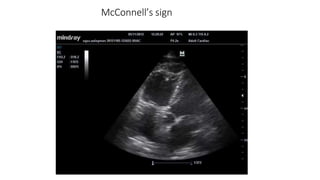
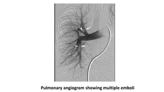
2. Introduction
• Pulmonary thromboembolic disease refers to the condition in which
thrombus or multiple thrombi migrate from the systemic circulation
to the pulmonary vasculature.
• Most of these blood clots arise from the “deep veins” of the lower
and upper extremities (deep venous thrombosis, DVT).
• From the clinical standpoint, DVT and pulmonary embolism can be
considered a continuum of the same disease, and the two terms are
often collectively referred to as venous thromboembolism (VTE).
3. Source of Emboli
• Thrombus originates in lower extrimities in 85 – 90% cases.
• Thrombus often begins at a site where blood flow is turbulent, such
as at a venous bifurcation, or behind a venous valve.
• Most thrombi originate in the deep veins of the calf and propagate
proximally to the popliteal and femoral veins.
• Other sources of emboli are pelvic vein, upper extremity, or may be
associated with thoracic outlet obstruction or effort thrombosis
(Paget von Schroetter syndrome)
4. • Air embolism is usually iatrogenic and typically enters the blood
stream accidentally through a central venous catheter.
• Fat embolism and amniotic fluid embolism are other causes of
pulmonary embolism.
• Pulmonary embolism in sickle cell disease is caused by “clumping” of
abnormal red blood cells in the setting of hypoxia and stress, and can
cause both acute respiratory distress as well as a more progressive
disease with secondary pulmonary hypertension.
5. Predisposing Factors
• Virchow identified three main factors contributing to the formation of
venous thrombosis (Virchow’s triad): venous stasis,
hypercoagulability, and injury to the venous wall (endothelium).
1. Inherited Conditions
• Antithrombin III deficiency
• Factor V Leiden mutation
2. Acquired Risk Factors:
• Surgery , Trauma
• Major medical risk factors include NYHA class III and IV congestive
heart failure, COPD, sepsis and other inflammatory disorders,
advanced age, stroke, critical illness, and prolonged bed rest.
6. • Any prolonged period of immobilization such as paralysis, bed rest,
and prolonged air travel.
• Economy class syndrome
• Pregnancy, OCP
• Obesity BMI > 29kg/m2
• Metabolic syndrome
• Cancer
• Hematological abnormalities
7. Pathophysiology
• Once detached from their point of origin, emboli travel via the
systemic venous system, through the right chambers of the heart, and
eventually reach the pulmonary arterial system.
• The physiologic effects and clinical consequences of pulmonary
thromboembolism vary widely, ranging from asymptomatic disease to
hemodynamic collapse and death. Major factors that determine the
outcome include:
• (1) size and location of emboli;
• (2) coexisting cardiopulmonary diseases;
• (3) secondary humoral mediator release and vascular hypoxic
responses;
• (4) the rate of resolution of emboli.
8. Hemodynamic Consequences
• The normal pulmonary arterial system is a low-pressure system capable of
accommodating substantial increases in blood flow with only modest increases in
pressure.
• With Pulmonary vascular Obstruction less than 20 % there is modest
hemodynamic abnormality.
• When the degree of pulmonary vascular obstruction exceeds 30 to 40 percent,
modest increases in right ventricular pressure occur, but cardiac output is
maintained through an increase in heart rate and myocardial contractility.
• Compensatory mechanisms begin to fail when the degree of pulmonary artery
obstruction exceeds 50 to 60 percent. Cardiac output begins to fall and right atrial
pressure increases dramatically.
• In patients without prior cardiopulmonary disease, the maximal mean pulmonary
artery pressure capable of being generated by the right ventricle appears to be 40
mmHg (pulmonary artery systolic pressure of approximately 70 mmHg).
9. Gas-Exchange Abnormalities
• Hypoxemia is the most common immediate physiologic consequence
of pulmonary embolism.
• Obstruction of the pulmonary vasculature re-directs the blood flow to
other parts of the pulmonary vascular bed. This results in an increase
in intra-pulmonary shunting, ventilation-perfusion (V/Q) inequality,
and decreases in the mixed venous O2 level, thereby magnifying the
effect of the normal venous admixture.
• Further shunting and alveolar dead space increases due to alveolar
hemorrhage, and atelactasis.
• Hypocapnia in PTE is due to hypoxia-induced intrapulmonary reflex
vagal stimulation, with resulting hyperventilation.
• Finally, hypoxemia may lead to an increase in sympathetic tone, which
in turn causes systemic vasoconstriction.
10. • One uncommon consequence of pulmonary embolism is pulmonary
infarction.
• Infarction is uncommon because the pulmonary parenchyma has
three potential sources of oxygen: the pulmonary arteries, bronchial
arteries, and airways.
• Two of these three sources apparently must be compromised before
infarction develops .
11. Clinical features
• The most common presenting symptom of acute embolism is the
sudden onset of dyspnea.
• Other symptoms include pleuritic chest pain, cough, leg swelling or
pain, and hemoptysis.
• The most common physical finding is unexplained tachypnea .
• Less frequent findings include inspiratory crepitations, tachycardia,
and loud S2.
• Fever may develop some hours after the event.
12. Investigations
• Increased Total Leukocyte Count but never more than
20,000cells/cumm
• Hypoxemia with hypocapnia with Normal (A-a)O2 gradient.
• ECG - The electrocardiogram is nonspecific in the diagnosis of
pulmonary embolism, and its major value may be in identifying other
clinical disorders that may be confused with pulmonary embolism.
• Findings in acute PE are generally nonspecific and include T- wave
changes, ST-segment abnormalities, and left- or right-axis deviation.
• The S1Q3T3 pattern, commonly considered to be specific for PE, is
seen in only a minority of patients
13.
14. Chest X ray
• Most patients with pulmonary embolism have abnormal but nonspecific
chest radiographic findings.
• Common radiographic findings include atelectasis, pleural effusion,
pulmonary infiltrates, and mild elevation of a hemidiaphragm.
• Classic findings of pulmonary infarction—such as Hampton’s hump or
decreased vascularity (Westermark’s sign)—are suggestive but infrequent.
• Hampton hump refers to a dome-shaped, pleurally-based opacification in
the lung.
• Pala sign is a prominent pulmonary artery that can be caused either
by pulmonary hypertension that develops or by distension of the vessel by
a large pulmonary embolus.
• Westermark’s sign – peripheral oligemia.
• A normal chest radiograph in a patient with otherwise unexplained acute
dyspnea or hypoxemia is strongly suggestive of embolism.
15.
16. Role of CT Pulmonary Angiogram- MDCT with
contrast
• At the present time, CT is considered confirmatory in excluding
embolism in patients with a low or intermediate likelihood of disease
and confirming embolism in patients with intermediate or high
probability of disease.
• When discordance exists between the clinical assessment and CT
findings, additional studies should be performed.
• It is possible this recommendation will change as studies with 64-
MDCT scanners are published.
17.
18. Ventilation-Perfusion Scanning
• ventilation and perfusion lung scanning can provide valuable
information if used and interpreted appropriately.
• A negative study rules out the diagnosis of pulmonary embolism with
the same degree of certainty as a negative pulmonary angiogram and
with a higher degree of certainty than can be achieved by a negative
CT scan.
• VQ scans are divided into 4 criteria high probability scan,
intermediate probability scan, low probability scan, and normal scan.
• The positive predictive value of a “high probability” scan (one
characterized by multiple, segmental-sized, mismatched defects)
approximates 88 percent.
19. The modified PIOPED criteria for diagnosis of PE determine the probability of
pulmonary emboli following a VQ scan .
20.
21.
22. Echocardiogram
• Presence of unexplained right ventricular volume or pressure
overload should suggest the possibility of embolism and lead to
confirmatory testing.
• Trans Esophageal Echocardiography has better sensitivity and
specificity especially in cases of proximal embolus, reaching up to
90%. Compared to 50% in transthoracic echo.
• McConnell’s sign :
24. • Duplex ultrasonography, which refers to the combination of Doppler
venous flow detection and real-time B-mode imaging is the
investigation of choice for diagnosing Venous Thrombosis of Limb.
• MR Angiography is being evaluated as diagnosing modality in PIOPED
III (Prospective Investigation of Pulmonary Embolism Diagnosis
III )study.
25. Pulmonary Angiogram- Gold Standard
• Pulmonary angiography remains the accepted “gold standard” for PE
diagnosis.
• Only two angiographic findings are diagnostic of acute embolism: the
filling defect and abrupt cutoff of a vessel.
• Catheter is inserted in the right heart and dye is injected into
pulmonary trunk. Filling is observed under fluoroscopy.
• Limitations of Pulmonary Angiogram
• It requires expertise in study performance and interpretation;
• it is invasive.
• High mortality due to procedure itself.
27. Clinical Assessment & Scoring
• Three scoring system has been developed and validated to use.
• These are
• 1 Wells score
• 2 Geneva Score
• 3 Modified Geneva score
30. • If readily available, duplex ultrasonography should be considered
prior to chest imaging.
• Although not confirming the diagnosis of embolism, a positive study
has the same therapeutic implication and avoids the need for contrast
administration and radiation exposure.
• A negative study, however, is incapable of excluding the disease.
31. A ventilation/perfusion (V/Q) scan based
approach
• It is used in settings such as pregnancy, contrast allergy, or renal
insufficiency.
• A negative V/Q scan is capable of excluding the diagnosis regardless
of the clinical assessment.
• A high probability scan is capable of confirming the diagnosis in
patients with a high clinical suspicion.
• All other circumstances require additional testing.
32. Classification of Acute pulmonary Embolism
• Massive PTE
• Sub massive PTE
• Small to moderate PTE
33.
34. Treatment
• I. Heparin
• Unfractionated heparin (UFH) : it is given as initial intravenous bolus
of 80 units of heparin per kilogram followed by a continuous infusion
initiated at 18 units per kilogram per hour.
• The heparin drip is adjusted to maintain aPTT ratio of 2.0 – 3.5
• UFH is given S/C in a dose of 333U stat followed by 250U S/C 12th
hourly
35. • LMWH has replaced UFH in treatment of PTE however UFH should be
used in
• renal insufficiency,
• Extremes of bodyweight,
• hypertensive crisis,
• and circumstances in which a rapid adjustment of anticoagulation is
needed, such as
• women in the late stage of pregnancy who may need Caesarian
sections,
• patients with recent surgery or recent history of bleeding, and
• hemodynamically unstable patients with VTE who may need surgical
procedures such as emergency embolectomy.
36. • II. Novel Agents
• 1.Fondaparinux - a synthetic pentasaccharide, represents the first in a
new class of antithrombotic agents.
• 2. Direct thrombin inhibitors - bivalirudin, lepirudin, argatroban.
37. • III Thrombolytic Therapy
• Thrombolytic agents available are streptokinase, recombinant tissue
plasminogen activator (rt-PA), and urokinase.
• Thrombolysis is indicated in following conditions were it is considered
lifesaving; that is, in patients
• with pulmonary embolism who present with hemodynamic
compromise,
• patients who develop hemodynamic compromise during conventional
therapy with heparin, and
• patients with embolism associated with intracavitary right heart
thrombi
38. Interventional Radiologic Techniques
• Interventional thrombus fragmentation represents a potential
alternative to systemic thrombolysis or surgical embolectomy.
• A wide variety of fragmentation and embolectomy devices designed
to either fragment and/or remove fresh embolic material have been
tested in patients with pulmonary embolism.
• In general, the devices use either pressured saline or a rotating
impeller to fragment central thrombi. The fragments are either
aspirated through a separate port on the catheter or allowed to
migrate distally.
• Most of the devices appear to be effective, safe, and potentially life-
saving in the presence of central, acute clots.
39. Pulmonary Embolectomy
• It is indicated in patients with persistent hypotension, shock, or
cardiac arrest who either failed thrombolysis or have
contraindications to thrombolytics.
• Its use has also been advocated in patients who are at high risk of
paradoxical embolism and who are not candidates for thrombolytics,
although further validation for this indication is needed.
40. Long term anticoagulation therapy
• Warfarin is drug of choice for this purpose.
• It is given in a dose to maintain INR between 2-3.
• It should be given for duration of 6 to 12 months and additional
prophylactic short course should be considered when predisposing
factor is encountered.
• In case of 2nd episode of VTE and inherited disorder lifelong
prophylactic therapy should be considered.
41. Vena Caval Filter
• It is indicated in patients who either have a contraindication to
anticoagulation or develop recurrent VTE while on adequate
anticoagulation.
• IVC filters has proved to have potentially life saving benefits. However,
long-term studies suggest that IVC filters, although capable of
preventing short-term embolic recurrence, they are associated with a
long term increase in the incidence of venous thromboembolism.
• To prevent long term side effects of IVC filters, retrievable filters are
developed.
• Four different retrievable vena caval filters have received approval by
the FDA (Gunther tulip filter, ALN filter, Recovery filter, OptEase filter).