Downloaded 196 times


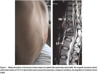


Pott's disease, or tuberculosis of the spine, is an extrapulmonary form of tuberculosis that infects the spine. It was first described in 1779 by Sir Percival Pott. Infection can spread to the spine hematogenously or contiguously from other sites. This leads to destruction of vertebral bodies and discs, causing spinal deformities like kyphosis. Advanced cases can cause paraplegia through cord compression. Diagnosis involves imaging and microbiological tests. Prompt treatment is needed to prevent neurological deficits and deformities.


![ONFH[AVN HIP] -TRIPLE REGIME -A NOVAL SURGICAL CONCEPT .pptx](https://cdn.slidesharecdn.com/ss_thumbnails/onfhavnhip2026koaconcalicutdrgokuldevdrmashraf-260210064517-213ec005-thumbnail.jpg?width=640&height=640&fit=bounds)


![PERI-PROSTHETIC FRACTURE NAIL-PLATE CONSTRUCT [NPC].pptx](https://cdn.slidesharecdn.com/ss_thumbnails/drarunkumardrmohamedashrafperiprostheticfrasturenail-plateconstructnpc-260209164459-7e9d15a1-thumbnail.jpg?width=640&height=640&fit=bounds)


