Bronchiectasis
Prepared by:
RN ARPANA BHUSAL
BNS
1
7/24/2022
Content
• Introduction
• Definition
• Risk factors
• Pathophysiology
2
7/24/2022
Cont…
• Clinical Features
• Diagnostic Evaluation
• Complications
• Management
• Nursing Management
3
7/24/2022
Introduction
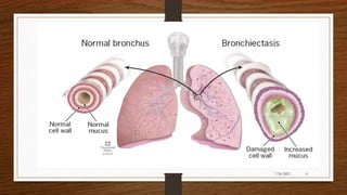
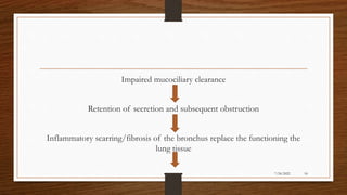
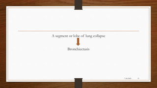
• Bronchiectasis is a chronic lung disease, defined pathologically as
irreversible dilation of the bronchi. The clinical course of the disease is
chronic and progressive and in most cases, causes lung damage over many
years. There is usually an initial event, which causes impairment of
mucociliary clearance of bronchial tree.
4
7/24/2022
Contd…
• The respiratory tract becomes colonized by bacteria that inhibit the cilliary
function ad promote further lung damage. the hall mark of bronchiectasis,
is a chronic cough with mucopurulent or purulent sputum, lasting for
months to years and may progress to chronic respiratory failure.
5
7/24/2022
6
7/24/2022
Definition
• Bronchiectasis is a condition in which the bronchial tubes in the lung
become damaged from inflammation or other causes and the smooth
muscles of the bronchial tubes are destroyed. In addition, elasticity of the
bronchi is often lost.
• Bronchiectasis may be acquired or have a genetic origin. Many clinicians
consider bronchiectasis to be a form of chronic obstructive pulmonary
disease(COPD),it includes chronic bronchitis and emphysema.
7
7/24/2022
Types of bronchiectasis
• Cylindrical bronchiectasis
• Saccular bronchiectasis
• Varicose bronchiectasis
8
7/24/2022
Cylindrical bronchiectasis
• The luminal dilation is uniform and the wall thickening is smooth and
there is failure of normal tapering of bronchi.
9
7/24/2022
Saccular Bronchiectasis
• Most severe form of bronchiectasis. The bronchi are severely dilated and
the bronchi end blindly in a dilated thick walled cyst.
10
7/24/2022
Varicose Bronchiectasis
• The Bronchi resembles like varicose veins and also like serpentine. The
luminal dilation is characterized by alternating areas of luminal dilation
and constriction, creating a beaded appearance, and the wall thickening is
irregular.
11
7/24/2022
12
7/24/2022
Risk factors
• People with cystic fibrosis
• Individuals with alpha-1 anti-proteinase(alpha-1 antitrypsin) deficiency or
an embryological defect termed immotile cilia syndrome
• Children that develop lung infections with lung tissue destruction are risk
for bronchiectasis to develop later in life
• People that abuse drugs and alcohol
13
7/24/2022
Contd…
• People that have recurrent lung infections, aspirate foreign bodies, have
had a history of tuberculosis or inflammatory bowel disease
• Individuals that are exposed to toxic gases or any substances that damage
lung tissue.
14
7/24/2022
Etiology
• Low Body Mass Index
• Toxic fumes, gases, smoke and other harmful substances
• Immunodeficiency
• Connective tissue diseases
• Exposure to chemical irritants
15
7/24/2022
Contd..
• Rheumatoid arthritis
• Childhood infections like pneumonia, tuberculosis, measles, whooping
cough
• Primary cilliary dyskinesia
• Exposure to chemical irritants
16
7/24/2022
Pathophysiology
Due to etiological factors
Infection and inflammation damaging the bronchial wall
Permanent distension and distortion of the bronchial wall
17
7/24/2022
Impaired mucociliary clearance
Retention of secretion and subsequent obstruction
Inflammatory scarring/fibrosis of the bronchus replace the functioning the
lung tissue
18
7/24/2022
A segment or lobe of lung collapse
Bronchiectasis
19
7/24/2022
Clinical manifestation
• Coughing up lots of sputum
• Foul smelling mucus
• Tiredness and poor concentration
• Wheeziness
20
7/24/2022
Cont..
• Chest pain and joint pain
• Recurring chest infections
• Abnormal chest sound
• weight loss
• A constant runny nose
21
7/24/2022
Cont..
• Fatigue
• Clubbing of fingers
• Chronic sinusitis
• Some people became breathless, particularly when exercising or exerting
themselves
22
7/24/2022
Diagnostic evaluation
• History collection
• Physical examination
• Chest CT scan: provides further information on disease location, presence
of mediastinal lesions, and the extent of segmental involvement.
• Chest x-ray: increase in size and loss of definition of bronchovascular
markings, crowding of bronchi, and loss of lung volume. Severe case:
honeycombing appearance
23
7/24/2022
24
7/24/2022
Cont…
• HR CT(High Resolution Computed Tomography) is gold standard for
diagnostic bronchiectasis, it will show either the presence or absence of
bronchial dilation
• Sputum culture: testing of mucous to identify any bacteria present
Pseudomonas
H. influenza
25
7/24/2022
Contd..
• Lung function tests
• Bronchoscopy
• Blood tests
26
7/24/2022
Complications
• Pneumonia
• Lung abscess
• Empyema
• Septicemia
27
7/24/2022
Cont..
• Corpulmonale
• Metastatic cerebral abscesses
• Secondary amyloidosis with nephrotic syndrome
• Recurrent pleurisy
• Purulent pericarditis
28
7/24/2022
Treatment
The goals of treatment are as follows:
1. Eliminate cause
2. Improve tracheobronchial clearance
3. Control infection
4. Reverse airflow obstruction
29
7/24/2022
Cont..
• Chest physiotherapy with percussion, postural drainage, expectorants or
bronchoscopy to remove bronchial secretions.
• Anti microbial therapy as guided by sputum sensitivity tests
• Bronchodilators, sympathomimetic(Beta-Adrenergic Receptors)
• Postural drainage: A technique used to mobilize large amounts of
secretions in people with respiratory conditions.
30
7/24/2022
31
7/24/2022
Cont..
• Mucolytics :helps in clearing mucus from the airways, e.g. bromhexine.
Mucine, erdosteine.
• Inhaled aerosolized aminoglycosides to prevent pseudomonas
colonization.
Antibiotics :
(the choice of antibiotics should be accurate by the results of sputum culture and drug
sensitivity test)
32
7/24/2022
Cont..
Initial treatment: Empiric coverage( amoxcilin, cotrimoxazole, levofloxacin)
is often given initially
• Pseudomonas- quinolone. Aminoglycoside, 3rd generation cephalosporin,
pipracilin.
33
7/24/2022
Cont..
 Supportive treatment:
• Smoking cessation
• Avoidance of second-hand smoke
• Adequate nutritional intake with supplementation, if necessary
• Immunizations for influenza and pneumococcal pneumonia
34
7/24/2022
• Confirmation of immunization for measles, rubella and pertussis
• Oxygen therapy is reserved for patients who are hypoxemic with severe
disease and end-stage complications, such as corpulmonale.
35
7/24/2022
Surgical management
• Segmental resection(segment of a lobe)
• Lobectomy (removal of lung lobe)
• Pneumonectomy (removal of the entire lung)
36
7/24/2022
37
7/24/2022
Nursing management
38
7/24/2022
Nursing Assessment
1. History or presence of supporting factors:
• Smoking
• Living or working in areas with severe air pollution
• History of allergies in family
• History of acid in childhood
39
7/24/2022
Cont..
2. History of the presence of triggering factors such exacerbations:
• Allergen (pollen, dust, skin, pollen or fungal)
• Excessive physical activity
• Air pollution
• Respiratory tract infections and emotional stress
40
7/24/2022
Cont..
3. Physical examination by focusing on the respiratory system include:
• Assess the frequency and respiratory rhythm
• Inspect color of skin and mucous membrane
• Auscultation of breath sounds
• Assess if there is chest pain on breathing
41
7/24/2022
Cont..
• Assess if symmetrical or asymmetrical chest expansion
• Assess cough(whether productive or nonproductive).
• Assess the level of consciousness
42
7/24/2022
Nursing diagnosis
1. Ineffective airway clearance related to the production of mucus and a
decreased ability to cough effectively
2. Impaired gas exchange related to ventilation perfusion inequality.
3. Ineffective breathing pattern related to shortness of breath, mucus,
bronchoconstriction
4. Self care deficit related to fatigue, increased work of breathing,
insufficient ventilation and oxygenation
43
7/24/2022
Cont…
5. Imbalanced nutrition less than body requirements related to nausea,
vomiting, sputum production
44
7/24/2022
Nursing interventions
1. Ineffective airway clearance related to the production of mucus and
a decreased ability to cough effectively
• Asses color, viscosity and mount of sputum
• Assist client in breathing exercises
• Teach how to cough effectively
• Adjust the position of the semi-fowler to the patient
45
7/24/2022
Cont…
• Maintain fluid intake at least 2500ml/day unless otherwise indicated
• Perform chest physiotherapy with postural drainage techniques,
percussion, chest vibrations
• Collaboration of bronchodilators: Nebulizer(via inhalation), collaboration
of mucolytic agent and expectorant
• Collaboration of corticosteroids
46
7/24/2022
Cont..
• Administer antibiotics as prescribed
• Encourage patient to be immunized against influenza and streptococcus
pneumonia
• If indicated, perform postural drainage with percussion and vibration in
the morning and at nights prescribed
47
7/24/2022
Cont..
• Instruct patient to avoid bronchial irritants such as cigarette smoke,
aerosols, extremes of temperature and fumes
• Teach early signs of infection;
Increased sputum production
Change in color of sputum
48
7/24/2022
Cont…
• Increased shortness of breath, tightness in chest or fatigue
• Increased cough
49
7/24/2022
2. Impaired gas exchange related to ventilation perfusion inequality
• Administer bronchodilators as prescribed
Inhalation is the preferred route
Educate regarding types of indoor and outdoor air pollution
• Instruct and encourage patient in diaphragmatic breathing and effective
coughing
50
7/24/2022
Cont…
• Administer oxygen by the prescribed method:
Evaluate effectiveness, observe for signs of hypoxemia
Notify physician if restlessness, anxiety, cyanosis and tachycardia is
present
Initiate pulse oximetry to monitor oxygen saturation
Explain that no smoking is permitted by patient or visitors while oxygen is
in use.
51
7/24/2022
Cont…
3. Ineffective breathing pattern related to shortness of breath, mucus,
bronchoconstriction
• Teach patient diaphragmatic and pursed lip breathing
• Encourage use of an inspiratory muscle trainer if prescribed.
• Encourage alternating activity with rest periods. Allow patient to make
some decisions(bath, saving) about care based on to tolerance level
52
7/24/2022
4. Self care deficit related to fatigue, insufficient ventilation and
oxygenation
• Teach postural drainage if indicated
• Encourage patient to begin to bathe themselves, dress self, walk, drink
fluids. Discuss energy conservation measures
• Teach patient to coordinate diaphragmatic breathing with activity( e.g,
walking, bending
53
7/24/2022
Cont..
• Maintain a supportive firm attitude. Allow patient sufficient time to
accomplish tasks
• Provide positive feedback for efforts and accomplishments
• Families can contribute to train and support client in the client’s ability to
self-care increases
• Place the patient in a comfortable position for feeding which reduces the
risk for aspiration
54
7/24/2022
5. Imbalanced nutrition less than body requirements related to nausea,
vomiting, sputum production
• Monitor input and output every 8 hours, the amount of food consumed and
body weight are weighed each week
• Create a fun atmosphere, an environment free of odor during mealtimes
• Ascertain client’s usual dietary pattern likes/dislikes
55
7/24/2022
Cont…
• Refer patient to a dietician to monitor food plan that will be consumed
• Encourage clients to drink at least 3 liters of fluid per day, if not given an
IV
• Instruct the client to avoid caffeinated beverages, junk foods
• Instruct the client to follow prescribed number of servings of the meals
included in the meal plan
56
7/24/2022
References
• Mandal G.N., textbook of medical surgical nursing published by makalu
publication house, 3rd edition, page no: 92-93
• https://www.google.com/search/Client=firefox-b-d=bronchictasis
@2021/07/12
• Nov2,2019.
Bronchiectasishttps//www.slideshare.net@Gamandeep2021/07/10 at 5pm
57
7/24/2022
58
7/24/2022
59
7/24/2022

Bronchiectasis

  • 1.
  • 2.
    Content • Introduction • Definition •Risk factors • Pathophysiology 2 7/24/2022
  • 3.
    Cont… • Clinical Features •Diagnostic Evaluation • Complications • Management • Nursing Management 3 7/24/2022
  • 4.
    Introduction • Bronchiectasis isa chronic lung disease, defined pathologically as irreversible dilation of the bronchi. The clinical course of the disease is chronic and progressive and in most cases, causes lung damage over many years. There is usually an initial event, which causes impairment of mucociliary clearance of bronchial tree. 4 7/24/2022
  • 5.
    Contd… • The respiratorytract becomes colonized by bacteria that inhibit the cilliary function ad promote further lung damage. the hall mark of bronchiectasis, is a chronic cough with mucopurulent or purulent sputum, lasting for months to years and may progress to chronic respiratory failure. 5 7/24/2022
  • 6.
  • 7.
    Definition • Bronchiectasis isa condition in which the bronchial tubes in the lung become damaged from inflammation or other causes and the smooth muscles of the bronchial tubes are destroyed. In addition, elasticity of the bronchi is often lost. • Bronchiectasis may be acquired or have a genetic origin. Many clinicians consider bronchiectasis to be a form of chronic obstructive pulmonary disease(COPD),it includes chronic bronchitis and emphysema. 7 7/24/2022
  • 8.
    Types of bronchiectasis •Cylindrical bronchiectasis • Saccular bronchiectasis • Varicose bronchiectasis 8 7/24/2022
  • 9.
    Cylindrical bronchiectasis • Theluminal dilation is uniform and the wall thickening is smooth and there is failure of normal tapering of bronchi. 9 7/24/2022
  • 10.
    Saccular Bronchiectasis • Mostsevere form of bronchiectasis. The bronchi are severely dilated and the bronchi end blindly in a dilated thick walled cyst. 10 7/24/2022
  • 11.
    Varicose Bronchiectasis • TheBronchi resembles like varicose veins and also like serpentine. The luminal dilation is characterized by alternating areas of luminal dilation and constriction, creating a beaded appearance, and the wall thickening is irregular. 11 7/24/2022
  • 12.
  • 13.
    Risk factors • Peoplewith cystic fibrosis • Individuals with alpha-1 anti-proteinase(alpha-1 antitrypsin) deficiency or an embryological defect termed immotile cilia syndrome • Children that develop lung infections with lung tissue destruction are risk for bronchiectasis to develop later in life • People that abuse drugs and alcohol 13 7/24/2022
  • 14.
    Contd… • People thathave recurrent lung infections, aspirate foreign bodies, have had a history of tuberculosis or inflammatory bowel disease • Individuals that are exposed to toxic gases or any substances that damage lung tissue. 14 7/24/2022
  • 15.
    Etiology • Low BodyMass Index • Toxic fumes, gases, smoke and other harmful substances • Immunodeficiency • Connective tissue diseases • Exposure to chemical irritants 15 7/24/2022
  • 16.
    Contd.. • Rheumatoid arthritis •Childhood infections like pneumonia, tuberculosis, measles, whooping cough • Primary cilliary dyskinesia • Exposure to chemical irritants 16 7/24/2022
  • 17.
    Pathophysiology Due to etiologicalfactors Infection and inflammation damaging the bronchial wall Permanent distension and distortion of the bronchial wall 17 7/24/2022
  • 18.
    Impaired mucociliary clearance Retentionof secretion and subsequent obstruction Inflammatory scarring/fibrosis of the bronchus replace the functioning the lung tissue 18 7/24/2022
  • 19.
    A segment orlobe of lung collapse Bronchiectasis 19 7/24/2022
  • 20.
    Clinical manifestation • Coughingup lots of sputum • Foul smelling mucus • Tiredness and poor concentration • Wheeziness 20 7/24/2022
  • 21.
    Cont.. • Chest painand joint pain • Recurring chest infections • Abnormal chest sound • weight loss • A constant runny nose 21 7/24/2022
  • 22.
    Cont.. • Fatigue • Clubbingof fingers • Chronic sinusitis • Some people became breathless, particularly when exercising or exerting themselves 22 7/24/2022
  • 23.
    Diagnostic evaluation • Historycollection • Physical examination • Chest CT scan: provides further information on disease location, presence of mediastinal lesions, and the extent of segmental involvement. • Chest x-ray: increase in size and loss of definition of bronchovascular markings, crowding of bronchi, and loss of lung volume. Severe case: honeycombing appearance 23 7/24/2022
  • 24.
  • 25.
    Cont… • HR CT(HighResolution Computed Tomography) is gold standard for diagnostic bronchiectasis, it will show either the presence or absence of bronchial dilation • Sputum culture: testing of mucous to identify any bacteria present Pseudomonas H. influenza 25 7/24/2022
  • 26.
    Contd.. • Lung functiontests • Bronchoscopy • Blood tests 26 7/24/2022
  • 27.
    Complications • Pneumonia • Lungabscess • Empyema • Septicemia 27 7/24/2022
  • 28.
    Cont.. • Corpulmonale • Metastaticcerebral abscesses • Secondary amyloidosis with nephrotic syndrome • Recurrent pleurisy • Purulent pericarditis 28 7/24/2022
  • 29.
    Treatment The goals oftreatment are as follows: 1. Eliminate cause 2. Improve tracheobronchial clearance 3. Control infection 4. Reverse airflow obstruction 29 7/24/2022
  • 30.
    Cont.. • Chest physiotherapywith percussion, postural drainage, expectorants or bronchoscopy to remove bronchial secretions. • Anti microbial therapy as guided by sputum sensitivity tests • Bronchodilators, sympathomimetic(Beta-Adrenergic Receptors) • Postural drainage: A technique used to mobilize large amounts of secretions in people with respiratory conditions. 30 7/24/2022
  • 31.
  • 32.
    Cont.. • Mucolytics :helpsin clearing mucus from the airways, e.g. bromhexine. Mucine, erdosteine. • Inhaled aerosolized aminoglycosides to prevent pseudomonas colonization. Antibiotics : (the choice of antibiotics should be accurate by the results of sputum culture and drug sensitivity test) 32 7/24/2022
  • 33.
    Cont.. Initial treatment: Empiriccoverage( amoxcilin, cotrimoxazole, levofloxacin) is often given initially • Pseudomonas- quinolone. Aminoglycoside, 3rd generation cephalosporin, pipracilin. 33 7/24/2022
  • 34.
    Cont..  Supportive treatment: •Smoking cessation • Avoidance of second-hand smoke • Adequate nutritional intake with supplementation, if necessary • Immunizations for influenza and pneumococcal pneumonia 34 7/24/2022
  • 35.
    • Confirmation ofimmunization for measles, rubella and pertussis • Oxygen therapy is reserved for patients who are hypoxemic with severe disease and end-stage complications, such as corpulmonale. 35 7/24/2022
  • 36.
    Surgical management • Segmentalresection(segment of a lobe) • Lobectomy (removal of lung lobe) • Pneumonectomy (removal of the entire lung) 36 7/24/2022
  • 37.
  • 38.
  • 39.
    Nursing Assessment 1. Historyor presence of supporting factors: • Smoking • Living or working in areas with severe air pollution • History of allergies in family • History of acid in childhood 39 7/24/2022
  • 40.
    Cont.. 2. History ofthe presence of triggering factors such exacerbations: • Allergen (pollen, dust, skin, pollen or fungal) • Excessive physical activity • Air pollution • Respiratory tract infections and emotional stress 40 7/24/2022
  • 41.
    Cont.. 3. Physical examinationby focusing on the respiratory system include: • Assess the frequency and respiratory rhythm • Inspect color of skin and mucous membrane • Auscultation of breath sounds • Assess if there is chest pain on breathing 41 7/24/2022
  • 42.
    Cont.. • Assess ifsymmetrical or asymmetrical chest expansion • Assess cough(whether productive or nonproductive). • Assess the level of consciousness 42 7/24/2022
  • 43.
    Nursing diagnosis 1. Ineffectiveairway clearance related to the production of mucus and a decreased ability to cough effectively 2. Impaired gas exchange related to ventilation perfusion inequality. 3. Ineffective breathing pattern related to shortness of breath, mucus, bronchoconstriction 4. Self care deficit related to fatigue, increased work of breathing, insufficient ventilation and oxygenation 43 7/24/2022
  • 44.
    Cont… 5. Imbalanced nutritionless than body requirements related to nausea, vomiting, sputum production 44 7/24/2022
  • 45.
    Nursing interventions 1. Ineffectiveairway clearance related to the production of mucus and a decreased ability to cough effectively • Asses color, viscosity and mount of sputum • Assist client in breathing exercises • Teach how to cough effectively • Adjust the position of the semi-fowler to the patient 45 7/24/2022
  • 46.
    Cont… • Maintain fluidintake at least 2500ml/day unless otherwise indicated • Perform chest physiotherapy with postural drainage techniques, percussion, chest vibrations • Collaboration of bronchodilators: Nebulizer(via inhalation), collaboration of mucolytic agent and expectorant • Collaboration of corticosteroids 46 7/24/2022
  • 47.
    Cont.. • Administer antibioticsas prescribed • Encourage patient to be immunized against influenza and streptococcus pneumonia • If indicated, perform postural drainage with percussion and vibration in the morning and at nights prescribed 47 7/24/2022
  • 48.
    Cont.. • Instruct patientto avoid bronchial irritants such as cigarette smoke, aerosols, extremes of temperature and fumes • Teach early signs of infection; Increased sputum production Change in color of sputum 48 7/24/2022
  • 49.
    Cont… • Increased shortnessof breath, tightness in chest or fatigue • Increased cough 49 7/24/2022
  • 50.
    2. Impaired gasexchange related to ventilation perfusion inequality • Administer bronchodilators as prescribed Inhalation is the preferred route Educate regarding types of indoor and outdoor air pollution • Instruct and encourage patient in diaphragmatic breathing and effective coughing 50 7/24/2022
  • 51.
    Cont… • Administer oxygenby the prescribed method: Evaluate effectiveness, observe for signs of hypoxemia Notify physician if restlessness, anxiety, cyanosis and tachycardia is present Initiate pulse oximetry to monitor oxygen saturation Explain that no smoking is permitted by patient or visitors while oxygen is in use. 51 7/24/2022
  • 52.
    Cont… 3. Ineffective breathingpattern related to shortness of breath, mucus, bronchoconstriction • Teach patient diaphragmatic and pursed lip breathing • Encourage use of an inspiratory muscle trainer if prescribed. • Encourage alternating activity with rest periods. Allow patient to make some decisions(bath, saving) about care based on to tolerance level 52 7/24/2022
  • 53.
    4. Self caredeficit related to fatigue, insufficient ventilation and oxygenation • Teach postural drainage if indicated • Encourage patient to begin to bathe themselves, dress self, walk, drink fluids. Discuss energy conservation measures • Teach patient to coordinate diaphragmatic breathing with activity( e.g, walking, bending 53 7/24/2022
  • 54.
    Cont.. • Maintain asupportive firm attitude. Allow patient sufficient time to accomplish tasks • Provide positive feedback for efforts and accomplishments • Families can contribute to train and support client in the client’s ability to self-care increases • Place the patient in a comfortable position for feeding which reduces the risk for aspiration 54 7/24/2022
  • 55.
    5. Imbalanced nutritionless than body requirements related to nausea, vomiting, sputum production • Monitor input and output every 8 hours, the amount of food consumed and body weight are weighed each week • Create a fun atmosphere, an environment free of odor during mealtimes • Ascertain client’s usual dietary pattern likes/dislikes 55 7/24/2022
  • 56.
    Cont… • Refer patientto a dietician to monitor food plan that will be consumed • Encourage clients to drink at least 3 liters of fluid per day, if not given an IV • Instruct the client to avoid caffeinated beverages, junk foods • Instruct the client to follow prescribed number of servings of the meals included in the meal plan 56 7/24/2022
  • 57.
    References • Mandal G.N.,textbook of medical surgical nursing published by makalu publication house, 3rd edition, page no: 92-93 • https://www.google.com/search/Client=firefox-b-d=bronchictasis @2021/07/12 • Nov2,2019. Bronchiectasishttps//www.slideshare.net@Gamandeep2021/07/10 at 5pm 57 7/24/2022
  • 58.
  • 59.