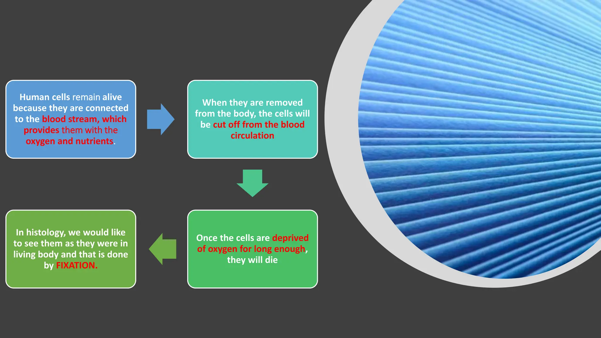
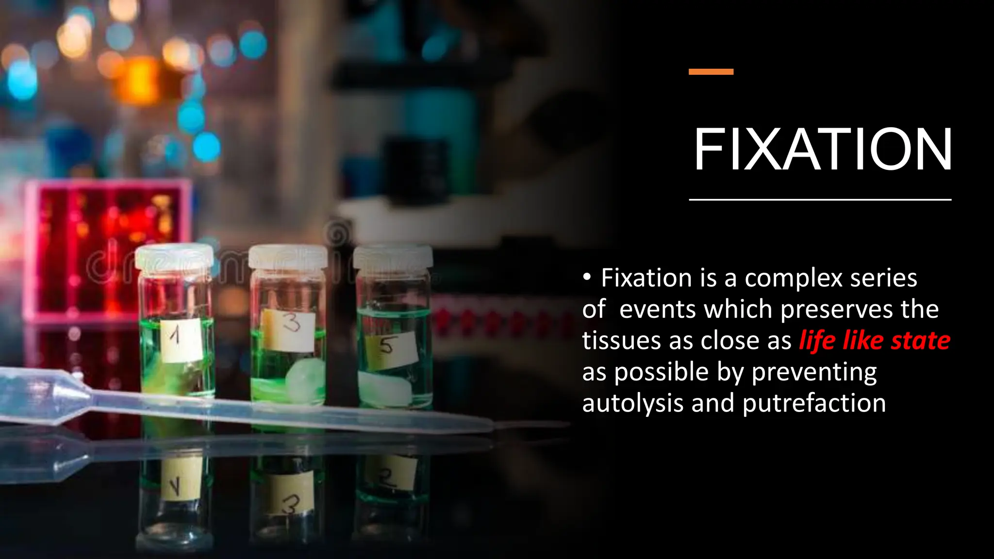
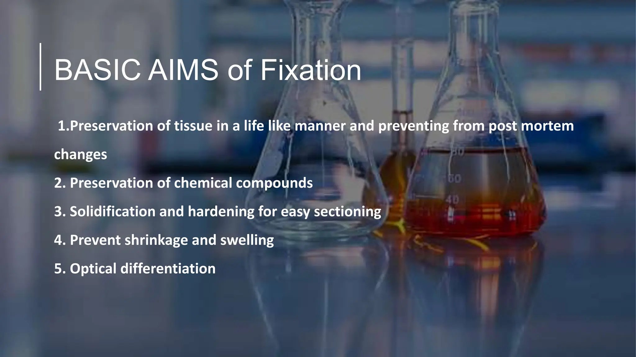
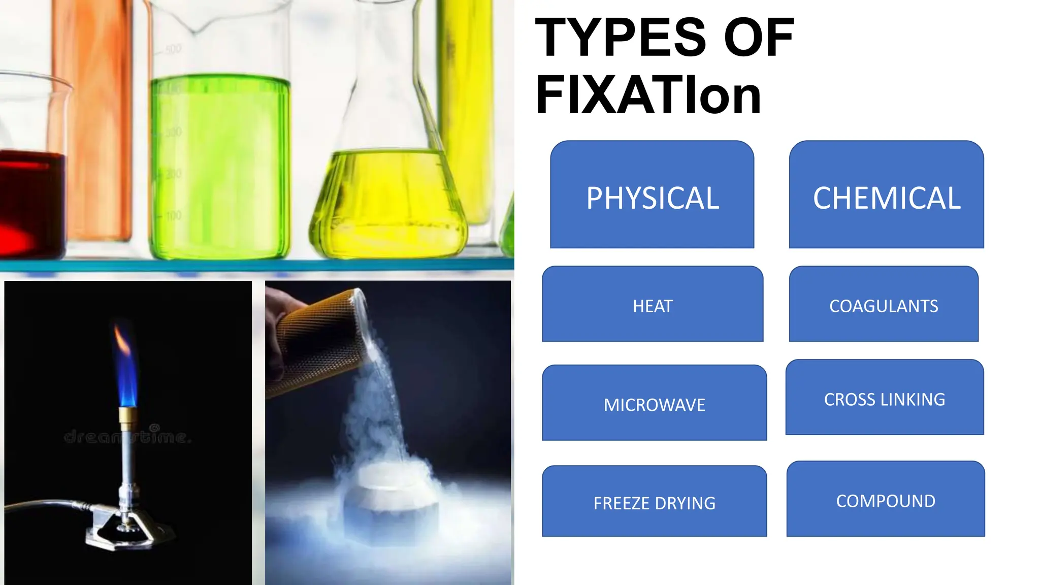
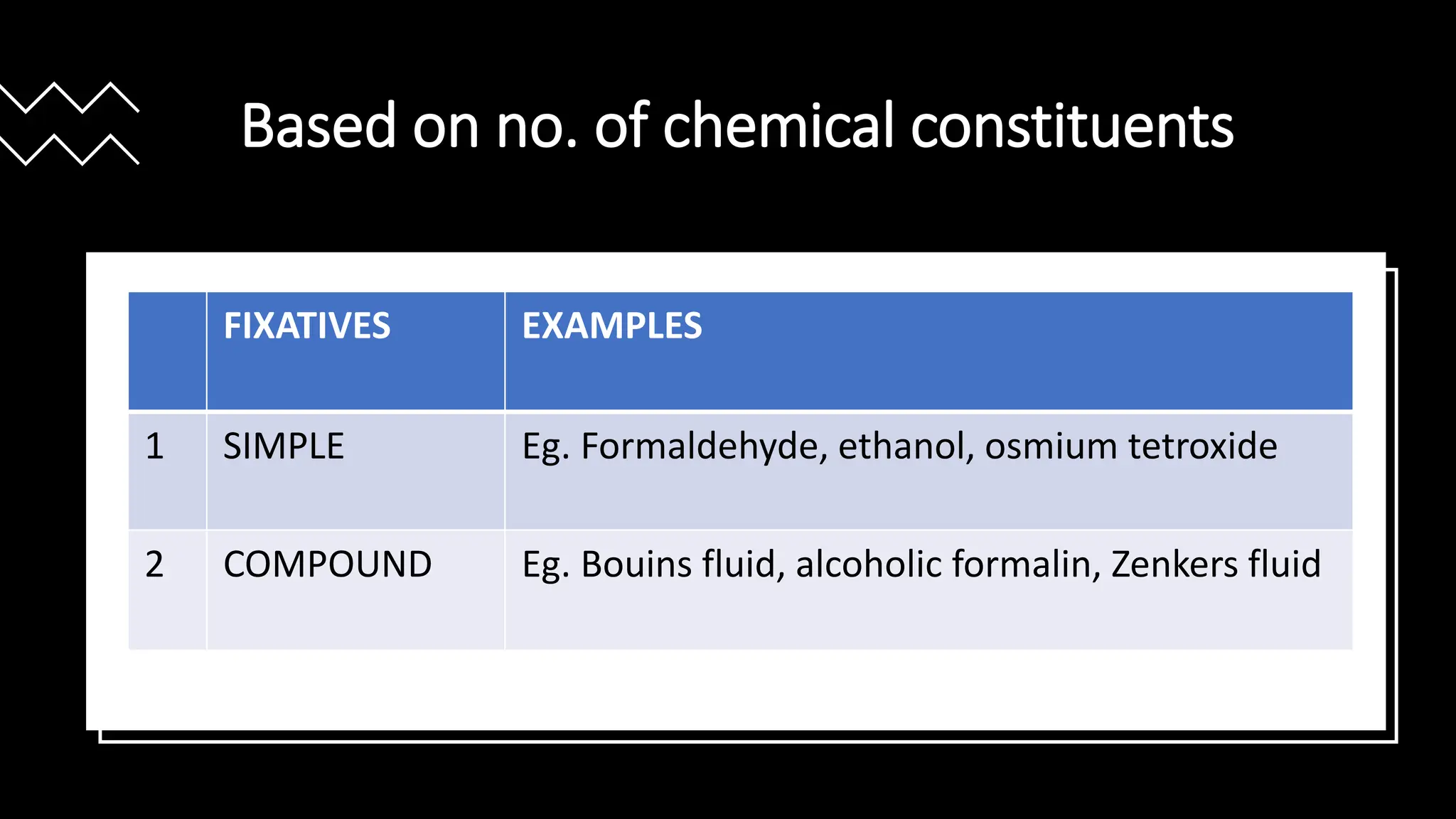
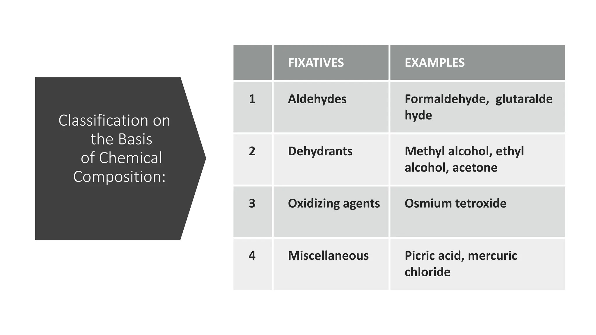
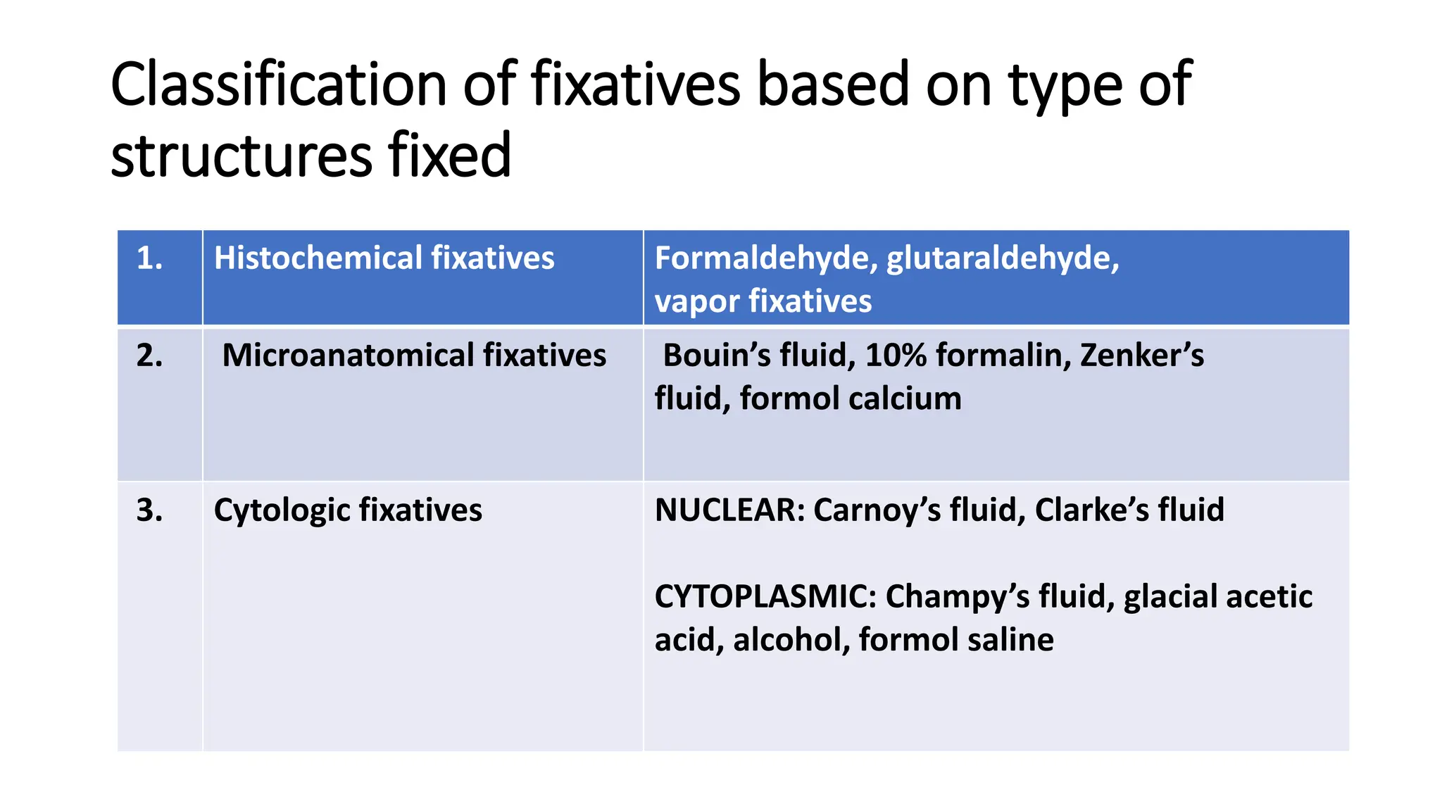
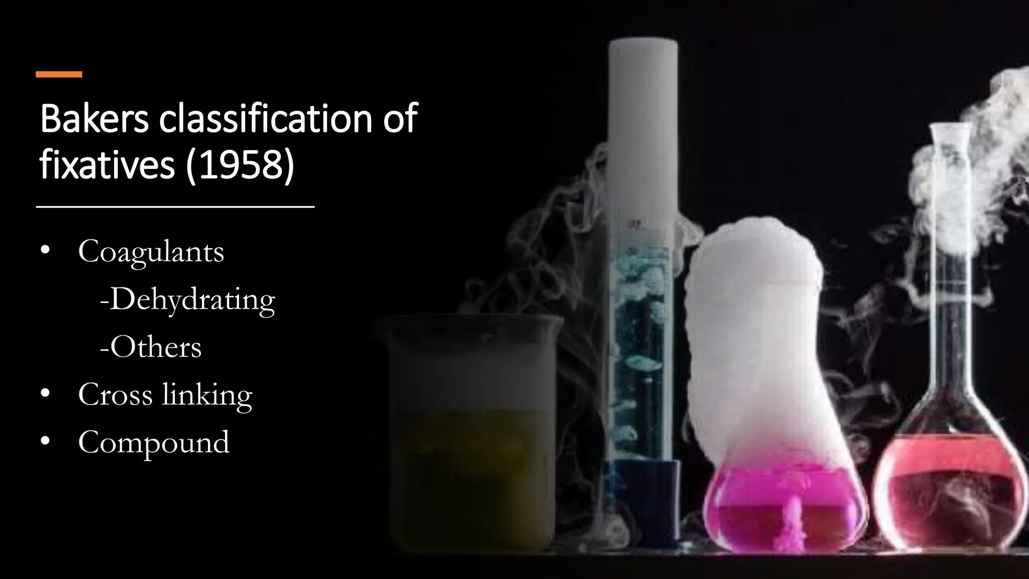
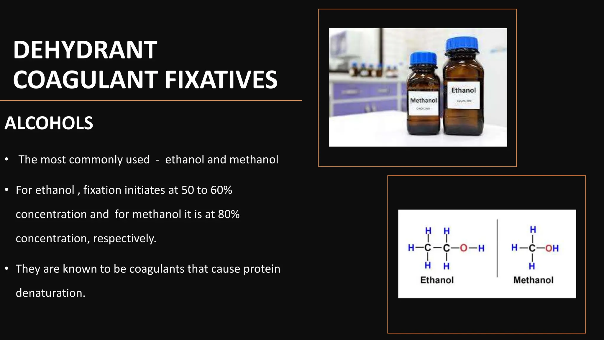
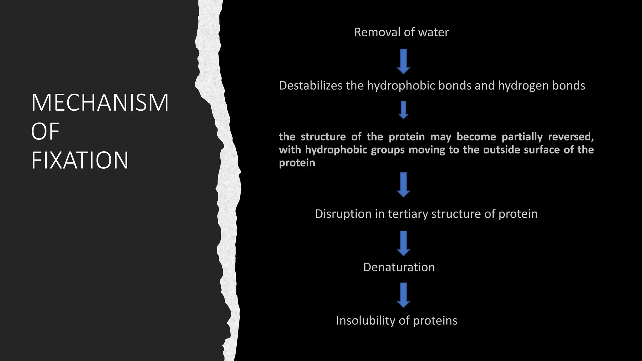
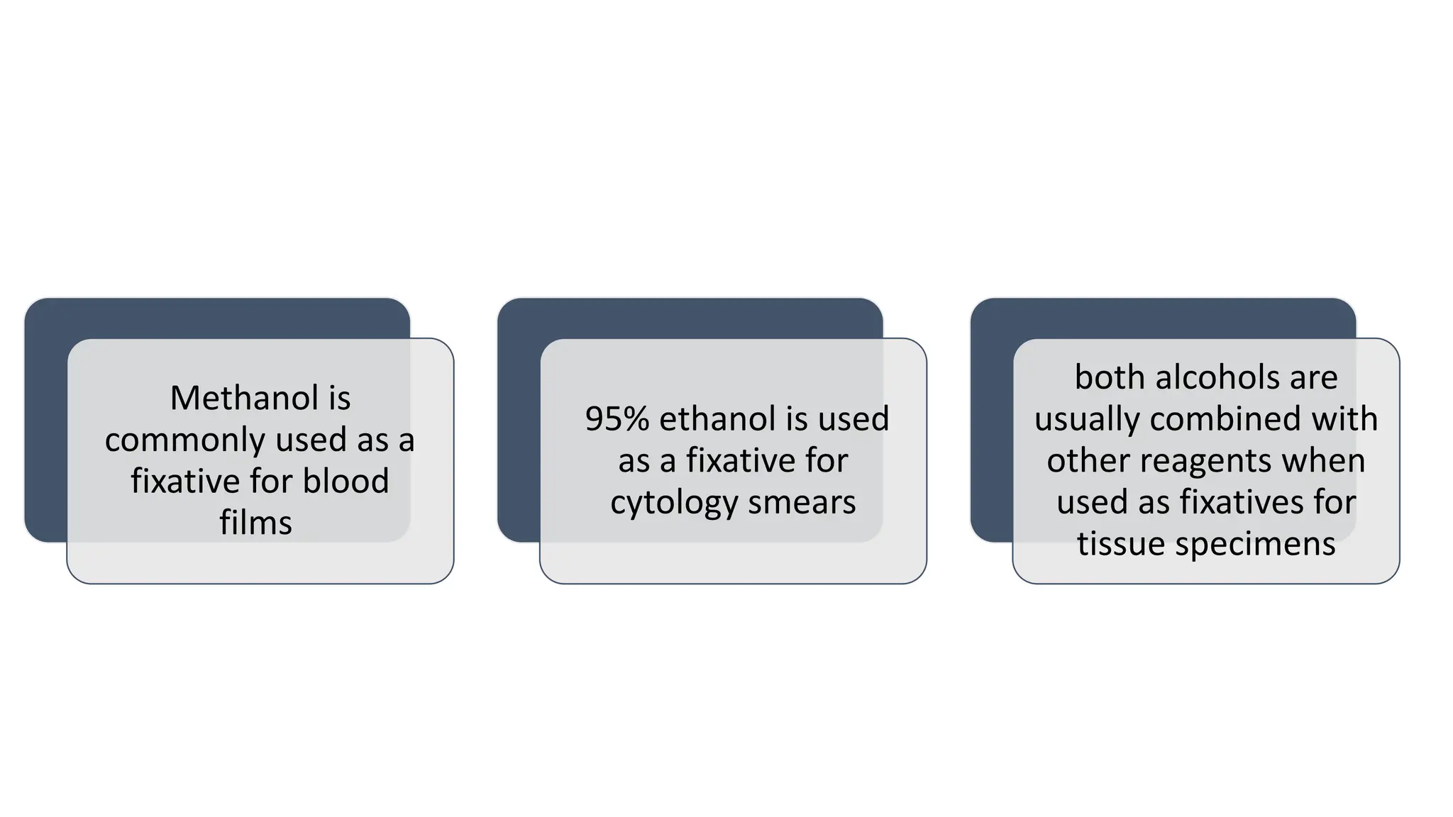
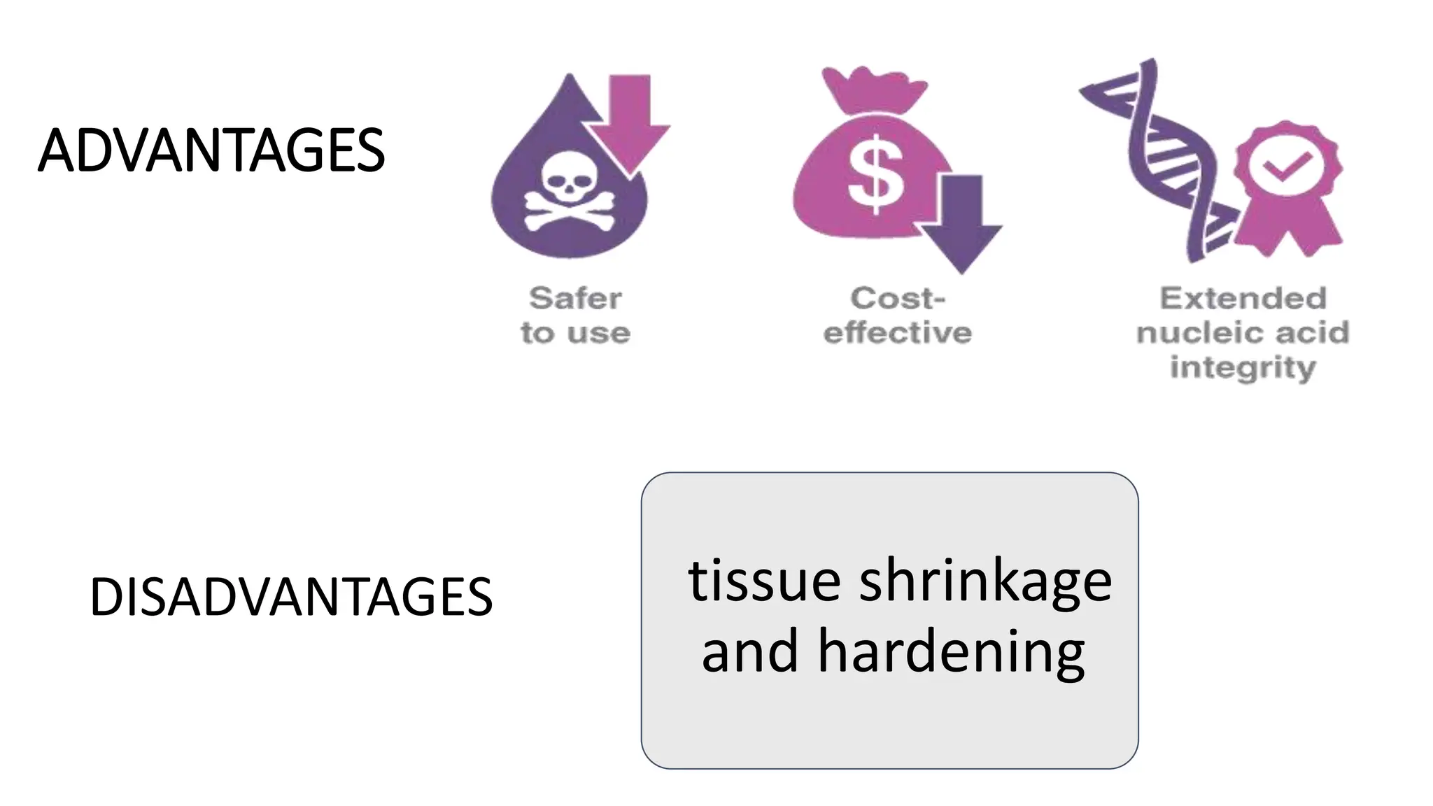
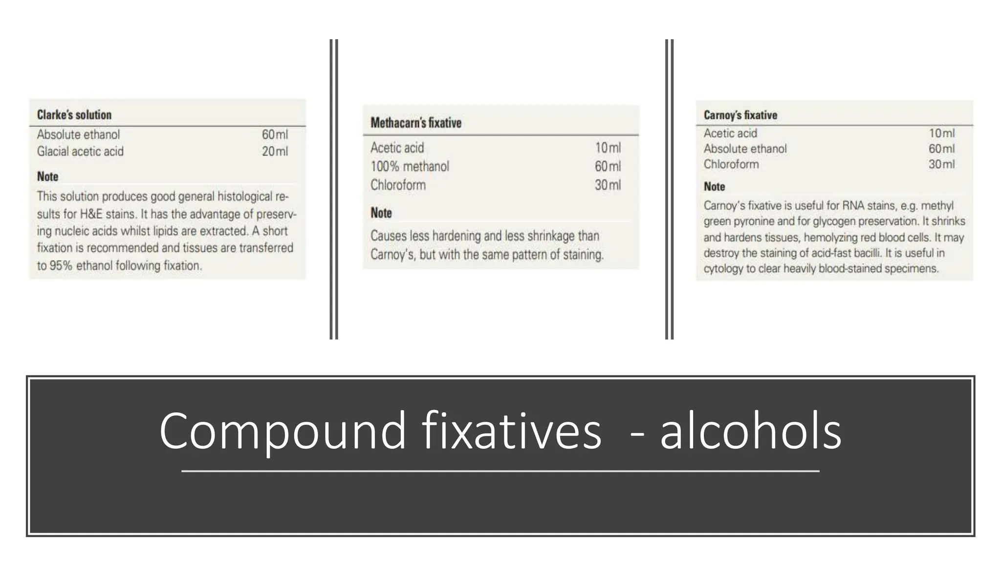
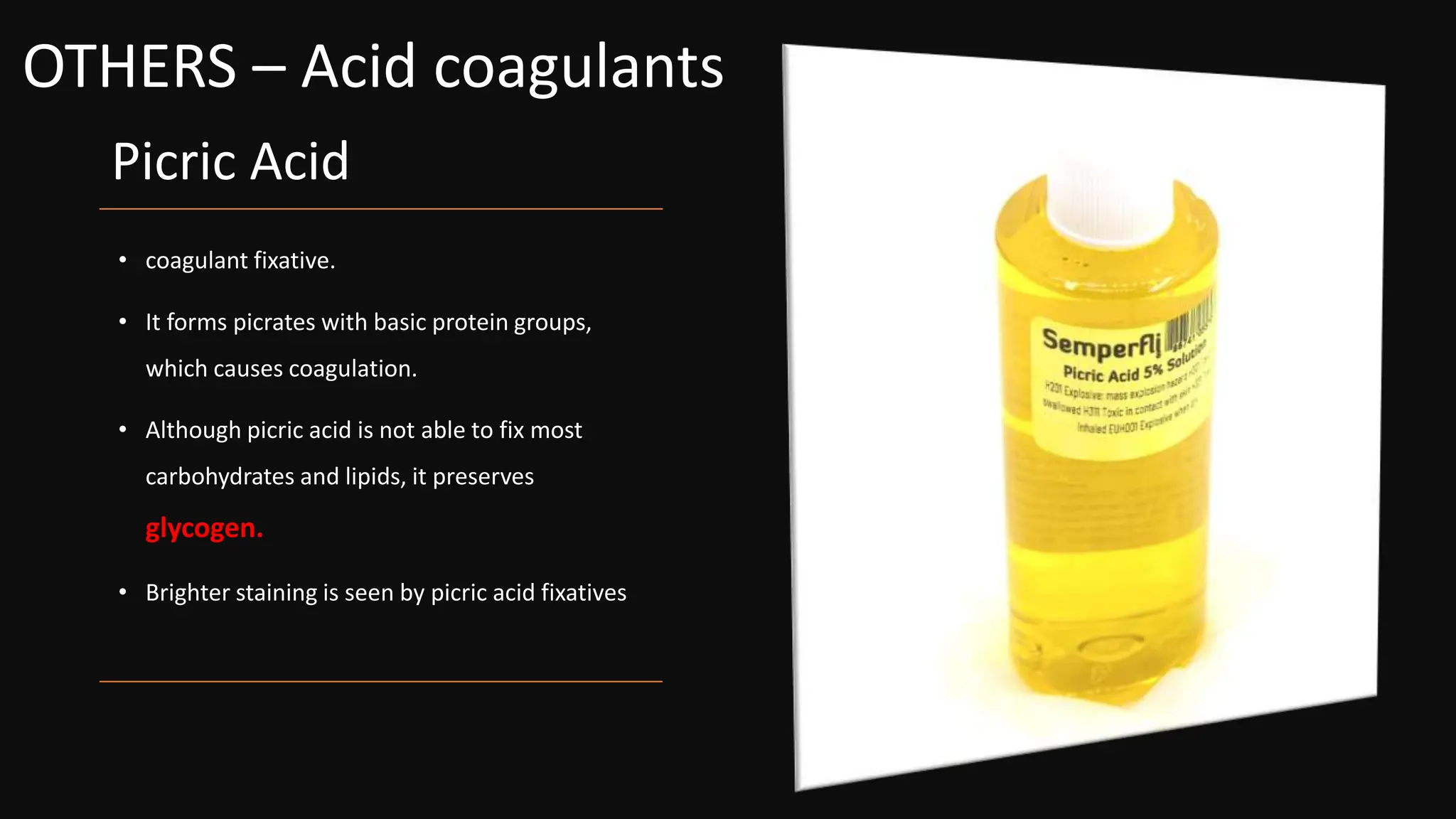
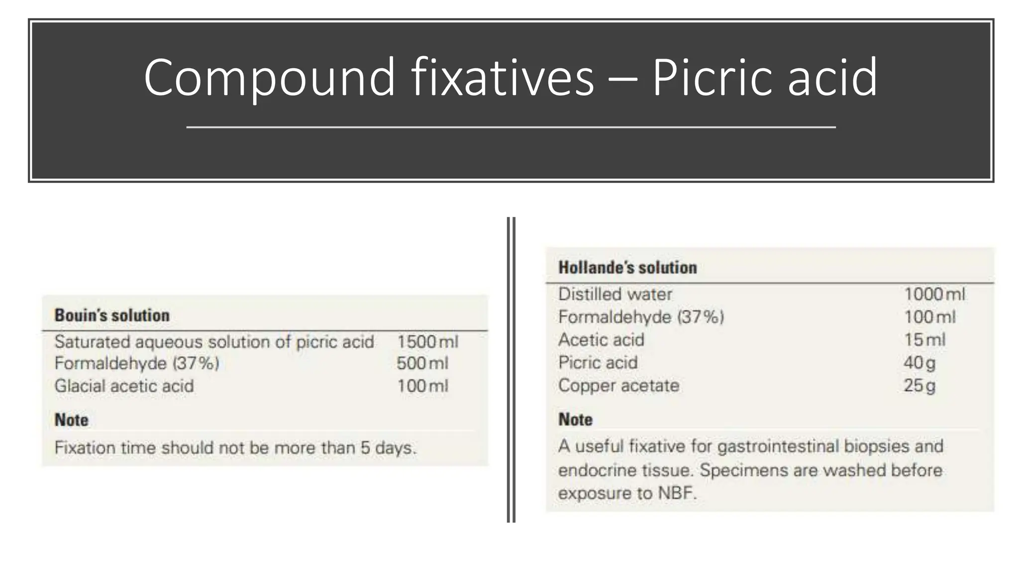
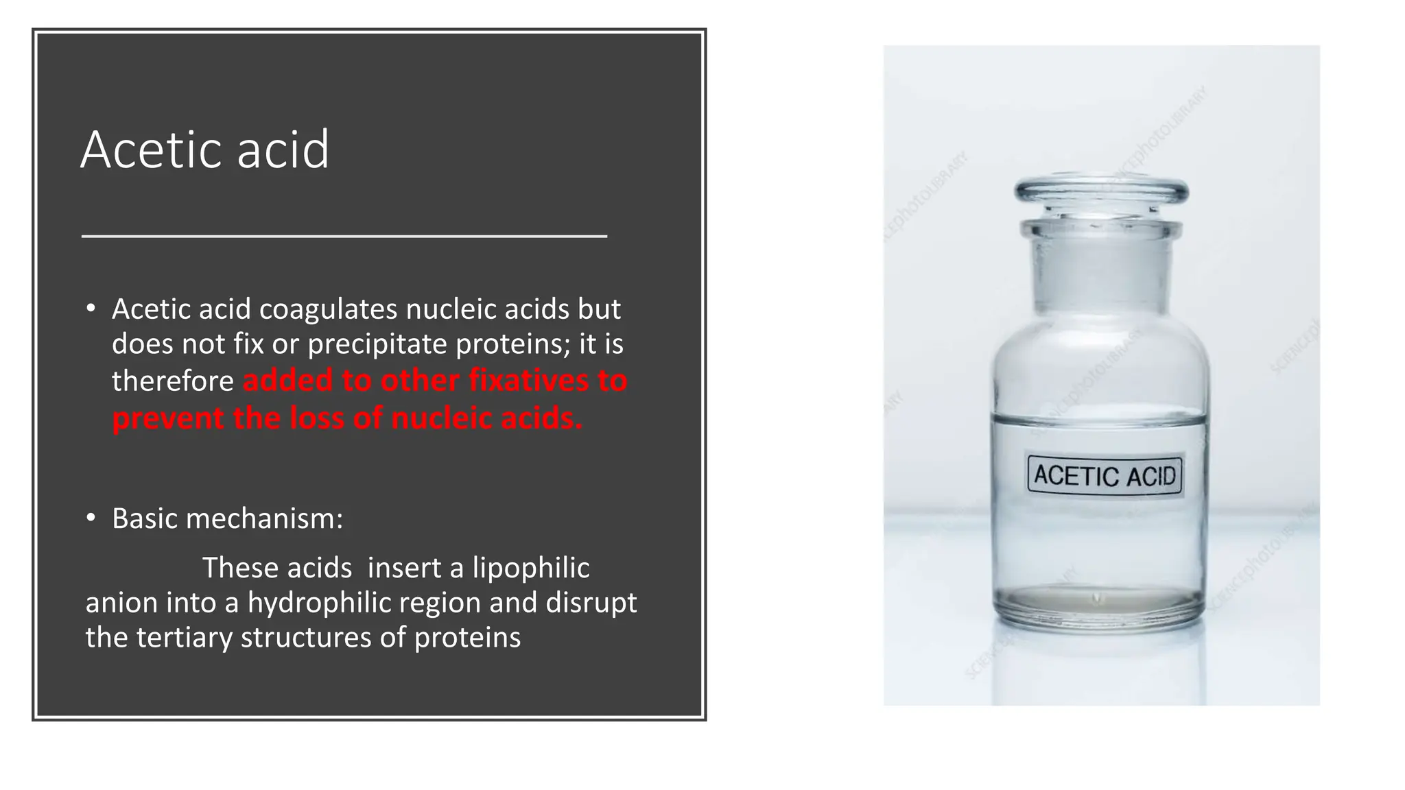
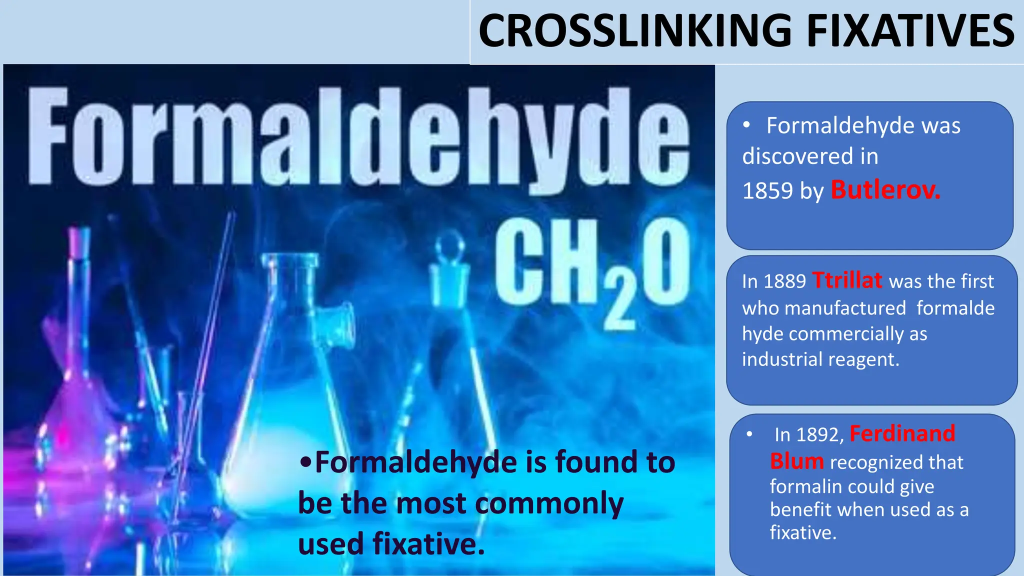
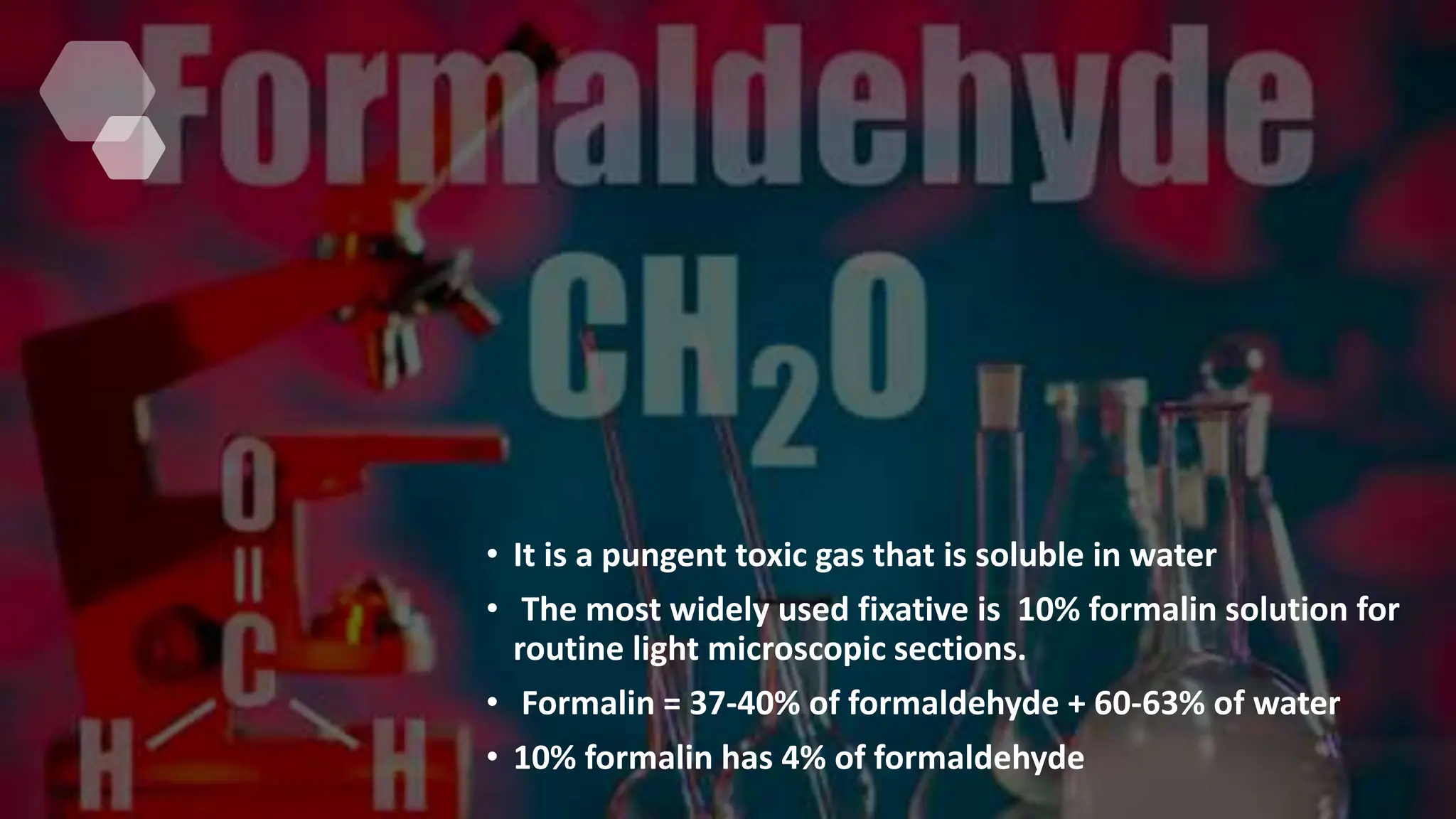
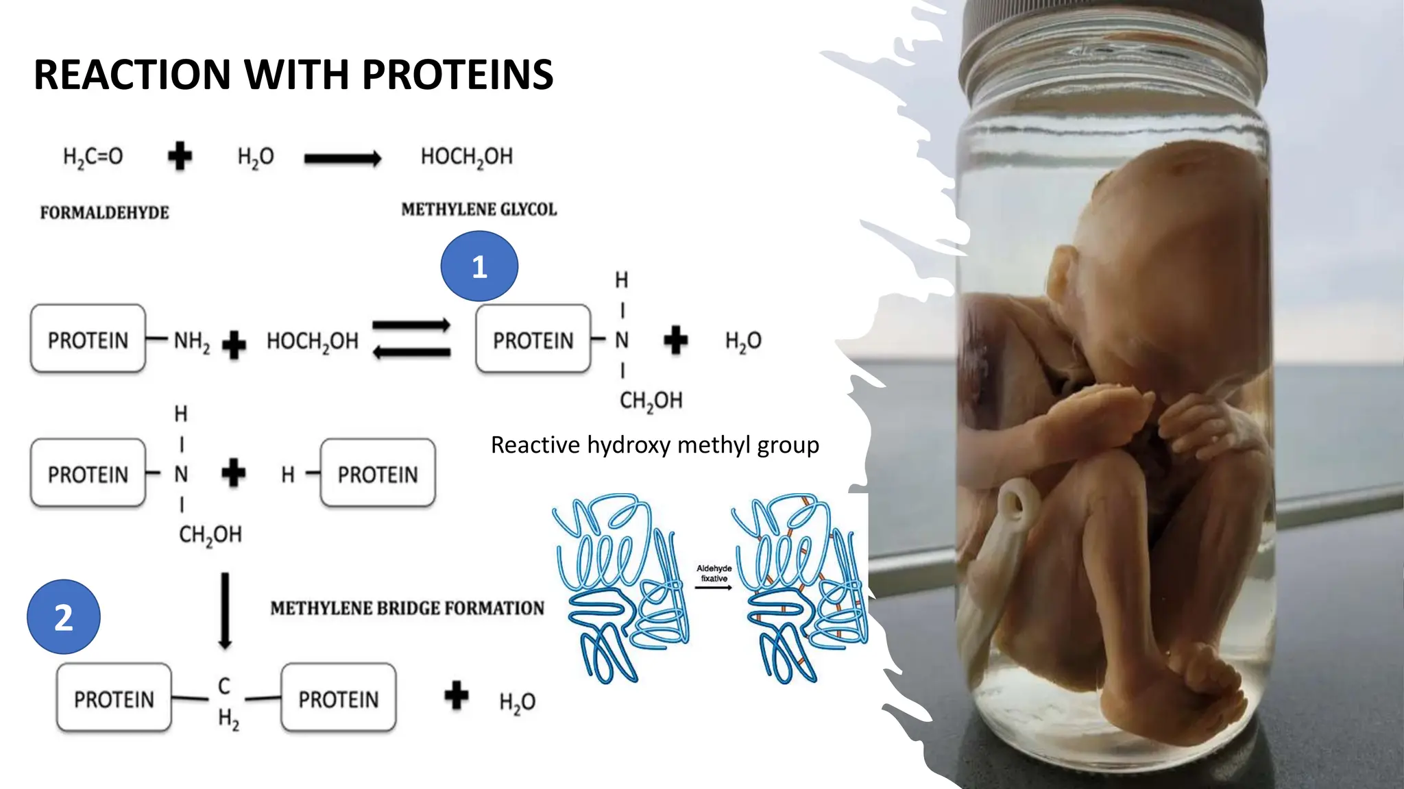
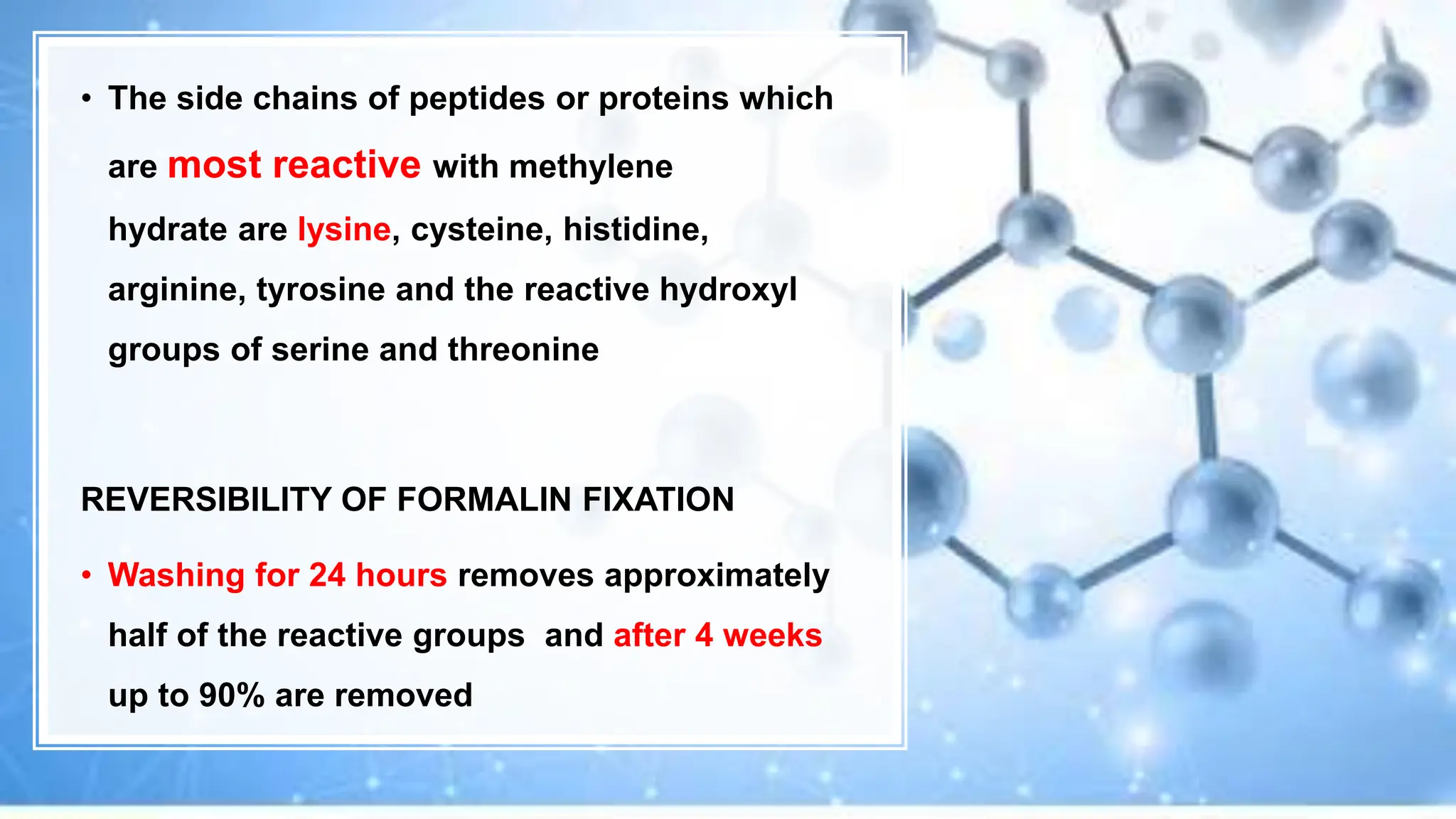
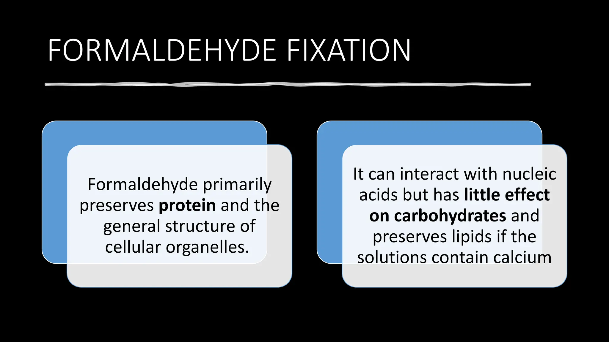
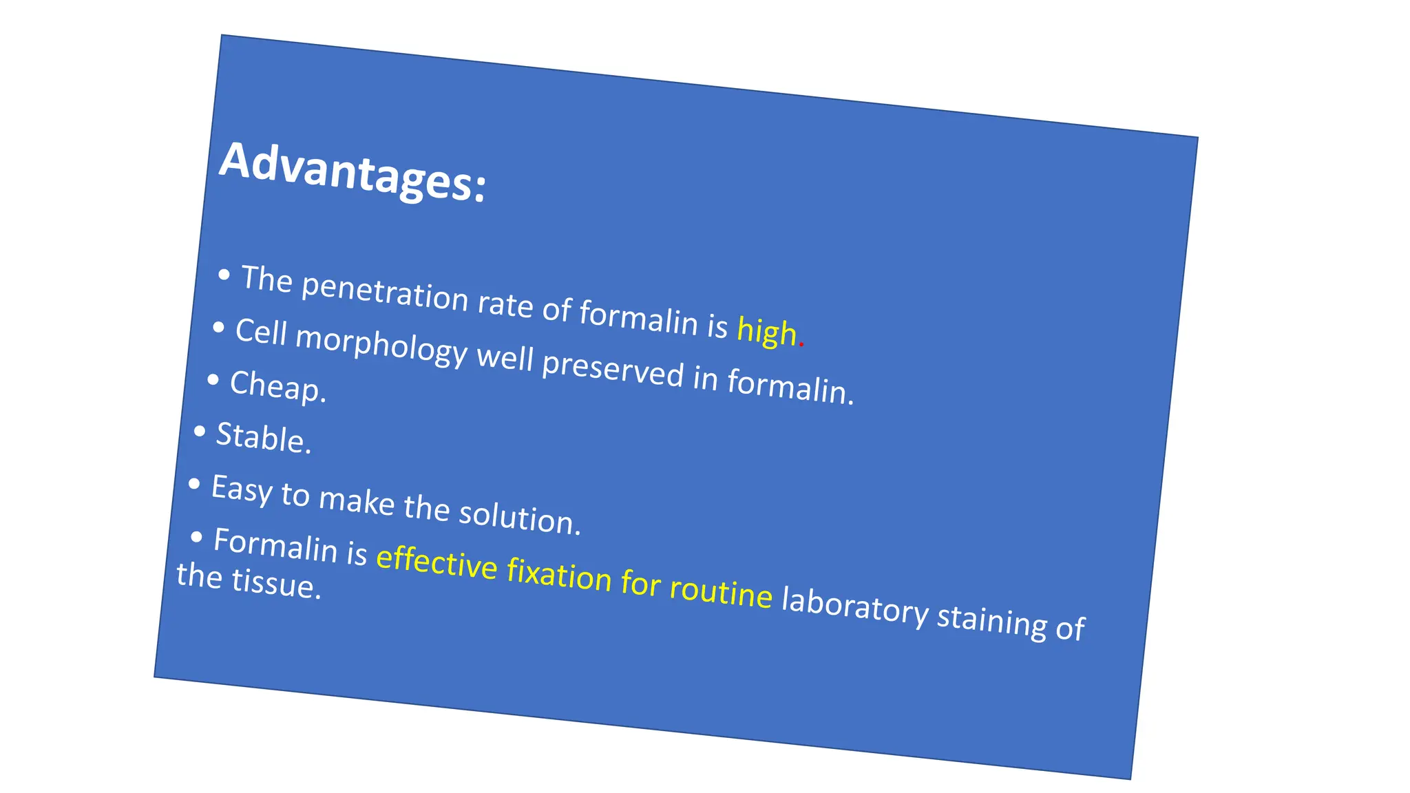
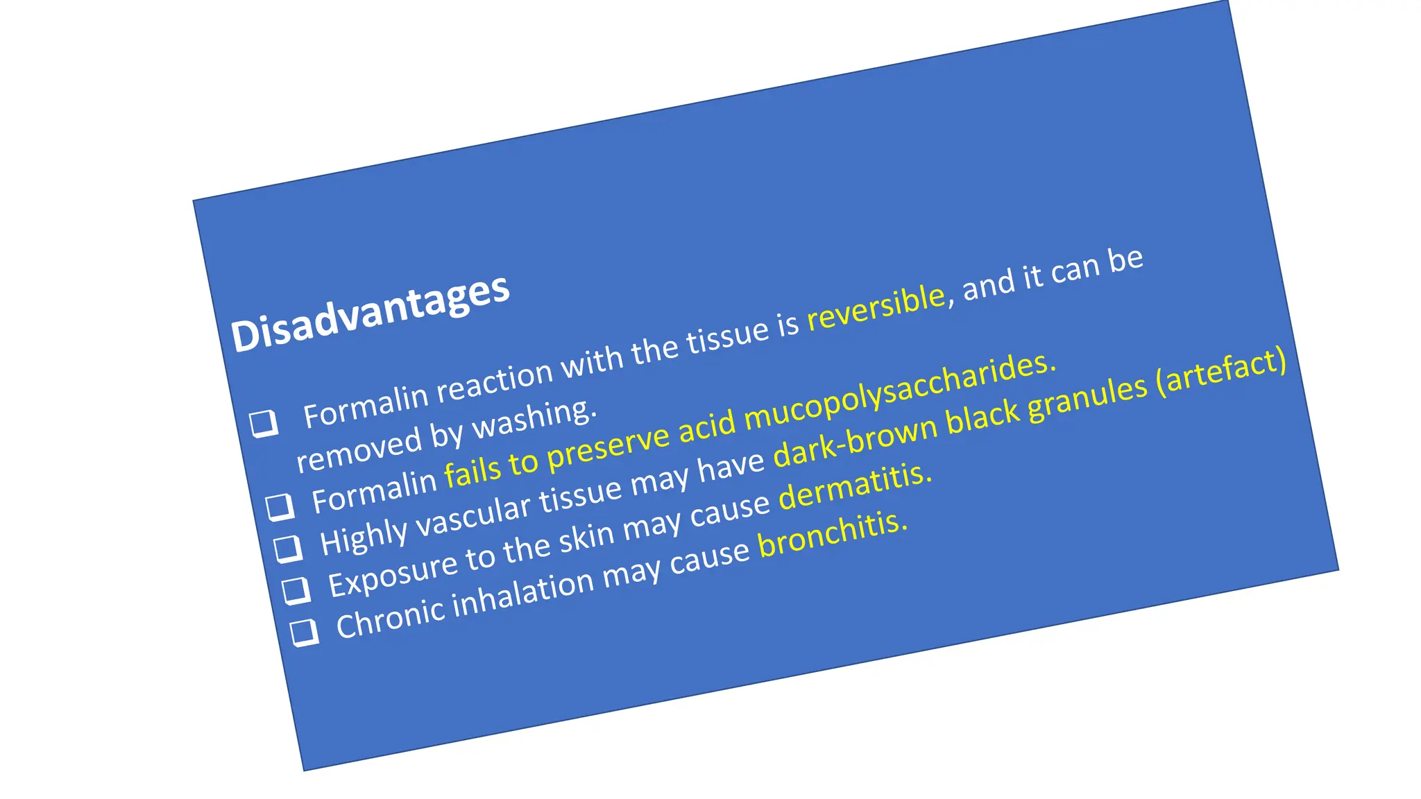
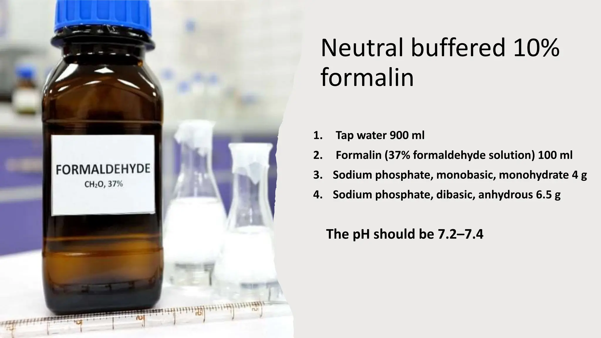
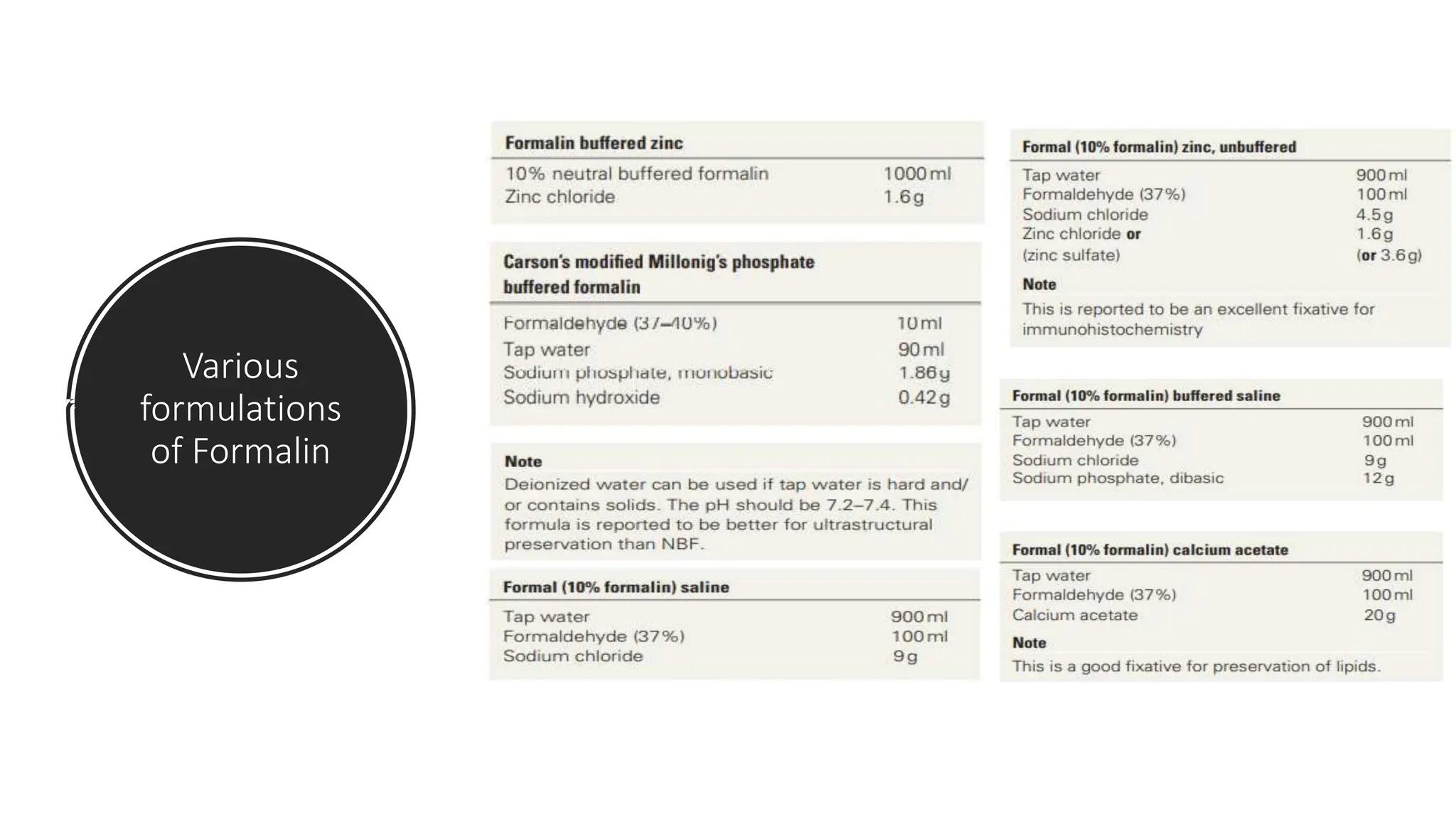
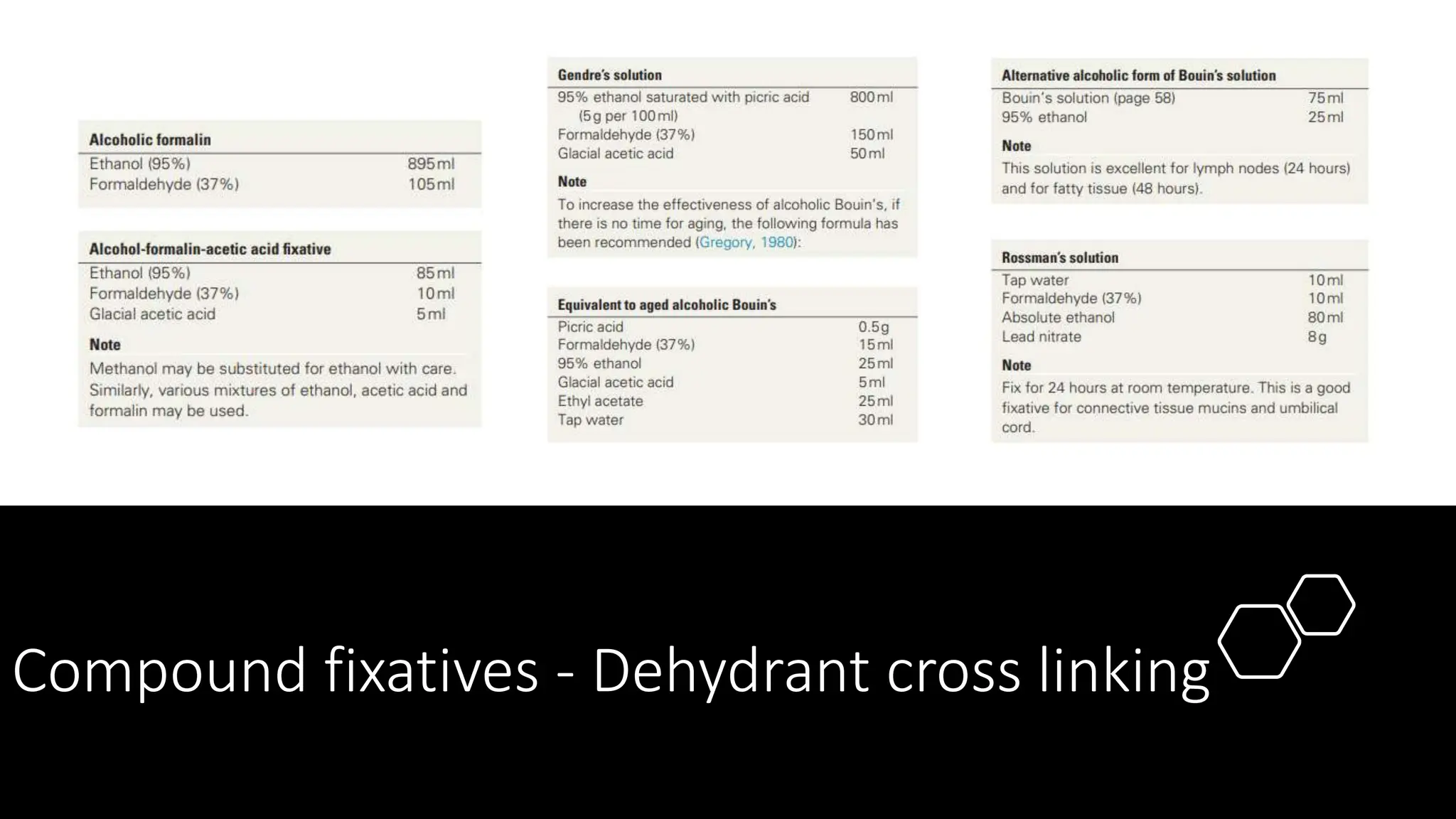
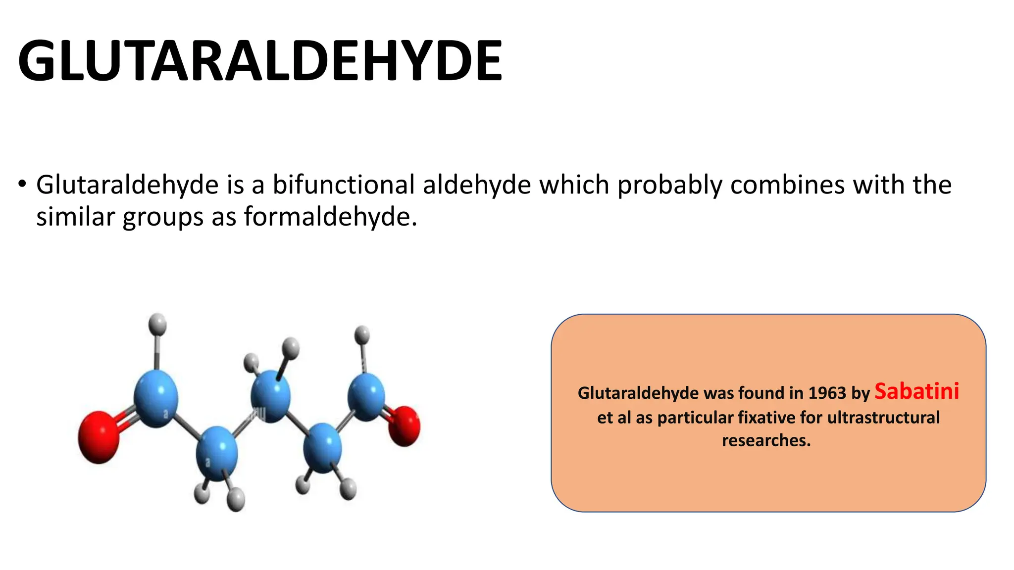
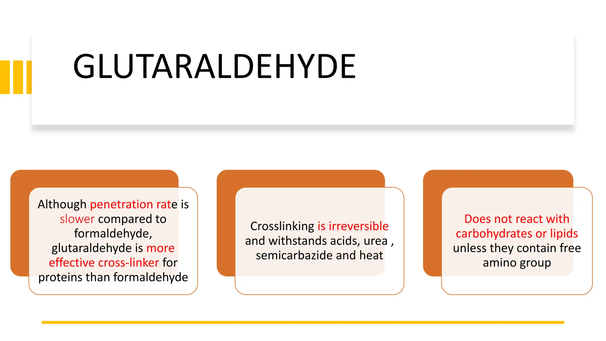
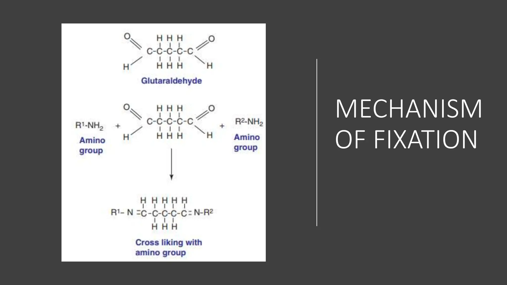
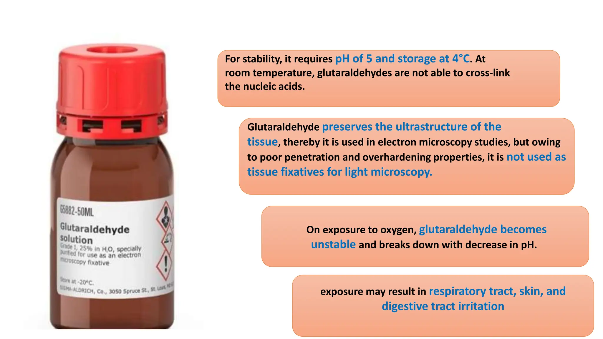
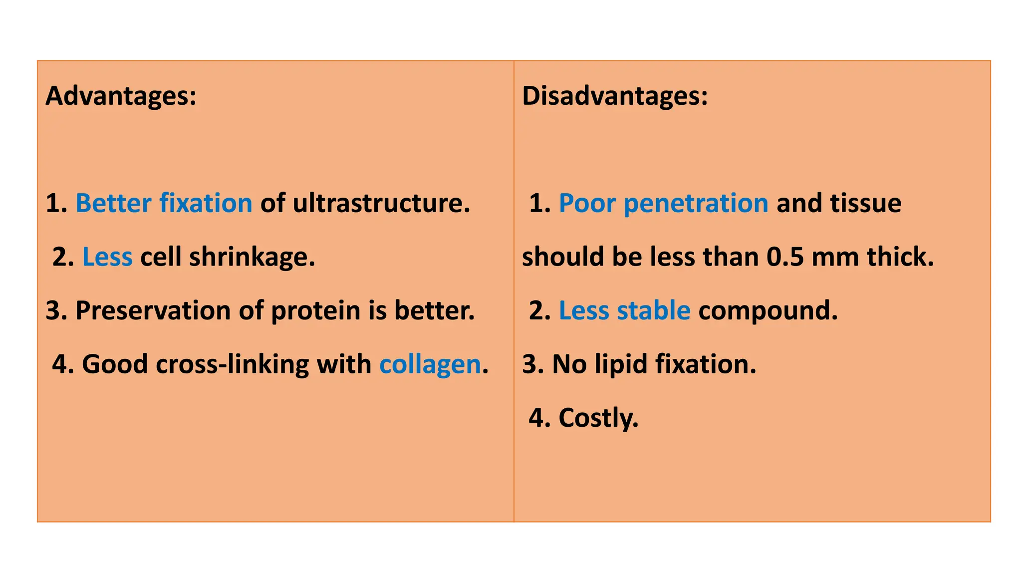
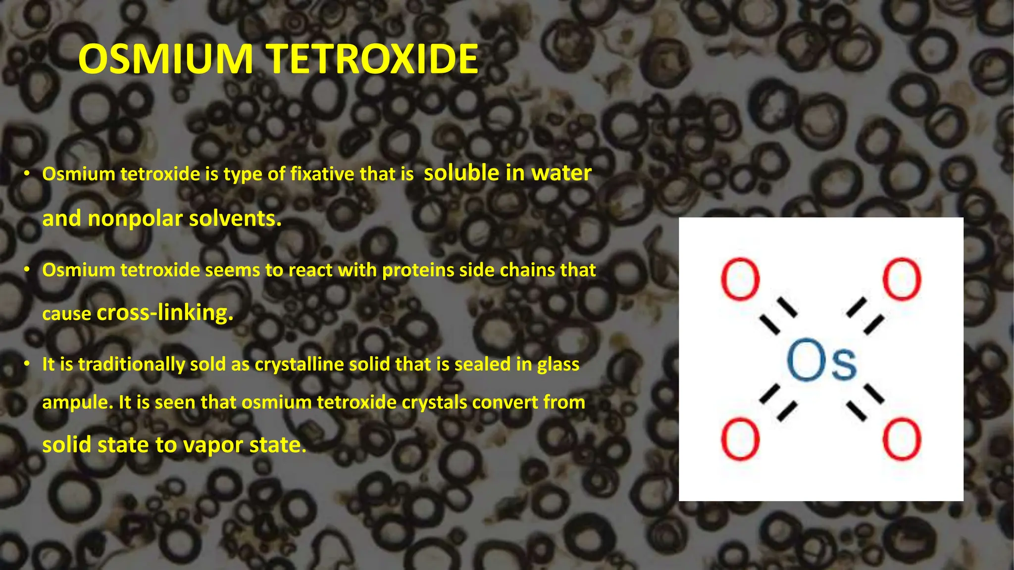
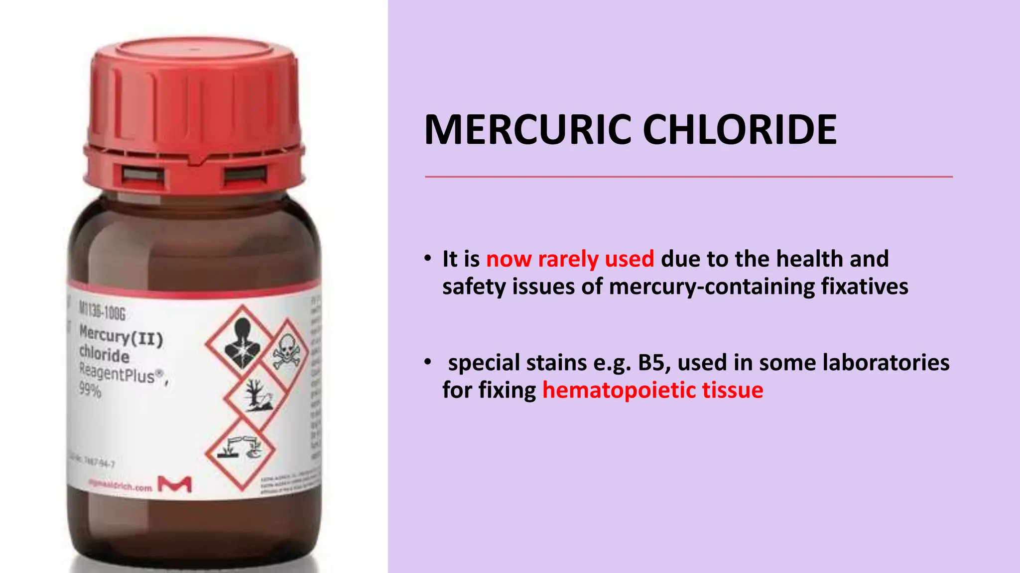
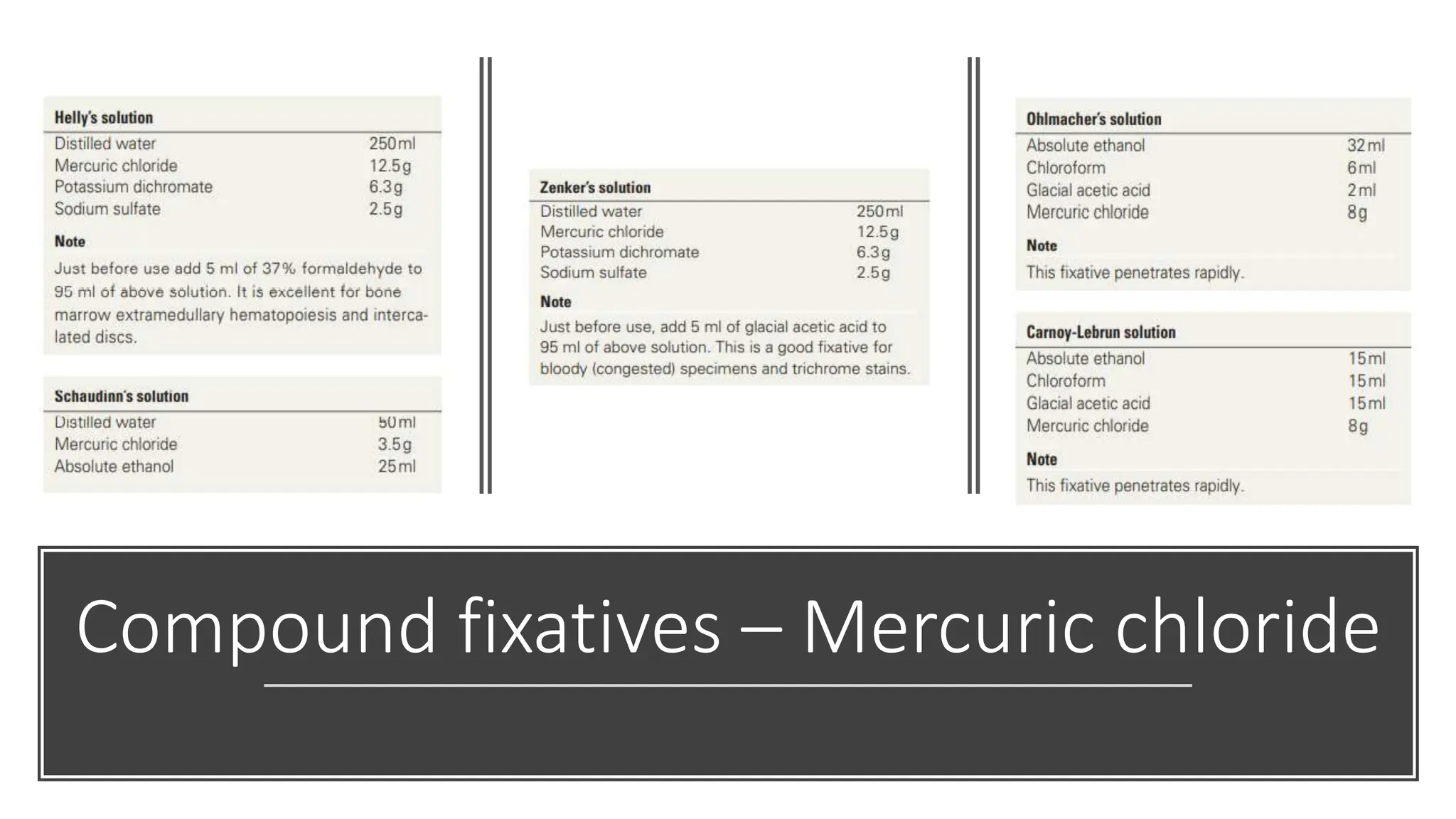
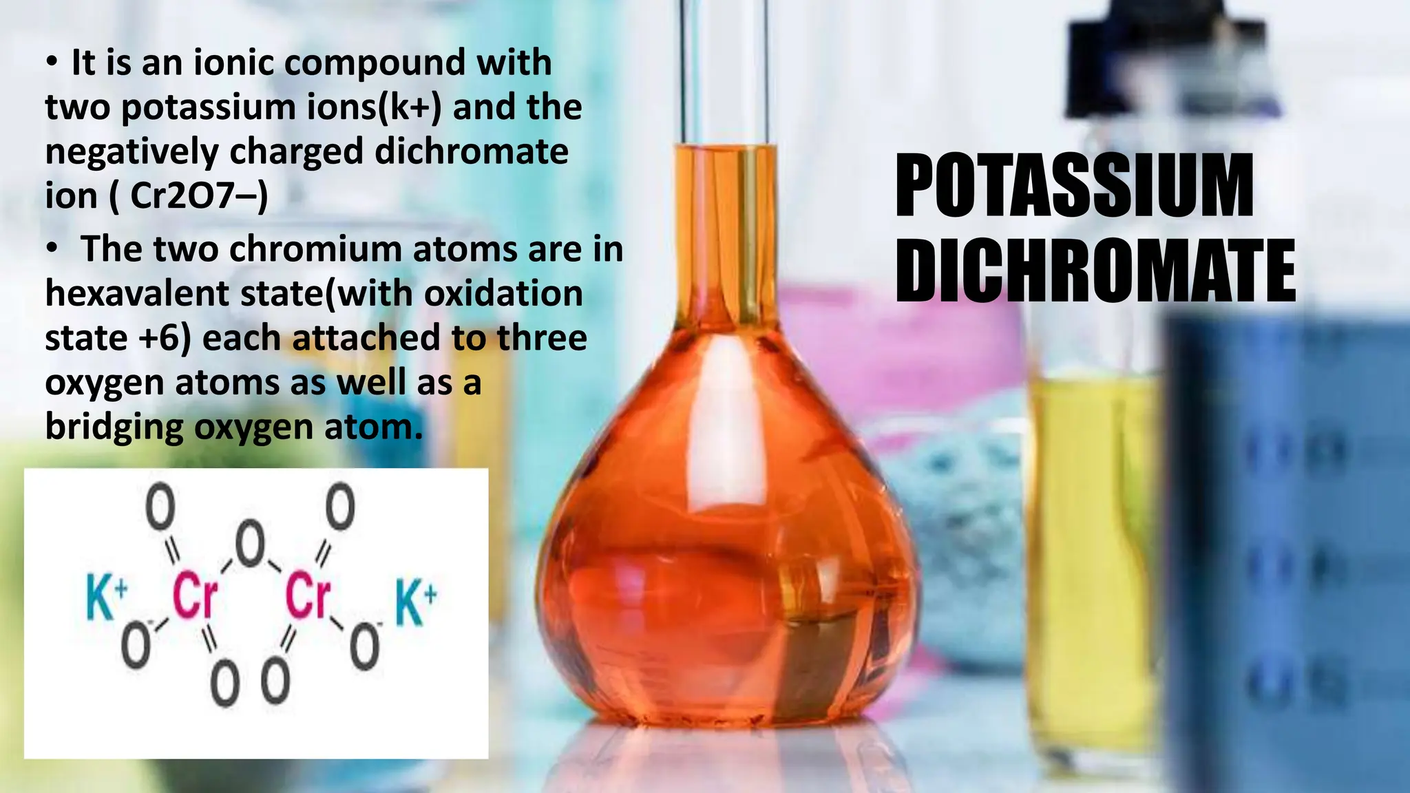
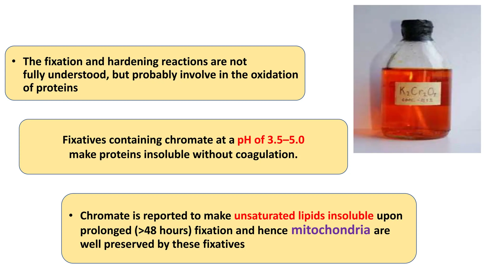
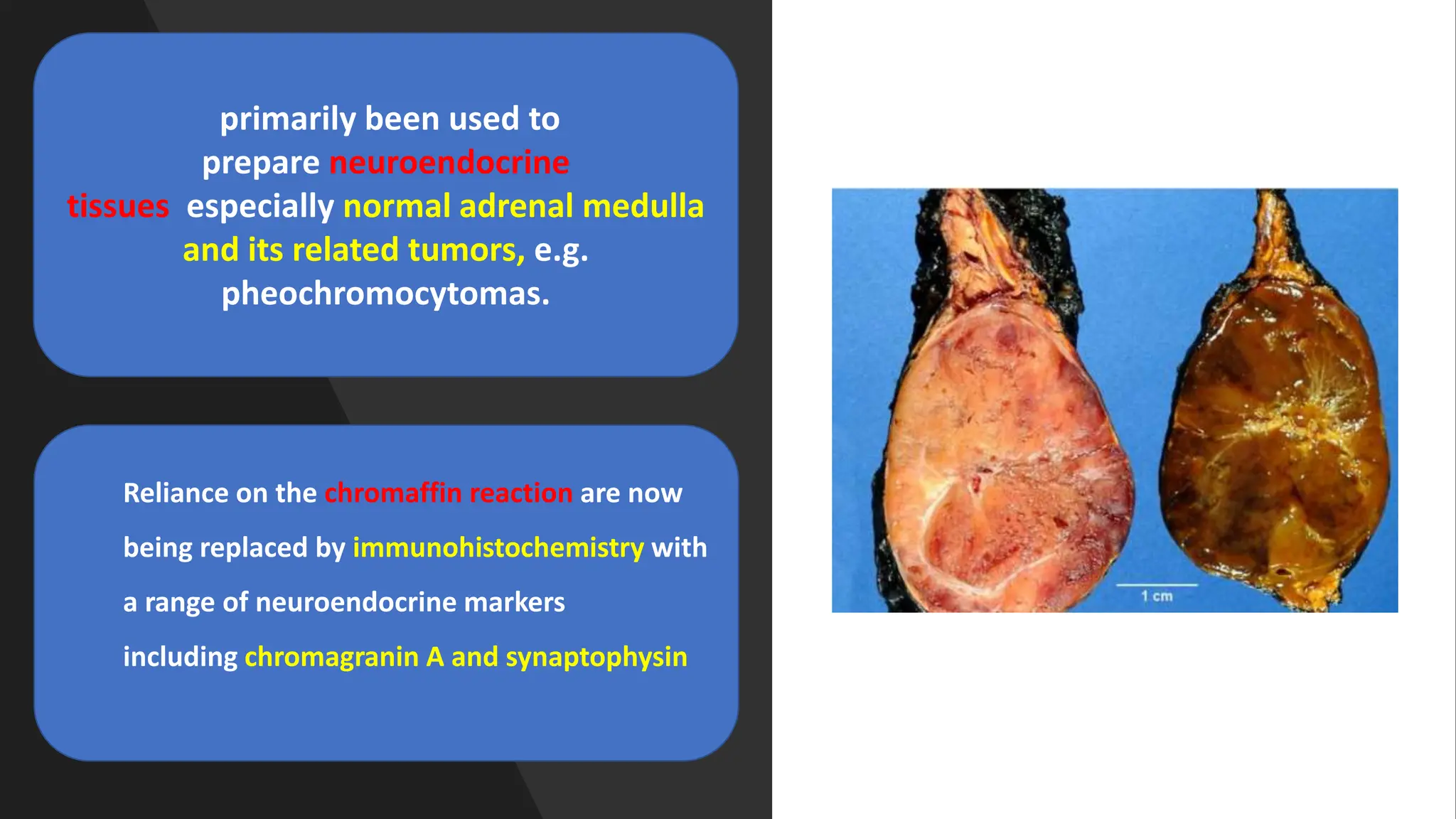
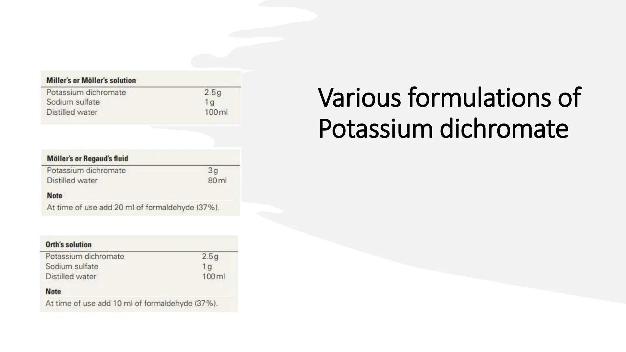
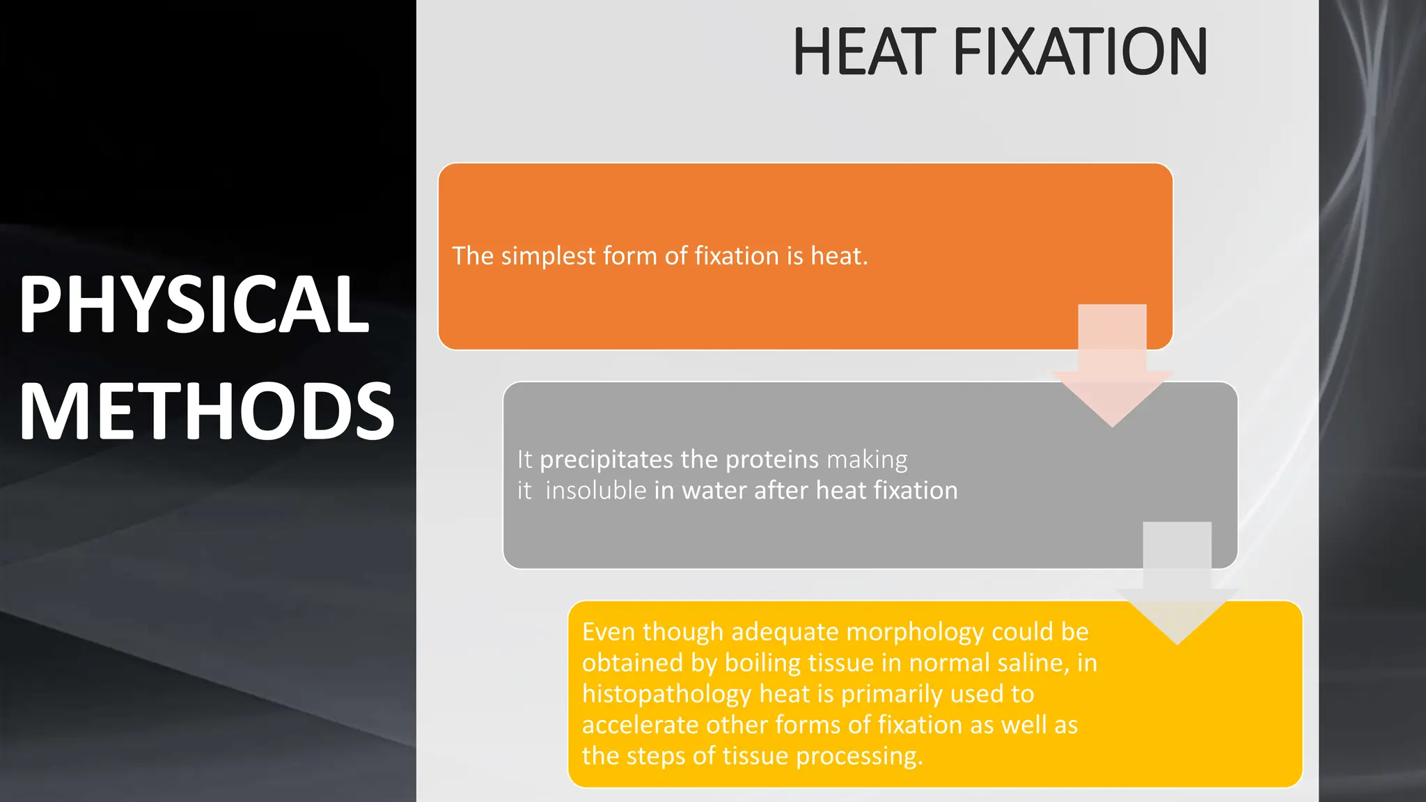
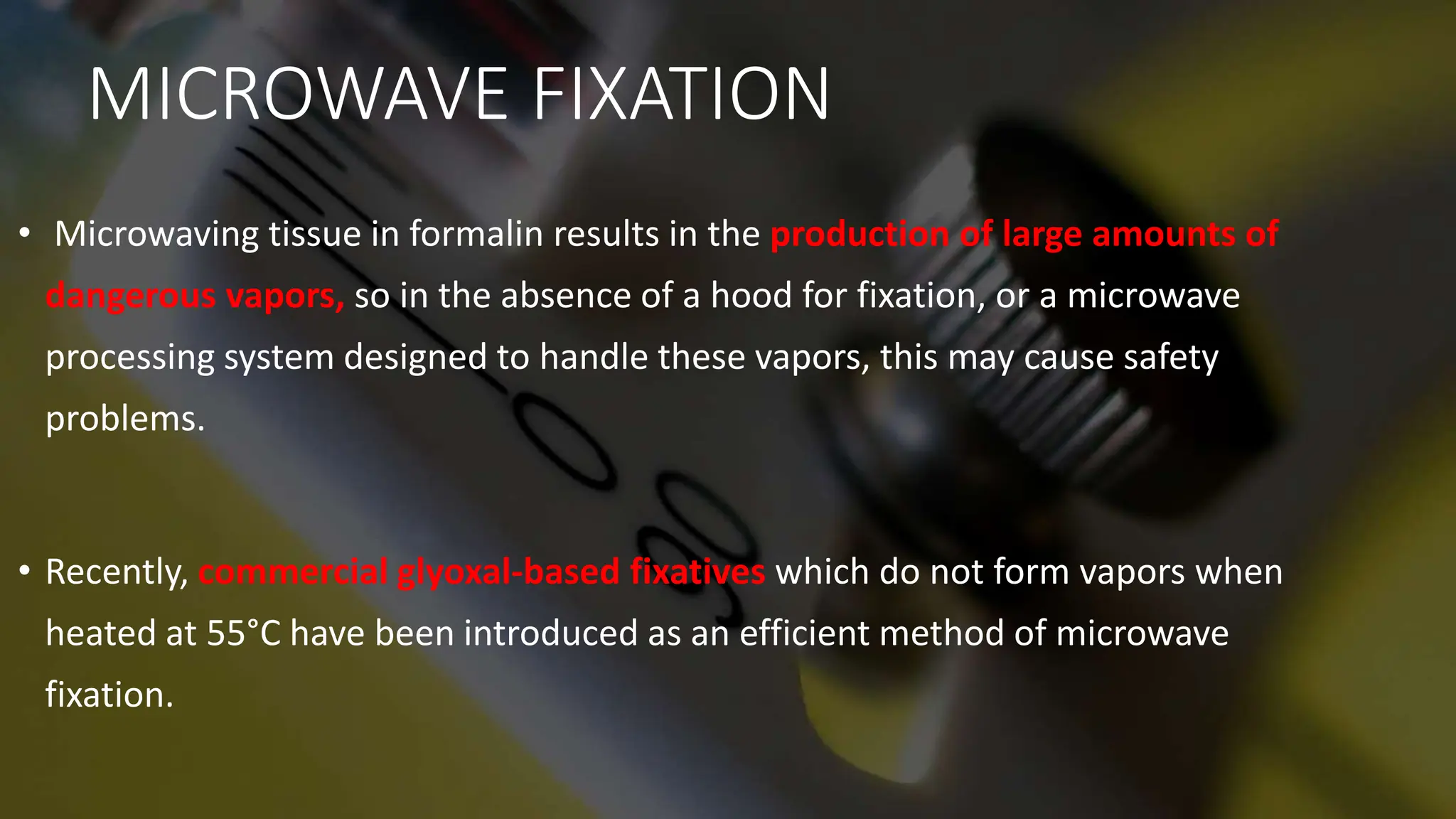
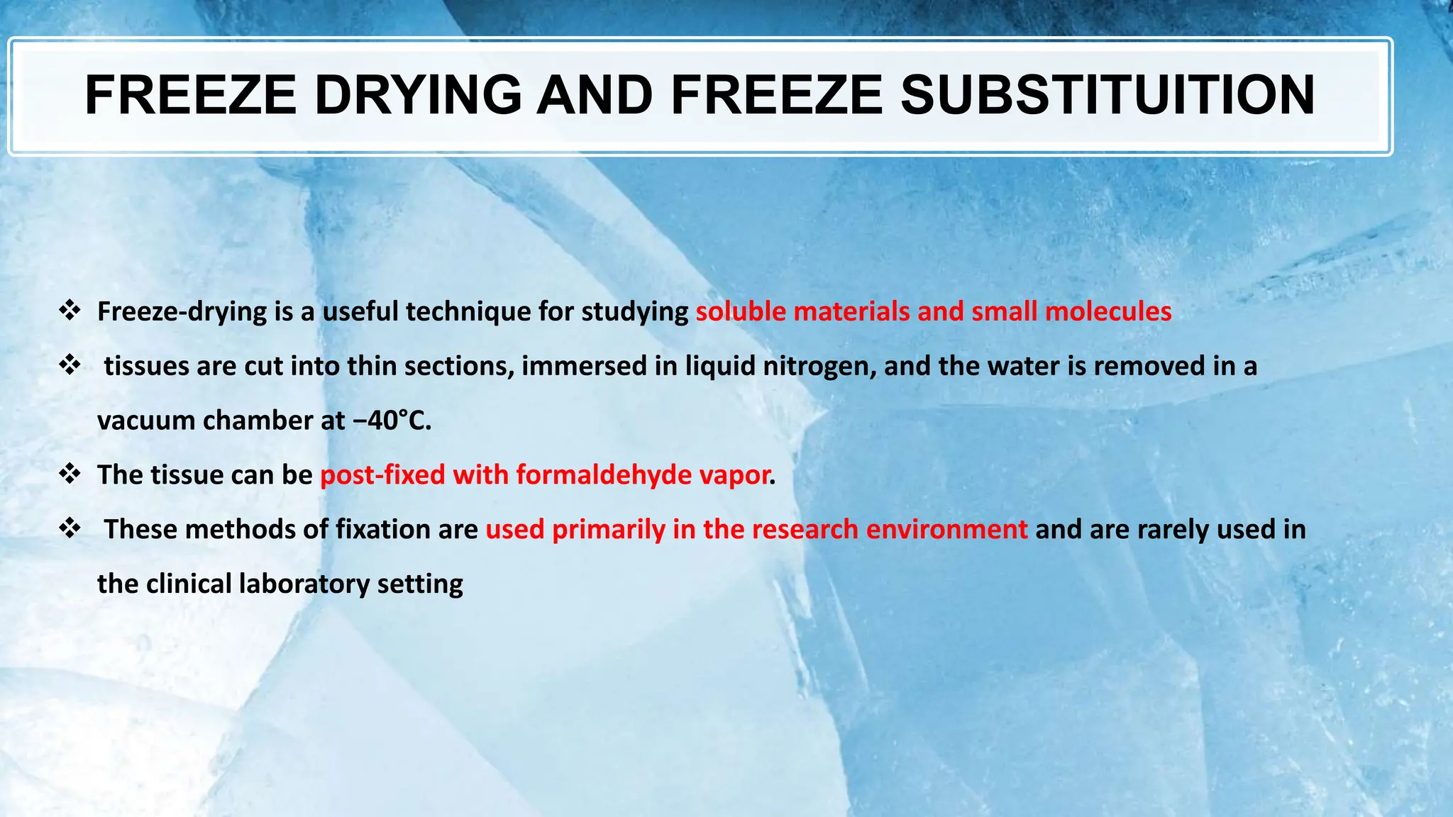
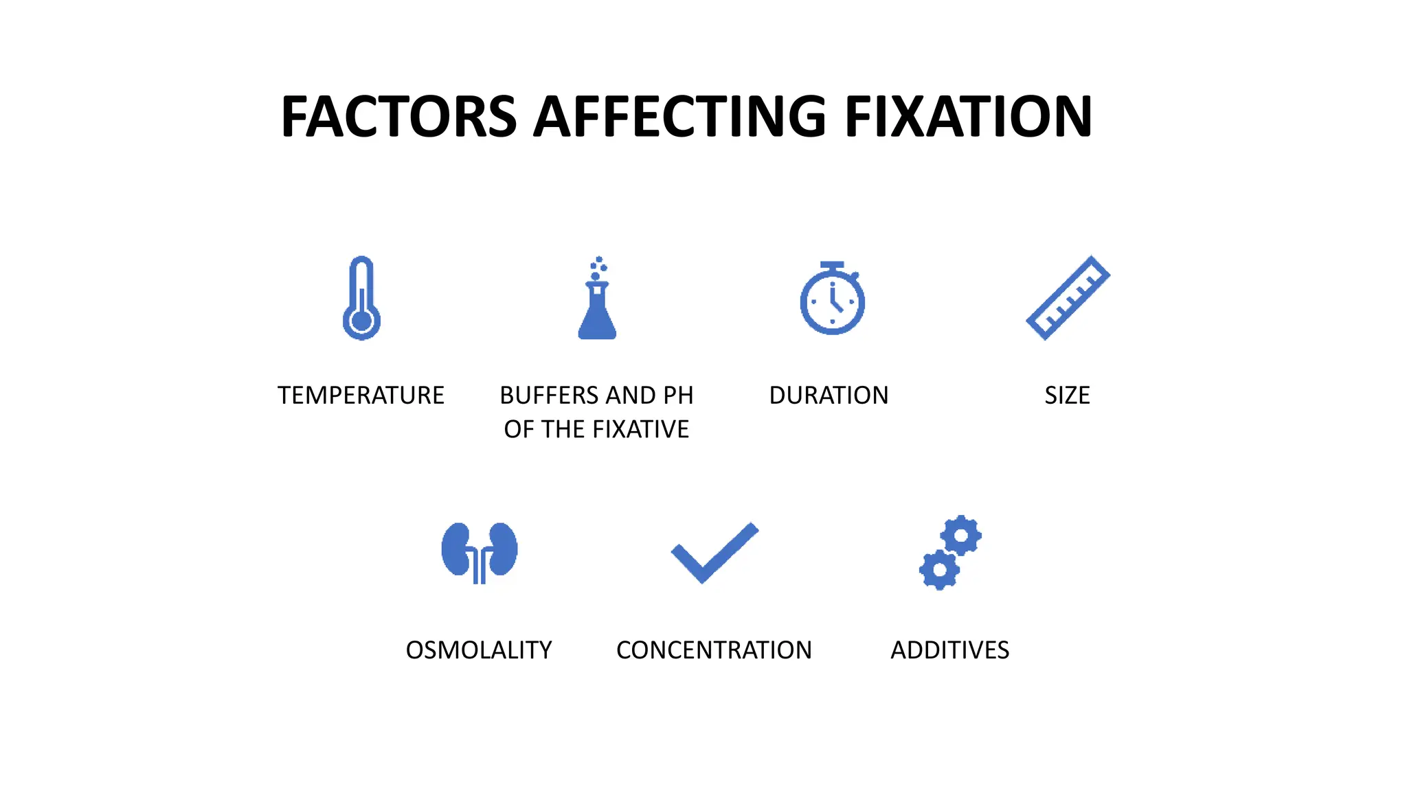
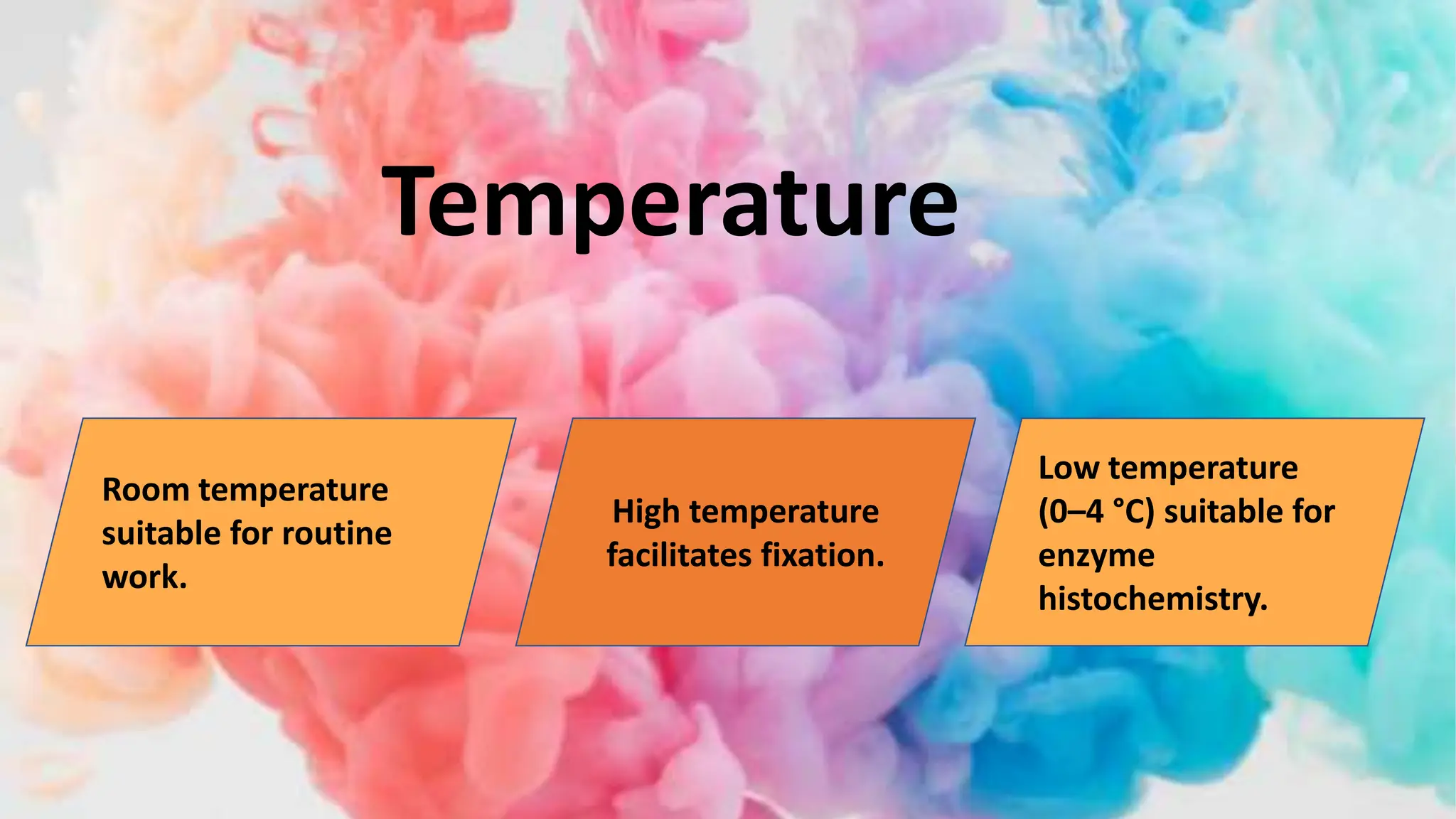
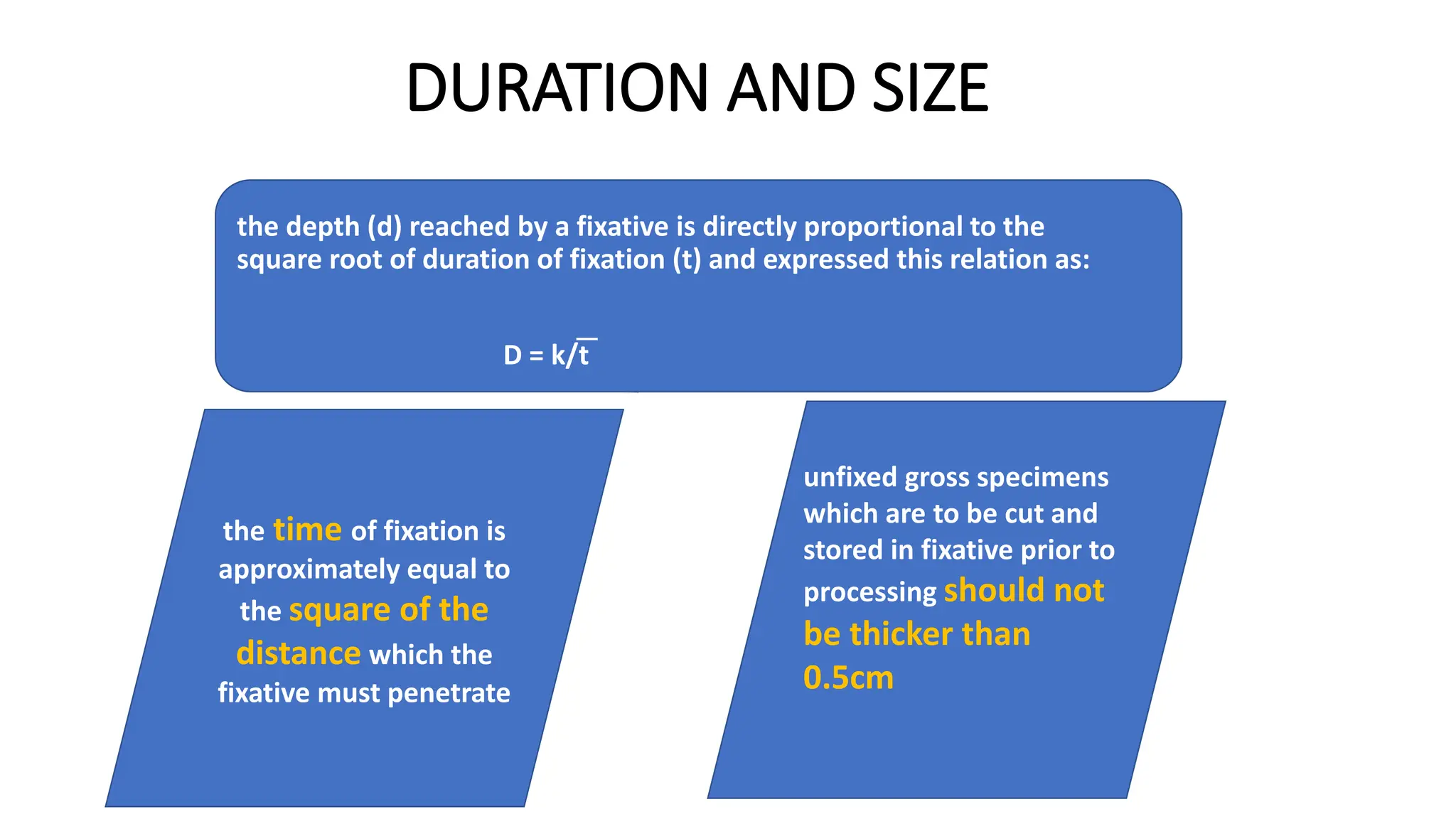
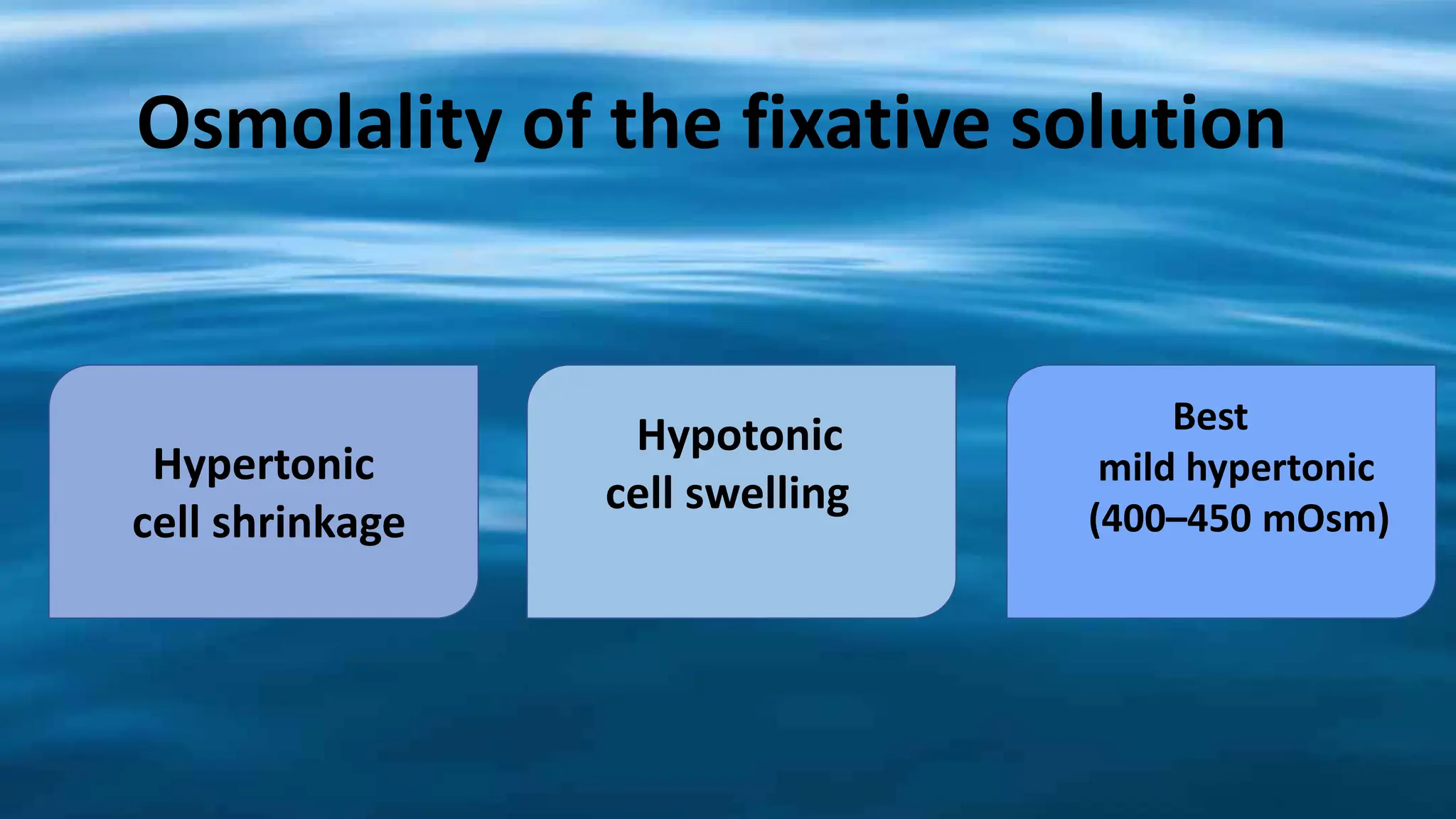
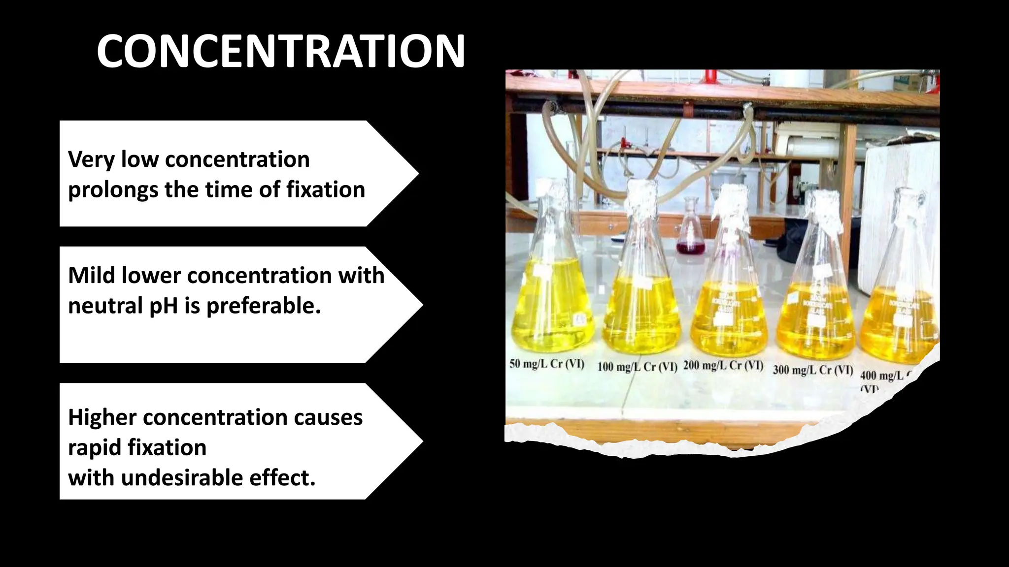
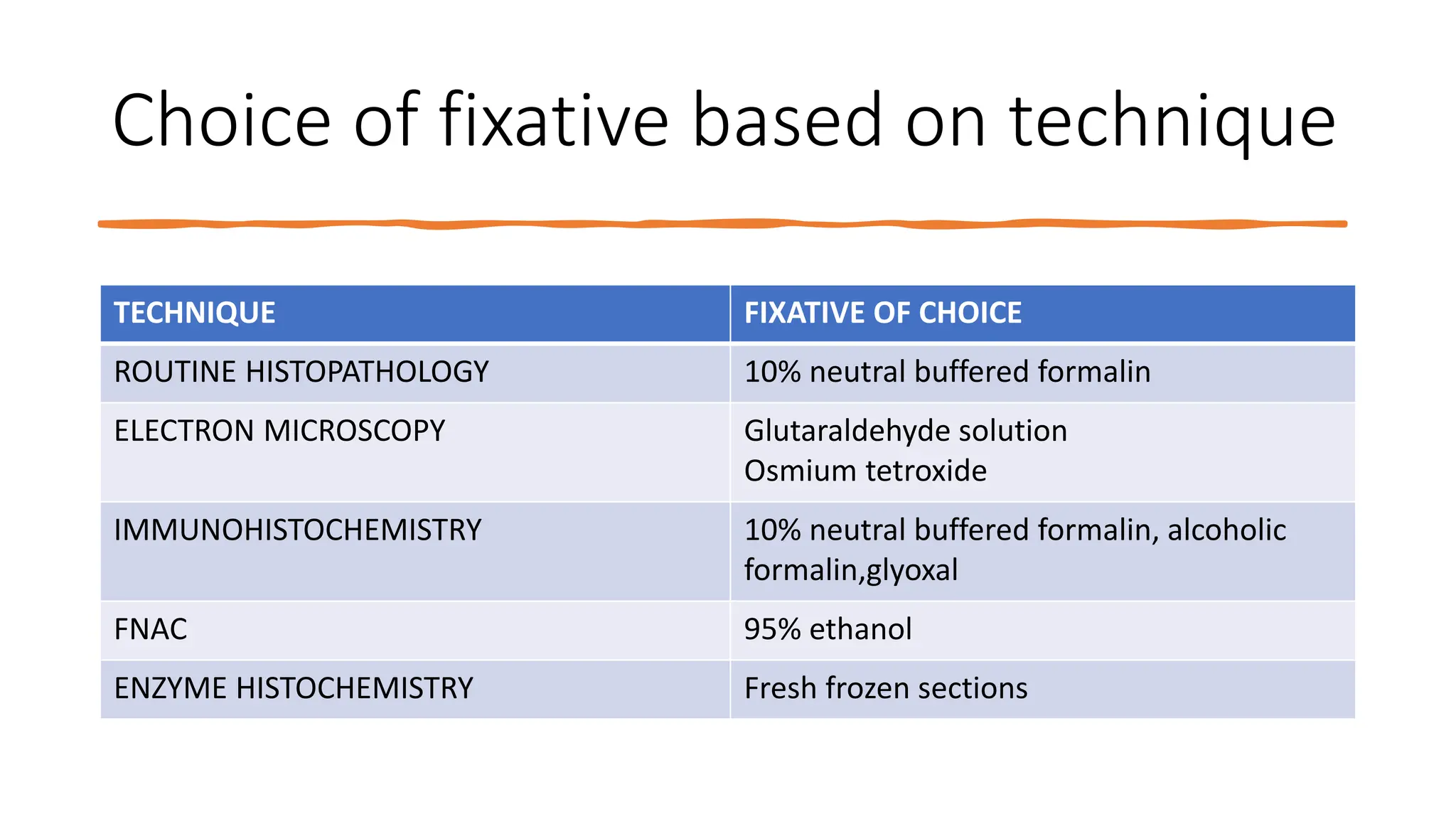
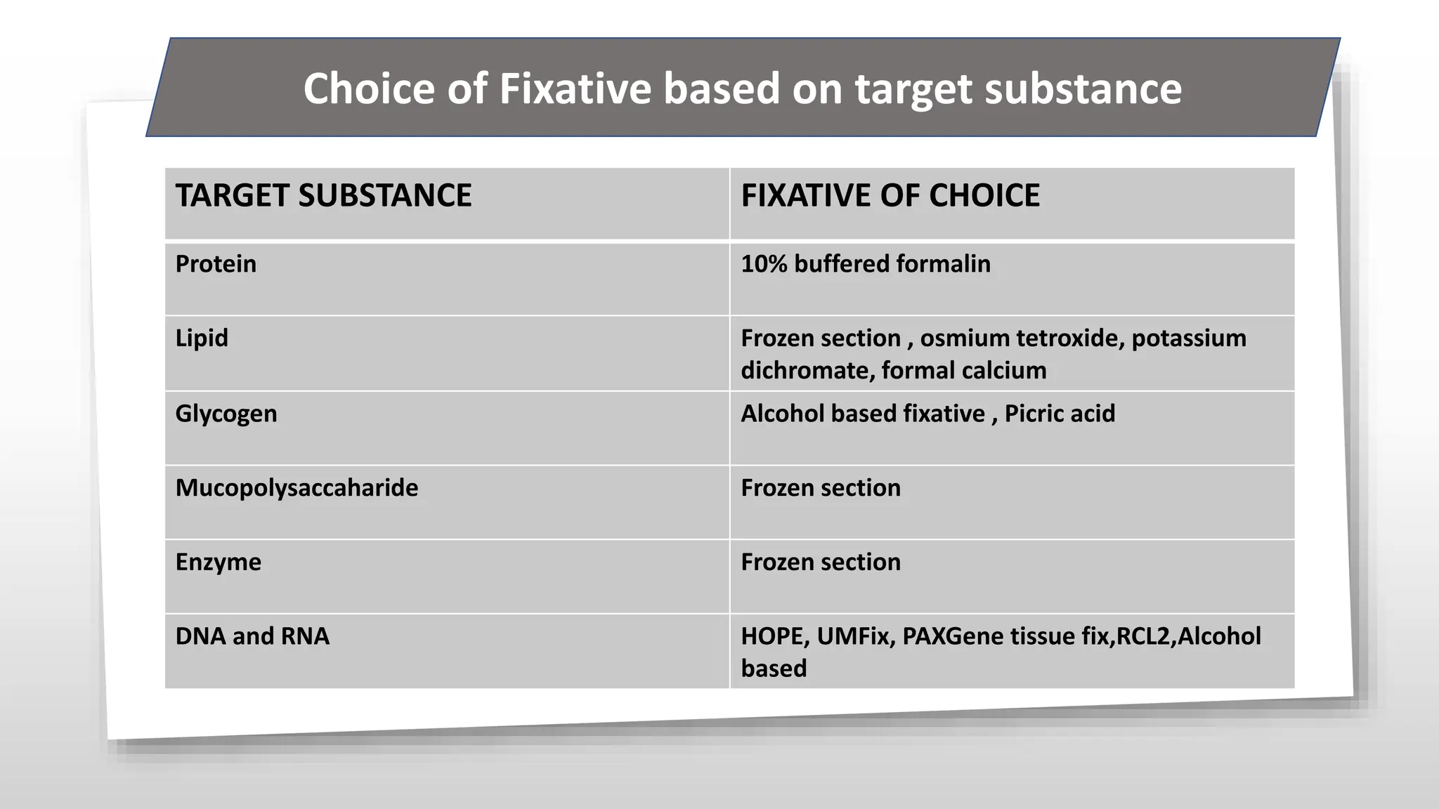
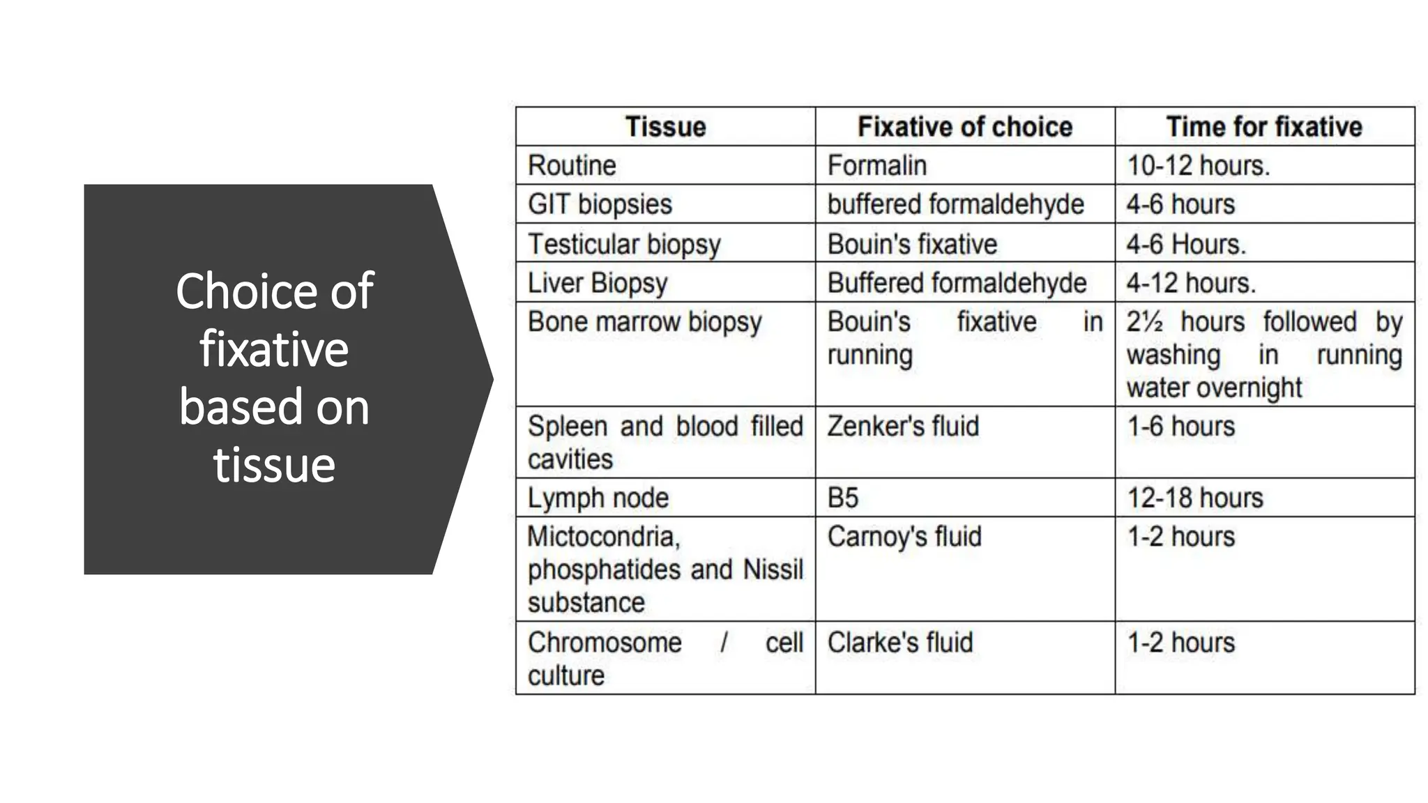
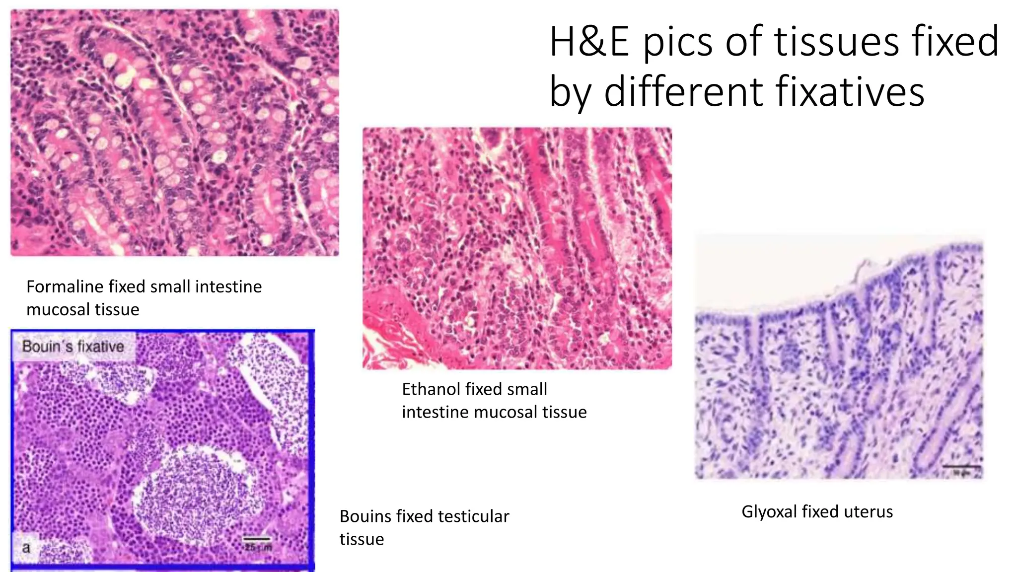
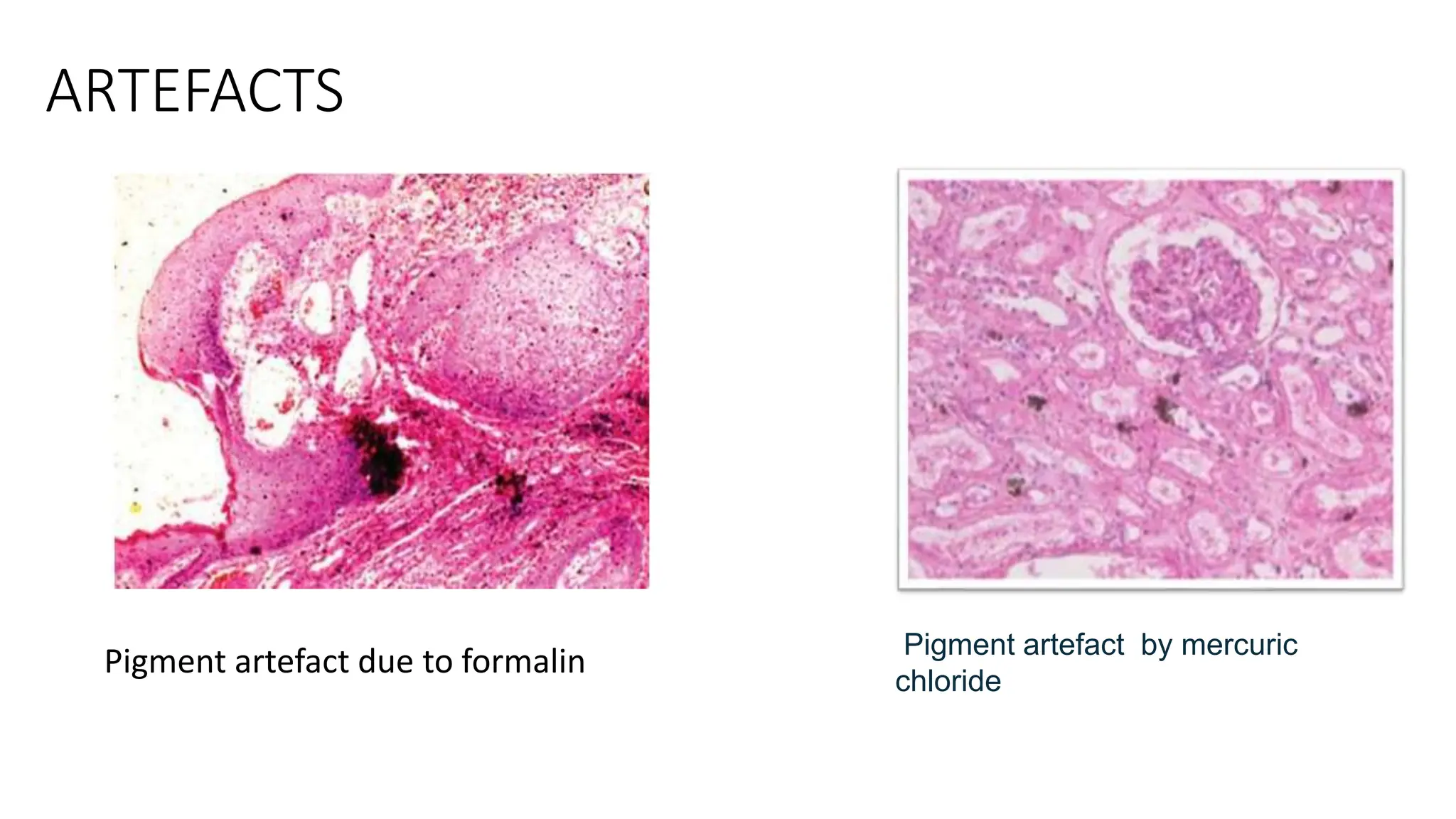
2. Human cells remain alive
because they are connected
to the blood stream, which
provides them with the
oxygen and nutrients.
When they are removed
from the body, the cells will
be cut off from the blood
circulation
Once the cells are deprived
of oxygen for long enough,
they will die
In histology, we would like
to see them as they were in
living body and that is done
by FIXATION.
3. FIXATION
• Fixation is a complex series
of events which preserves the
tissues as close as life like state
as possible by preventing
autolysis and putrefaction
4. BASIC AIMS of Fixation
1.Preservation of tissue in a life like manner and preventing from post mortem
changes
2. Preservation of chemical compounds
3. Solidification and hardening for easy sectioning
4. Prevent shrinkage and swelling
5. Optical differentiation
5. A PERFECT FIXATIVE NEEDS
Penetrate tissues quickly and evenly
Prevent autolysis and putrefaction.
Not add any extraneous material to the
tissue.
Not swell or shrink the tissue.
6. v Prepare the tissue for later treatments such
as staining and not prevent any later
investigation that might be needed.
Prevent desiccation and drying of tissue, which
would cause shrinkage and distortion.
Be safe to use (non-flammable).
Be reasonably priced.
Be convenient to use (shelf life, storage, etc.).
A PERFECT FIXATIVE NEEDS
13. DEHYDRANT
COAGULANT FIXATIVES
ALCOHOLS
• The most commonly used - ethanol and methanol
• For ethanol , fixation initiates at 50 to 60%
concentration and for methanol it is at 80%
concentration, respectively.
• They are known to be coagulants that cause protein
denaturation.
15. Methanol is
commonly used as a
fixative for blood
films
95% ethanol is used
as a fixative for
cytology smears
both alcohols are
usually combined with
other reagents when
used as fixatives for
tissue specimens
18. Picric Acid
• coagulant fixative.
• It forms picrates with basic protein groups,
which causes coagulation.
• Although picric acid is not able to fix most
carbohydrates and lipids, it preserves
glycogen.
• Brighter staining is seen by picric acid fixatives
OTHERS – Acid coagulants
20. Acetic acid
• Acetic acid coagulates nucleic acids but
does not fix or precipitate proteins; it is
therefore added to other fixatives to
prevent the loss of nucleic acids.
• Basic mechanism:
These acids insert a lipophilic
anion into a hydrophilic region and disrupt
the tertiary structures of proteins
21. • Formaldehyde was
discovered in
1859 by Butlerov.
In 1889 Ttrillat was the first
who manufactured formalde
hyde commercially as
industrial reagent.
• In 1892, Ferdinand
Blum recognized that
formalin could give
benefit when used as a
fixative.
•Formaldehyde is found to
be the most commonly
used fixative.
CROSSLINKING FIXATIVES
22. • It is a pungent toxic gas that is soluble in water
• The most widely used fixative is 10% formalin solution for
routine light microscopic sections.
• Formalin = 37-40% of formaldehyde + 60-63% of water
• 10% formalin has 4% of formaldehyde
24. • The side chains of peptides or proteins which
are most reactive with methylene
hydrate are lysine, cysteine, histidine,
arginine, tyrosine and the reactive hydroxyl
groups of serine and threonine
REVERSIBILITY OF FORMALIN FIXATION
• Washing for 24 hours removes approximately
half of the reactive groups and after 4 weeks
up to 90% are removed
25. FORMALDEHYDE FIXATION
Formaldehyde primarily
preserves protein and the
general structure of
cellular organelles.
It can interact with nucleic
acids but has little effect
on carbohydrates and
preserves lipids if the
solutions contain calcium
26.
27.
28. Neutral buffered 10%
formalin
1. Tap water 900 ml
2. Formalin (37% formaldehyde solution) 100 ml
3. Sodium phosphate, monobasic, monohydrate 4 g
4. Sodium phosphate, dibasic, anhydrous 6.5 g
The pH should be 7.2–7.4
31. GLUTARALDEHYDE
• Glutaraldehyde is a bifunctional aldehyde which probably combines with the
similar groups as formaldehyde.
Glutaraldehyde was found in 1963 by Sabatini
et al as particular fixative for ultrastructural
researches.
32. Although penetration rate is
slower compared to
formaldehyde,
glutaraldehyde is more
effective cross-linker for
proteins than formaldehyde
Crosslinking is irreversible
and withstands acids, urea ,
semicarbazide and heat
Does not react with
carbohydrates or lipids
unless they contain free
amino group
GLUTARALDEHYDE
34. exposure may result in respiratory tract, skin, and
digestive tract irritation
On exposure to oxygen, glutaraldehyde becomes
unstable and breaks down with decrease in pH.
Glutaraldehyde preserves the ultrastructure of the
tissue, thereby it is used in electron microscopy studies, but owing
to poor penetration and overhardening properties, it is not used as
tissue fixatives for light microscopy.
For stability, it requires pH of 5 and storage at 4°C. At
room temperature, glutaraldehydes are not able to cross-link
the nucleic acids.
35. • For the purpose of electron
microscopy:
• Glutaraldehyde is used 2.5% glutaraldehyde
in 100 mM phosphate buffer at pH 7.0.
• Glutaraldehyde comes commercially as 25%
or 50% solutions in 10 ml.
36. Advantages:
1. Better fixation of ultrastructure.
2. Less cell shrinkage.
3. Preservation of protein is better.
4. Good cross-linking with collagen.
Disadvantages:
1. Poor penetration and tissue
should be less than 0.5 mm thick.
2. Less stable compound.
3. No lipid fixation.
4. Costly.
37. OSMIUM TETROXIDE
• Osmium tetroxide is type of fixative that is soluble in water
and nonpolar solvents.
• Osmium tetroxide seems to react with proteins side chains that
cause cross-linking.
• It is traditionally sold as crystalline solid that is sealed in glass
ampule. It is seen that osmium tetroxide crystals convert from
solid state to vapor state.
38. • During fixation by osmium tetroxide, either due to
slow rate of reaction or due to restricted
penetration of osmium tetroxide into tissue, large
amounts of carbohydrates as well as proteins are
eradicated.
• Osmium tetroxide is helpful for staining of lipids
in frozen sections.
• In addition to its use as a secondary fixative for
electron microscope examinations, OsO4 can be
used to stain lipids in frozen sections.
39. • It is observed that fixation by osmium
tetroxide causes swelling in tissue,
which can be decreased by adding
sodium chloride or calcium chloride to
fixatives.
• Continued exposure to osmium
tetroxide vapors can cause deposition
into cornea, which eventually leads to
blindness.
40. Advantages:
1. This is a very good fixative for lipid.
2. It preserves cytoplasmic organelles such as Golgi
bodies and mitochondria,
3. Does not make the tissue hard
41.
42. OSMIUM
TETROXIDE
• It is commercially available in
sealed vial 0.1–1 g.
• Aqueous solution of 4% OsO4 is
made.
• This should be stored in clean glass
vial away from sunlight.
• In laboratory 2–4% OsO4 in buffer
solution of pH 7.2 is used.
43. MERCURIC CHLORIDE
• It is now rarely used due to the health and
safety issues of mercury-containing fixatives
• special stains e.g. B5, used in some laboratories
for fixing hematopoietic tissue
44. MERCURIC CHLORIDE
• These fixatives have slow
penetration capacity, so
the sections of specimens should
be thin.
The chemistry of fixation is not well
understood but it is known that it reacts
with ammonium salts, amines, amides,
amino acids and sulfydryl groups to
harden tissues.
46. POTASSIUM
DICHROMATE
• It is an ionic compound with
two potassium ions(k+) and the
negatively charged dichromate
ion ( Cr2O7–)
• The two chromium atoms are in
hexavalent state(with oxidation
state +6) each attached to three
oxygen atoms as well as a
bridging oxygen atom.
47. • The fixation and hardening reactions are not
fully understood, but probably involve in the oxidation
of proteins
• Chromate is reported to make unsaturated lipids insoluble upon
prolonged (>48 hours) fixation and hence mitochondria are
well preserved by these fixatives
Fixatives containing chromate at a pH of 3.5–5.0
make proteins insoluble without coagulation.
48. • During this procedure
the chromium attaches
to the lipid and may
be used
as a mordant for some
dyes, including
hematoxylin.
• Potassium dichromate is a
very valuable fixing agent
where lipids are concerned.
This is best observed with
the postchroming procedure,
which makes many
unsaturated lipids insoluble
which take weeks to be fully
effective.
For faster demonstration of
lipids in paraffin
sections, post osmication
should be used.
49. primarily been used to
prepare neuroendocrine
tissues especially normal adrenal medulla
and its related tumors, e.g.
pheochromocytomas.
Reliance on the chromaffin reaction are now
being replaced by immunohistochemistry with
a range of neuroendocrine markers
including chromagranin A and synaptophysin
51. HEAT FIXATION
The simplest form of fixation is heat.
It precipitates the proteins making
it insoluble in water after heat fixation
Even though adequate morphology could be
obtained by boiling tissue in normal saline, in
histopathology heat is primarily used to
accelerate other forms of fixation as well as
the steps of tissue processing.
PHYSICAL
METHODS
52. MICROWAVE FIXATION
• Microwaves are electromagnetic wave with frequencies between 300 MHz and
300 GHz
• Microwave creates electromagnetic field, and the molecules rapidly oscillate
generating heat by motion.
• Microwave heating speeds fixation and can reduce times for fixation of some
gross specimens and histological sections from more than 12 hours to less
than 20 minutes.
53. MICROWAVE FIXATION
• Microwaving tissue in formalin results in the production of large amounts of
dangerous vapors, so in the absence of a hood for fixation, or a microwave
processing system designed to handle these vapors, this may cause safety
problems.
• Recently, commercial glyoxal-based fixatives which do not form vapors when
heated at 55°C have been introduced as an efficient method of microwave
fixation.
54. FREEZE DRYING AND FREEZE SUBSTITUITION
Freeze-drying is a useful technique for studying soluble materials and small molecules
tissues are cut into thin sections, immersed in liquid nitrogen, and the water is removed in a
vacuum chamber at −40°C.
The tissue can be post-fixed with formaldehyde vapor.
These methods of fixation are used primarily in the research environment and are rarely used in
the clinical laboratory setting
57. pH of the fixative
Buffers
High acidity or alkalinity
interferes fixation.
pH 6–8 is the best range.
Neutral pH is
preferable.
The pH for the
ultrastructure preservation
should be buffered between
7.2 to 7.4.
•phosphates,
• cacodylate,
• bicarbonate,
• Tris, and
• acetate.
58. DURATION AND SIZE
the depth (d) reached by a fixative is directly proportional to the
square root of duration of fixation (t) and expressed this relation as:
D = k/t
the time of fixation is
approximately equal to
the square of the
distance which the
fixative must penetrate
unfixed gross specimens
which are to be cut and
stored in fixative prior to
processing should not
be thicker than
0.5cm
59. Osmolality of the fixative solution
Hypertonic
cell shrinkage
Hypotonic
cell swelling
Best
mild hypertonic
(400–450 mOsm)
60. CONCENTRATION
Mild lower concentration with
neutral pH is preferable.
Very low concentration
prolongs the time of fixation
Higher concentration causes
rapid fixation
with undesirable effect.
61. CONCENTRATION OF FIXATIVES
Concentrations of
formalin above 10%
tend to cause
increased hardening
and shrinkage.
• Ethanol
concentrations
below 70% do not
remove free water
from tissues
efficiently
In addition, higher
concentrations result in
formalin being present
in its polymeric form,
which can be deposited
as white precipitate.
62. Additives
improves the morphology of
the fixed tissue.
The electrolytes may react either directly with proteins
causing denaturation, or with the fixatives and cellular
constituents
calcium chloride
potassium thiocyanate
ammonium sulfate
potassium
dihydrogen phosphate.
63. • The choice of electrolytes to be
added to fixatives used on
a tissue processor may vary.
• electrolytes such as phosphates
may cause problems with some
processors due to precipitation of
the salts.
• non-electrolyte substances such as
sucrose, dextran, and detergent has
also been reported to
improve fixation
Additives
64. Choice of fixative based on technique
TECHNIQUE FIXATIVE OF CHOICE
ROUTINE HISTOPATHOLOGY 10% neutral buffered formalin
ELECTRON MICROSCOPY Glutaraldehyde solution
Osmium tetroxide
IMMUNOHISTOCHEMISTRY 10% neutral buffered formalin, alcoholic
formalin,glyoxal
FNAC 95% ethanol
ENZYME HISTOCHEMISTRY Fresh frozen sections
65. TARGET SUBSTANCE FIXATIVE OF CHOICE
Protein 10% buffered formalin
Lipid Frozen section , osmium tetroxide, potassium
dichromate, formal calcium
Glycogen Alcohol based fixative , Picric acid
Mucopolysaccaharide Frozen section
Enzyme Frozen section
DNA and RNA HOPE, UMFix, PAXGene tissue fix,RCL2,Alcohol
based
Choice of Fixative based on target substance
To preserve a tissue live and prevent from post mortem changes like autolysis and putrefaction
2. Preservation of chemical compounds and microanatomic constituents so that further histochemistry is possible.
3. Solidification: The semifluid consistency of cells (gel) is changed into an irreversible semisolid consistency (solid).
Hardening: Easy manipulation of soft tissue like brain, intestines etc. is possible and maximally explored through Hardening via fixatives.
5. Optical differentiation: It alters to varying degrees of the refractive indices of the various components of cells and tissues so that unstained components are more easily visualized than when unfixed.
6. Effects of staining by certain fixatives intensifies the staining character of tissues.
First we can see about chemical fixatives since we use it in routine life.
Amount of fixative: The fixative should be atleast 15-20 times the bulk of tissue. For museum specimens the volume of fixative is > 50 times.
Zenkers = potassium dichromate6.3 g+mercuric chloride12.5 g
Bouins= picric acid 1500 ml and formaldehyde500 ml+gaa 100 ml
Alcoholic formalin = ethanol 895ml+ formalin 100ml
Water molecules surround hydrophobic areas of proteins and, by repulsion, force hydrophobic chemical groups into closer contact with each other stabilizing hydrophobic bonding. By removing water, the opposite principle weakens hydrophobic bonding.
Similarly, molecules of water participate in hydrogen bonding in hydrophilic areas of proteins, and therefore removal of water destabilizes this hydrogen bonding
Water molecules surround hydrophobic areas of proteins and, by repulsion, force hydrophobic chemical groups into closer contact with each other stabilizing hydrophobic bonding. By removing water, the opposite principle weakens hydrophobic bonding. Similarly, molecules of water participate in hydrogen bonding in hydrophilic areas of proteins, and therefore removal of water destabilizes this hydrogen bonding.
Ethanol is sometimes used to preserve glycogen but will cause distortion of nuclear and cytoplasmic detail.
Excessive shrinkage
Reactions of formaldehyde are complex and numerous.
1- formation of these reactive groups is the primary and characteristic reaction ( timing hours to days) 24 hrs
2- when combine hydrogen groups or with each recative groups , these form methylene bridges.( weeks)
In naked and free DNA, the cross-linking reactions are believed to start at adenine-thymidine rich regions and cross-linking increases with rising temperatures
The principle type of cross link studied is between lysine and reactive hydroxymethyl group
Penetrating rate: 1 mm penetration of formalin per hour for first hour, then 1mm penetration per 3 hours for subsequent thickness at room temperature.
Precaution: Formaldehyde is irritant to the eye and skin and toxic for inhalation. It is a carcinogenic element.
. 1 part of the stock formalin with 9 parts water, preferably distilled. This makes an unbuffered formalin solution, which will have a pH of 3-4. If used unbuffered the acidity can react with haemoglobin in the tissues to produce dark brown acid formaldehyde haematin precipitates, which complicate histological interpretation.
Deionized water- all minerals are removed along with impurities
Distilled water – impurities removed by boiling folloed by condensation eg.chlorine
Alcoholic formalin advantageous in large specimen embedded in fat since it extract fats so lymph nodes if present can be visualized
Alcoholic bouins better reservation of glycogen
Osmium tetroxide gives off fumes which cause a piercing headache
This vapor is toxic to the eye and respiratory tract.
Special formulations of zinc sulfate in formaldehyde replacing mercuric chloride in B5 may give better nuclear detail than formaldehyde alone and improve tissue penetration
B5 fixative Stock solution: Mercuric chloride 12 g Sodium acetate 2.5 g Distilled water 200 ml Add 2 ml of formaldehyde (37%) to 20 ml of stock solution just before use.
Frequently used for bone marrow, lymph nodes, spleen, and other hematopoietic tissues.
Hellys : It is excellent for bone marrow extramedullary hematopoiesis and intercalated discs
Zenkers: . This is a good fixative for bloody (congested) specimens and trichrome stains.
3+ state may destroy animal tissue
. Chromium ions specifically interact with the carboxyl and hydroxyl side chains of proteins and chromic acid interacts with disulfide bridges and attacks lipophilic residues such as tyrosine and methionine
post chroming: It is the practice of treating tissues fixed in a primary fixative, usually one containing formalin or potassium dichromate, with a simple solution of potassium dichromate for an extended period, usually days and sometimes weeks.
The purpose is to improve the preservation of lipid and lipoprotein materials that would otherwise be removed by processing reagents. The process does not well preserve triglycerides and, for them, frozen sections or post osmication should be used.
Mordant: metal ion cationic complex that confers net positive charge to the dye mordant complex enabling to bind to anionic sites like nucleus.
chromaffin reaction: oxidation of stored catecholamines synthesized by the tumor when reacted with potassium dichromate because of which it appears yellowish brown in colour
Tissue should be washed after fixation and transferred to 70% ethanol. Failure to wash the tissue after fixation precipitates pigments.
Excessive shrinkage occurs when tissues are processed tp paraffin blocks
Glyoxal – oxalaldehyde. Bifunctional. Doesn’t evoprate so no vapors produced. Fixed tissues produce good cellular details .
The diffusion of molecules increases with rising temperature due to their more rapid movement and vibration
Closed tissue processors have their processing retort directly above the paraffin holding stations which are held at 60–65°C, making the retort slightly warmer than room temperature.
Microwaves nowadays have been used -vapor levels-safety problem- so glyoxal with no vapors
Buffers: the solution of reserve acidity or alkalinity which resists change of pH upon the addition of a small amount of acid or alkali.
Contains weak acid and base that resist mild ph changes when comes in contact with acidic or basic solution.
Specimens should not rest in the bottom of the container it should be separated by a paper allowing even penetration of fixatives.
Osmolality is a measure of the number of dissolved particles in a solution.
It is typically expressed in units of osmoles per liter (osmol/L). The most common type of osmolality measurement is blood osmolality, which is used to assess the concentration of various electrolytes in the blood.
Osmolality can also be used to measure the amount of solute in other bodily fluids, such as urine or sweat. In general, solutions with a higher osmolality are considered more concentrated than those with a lower osmolality.
Treatment of specimens for mercuric chrolide pigments with iodine (Lugol’s iodine) during processing or sections prior to staining, will produce mercuric iodide which can be washed out of the tissues. A subsequent treatment with sodium thiosulphate then removes residual iodine.
For formalin pigments treat it with 10% ammonium hydroxide or 1.8% picric acid in ethyl alcohol