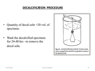
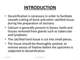
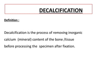
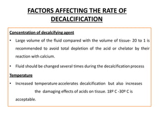
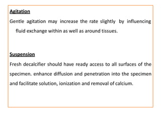
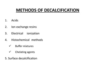
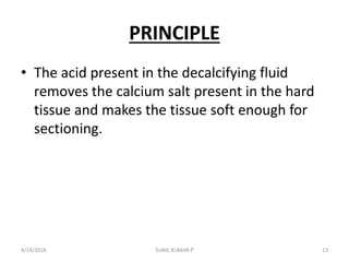
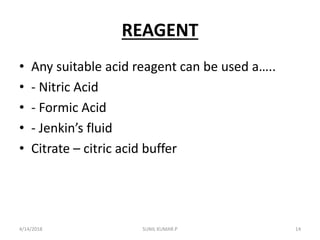
SlideShare a Scribd company logo
DECALCIFICATION
SUNIL KUMAR.P
ST.JOHN’S MEDICAL COLLEGE
BANGALORE
4/14/2018 1SUNIL KUMAR.P
4/14/2018 2SUNIL KUMAR.P
4/14/2018 SUNIL KUMAR.P 3
Contents……………….
• - Introduction
• -Definition
• - Properties of ideal Decalcifi…. agents
• - Methods of Decalcification
• - principle
• -Types of decalcifying agents
• - Compositions, Action, Advantages
• -procedure of Decalcification
4/14/2018 4SUNIL KUMAR.P
INTRODUCTION
• Decalcification is necessary in order to facilitate
smooth cutting of bone and other calcified tissue
during the preparation of sections.
• Calcium is generally present in bones, teeth and
tissues removed from glands such as tubercular
and lymphatic.
• The calcified hard tissue is cut into small pieces.
• The tissue should be thoroughly washed, to
remove excess of fixative before the specimen is
subjected to decalcification.
4/14/2018 5SUNIL KUMAR.P
DECALCIFICATION
Definition :
Decalcification is the process of removing inorganic
calcium (mineral) content of the bone /tissue
before processing the specimen after fixation.
Criteria of a good decalcifying Agent
1. Complete removal of calcium
2. Absence of damage to tissue cells or fibres
3. Non impairment of subsequent staining
technique
4. Reasonable speed of decalcification
FACTORS AFFECTING THE RATE OF
DECALCIFICATION
• 1. Concentration of decalcifying agent
• 2. Temperature
• 3. Agitation
• 4. Suspension
4/14/2018 SUNIL KUMAR.P 8
FACTORS AFFECTING THE RATE OF
DECALCIFICATION
Concentration of decalcifying agent
• Large volume of the fluid compared with the volume of tissue- 20 to 1 is
recommended to avoid total depletion of the acid or chelator by their
reaction with calcium.
• Fluid should be changed several times during the decalcificationprocess
Temperature
• Increased temperature accelerates decalcification but also increases
the damaging effects of acids on tissue. 18º C -30º C is
acceptable.
Agitation
Gentle agitation may increase the rate slightly by influencing
fluid exchange within as well as around tissues.
Suspension
Fresh decalcifier should have ready access to all surfaces of the
specimen. enhance diffusion and penetration into the specimen
and facilitate solution, ionization and removal of calcium.
METHODS OF DECALCIFICATION
1. Acids
2. Ion exchange resins
3. Electrical ionization
4. Histochemical methods
 Buffer mixtures
 Chelating agents
5. Surface decalcification
METHOD
Acid Decalcification method
4/14/2018 12SUNIL KUMAR.P
PRINCIPLE
• The acid present in the decalcifying fluid
removes the calcium salt present in the hard
tissue and makes the tissue soft enough for
sectioning.
4/14/2018 13SUNIL KUMAR.P
REAGENT
• Any suitable acid reagent can be used a…..
• - Nitric Acid
• - Formic Acid
• - Jenkin’s fluid
• Citrate – citric acid buffer
4/14/2018 14SUNIL KUMAR.P
TYPES
• Two types
• Strong Inorganic Acids
• Weak Organic Acids
4/14/2018 15SUNIL KUMAR.P
Strongacids
• It Is An InorganicAcid
• Eg: Nitric Acid, HydrochloricAcid
• Recommended Concentration - 5-10%
• They Decalcify Rapidly By Dissolving Calcium
4/14/2018 16SUNIL KUMAR.P
MINERAL ACID DECALCIFIERS
4/14/2018 17SUNIL KUMAR.P
a)Nitric acid
CHEMICAL QUANTITY
Nitric acid 5-10ml
Distilled water 100ml
4/14/2018 18SUNIL KUMAR.P
1. Fix the selected block of bone for 2-3 days in buffered
neutral formalin.
2. Place a mixture of 95ml distilled water and 5ml of
nitric acid.
3. Change nitric acid solutions daily until bubbles cease
to evolve from the tissues(1-3 days,depending on the
size and consistency of the bone block)
4.Wash in 3 changes of 90% alcohol.
5.Dehydrate,clear in xylene or benzene and embed in
paraffin
4/14/2018 19SUNIL KUMAR.P
• Formation of nitrous acid checked temporarily
by addition of 0.1% urea to the conc nitric acid
• It’s the fastest decalcifier, but end point must
be carefully watched .
• Yellow discolouration owing to formation of
nitrous acid, this accelerates decalcification but
also stains and damage tissues
4/14/2018 20SUNIL KUMAR.P
Rapid in action
Gives better nuclear staining
Causes very little hydrolysis
For Needle & Small Biopsy Specimens To Permit
Rapid Diagnosis .
Tissue left for long time causes damage to tissue
Urea is added to remove yellow color of tissue
4/14/2018 21SUNIL KUMAR.P
WEAK ACID DECALICIFYING
REAGENTS
ACETICACID
PICRIC ACID4/14/2018 22SUNIL KUMAR.P
Weak, organic acids
e.g. formic, acetic, picric.
•Acetic & picric acid cause tissue swelling & are not used alone
as primary decalcifiers but are found as components of Carnoy’s
& Bouin’s fixatives
4/14/2018 23SUNIL KUMAR.P
•Formicacidis the only weak acid used extensively
as a decalcifier
•Formic acid solutions are either
•aqueous (5-10%)
•buffered or combined with formalin.
4/14/2018 24SUNIL KUMAR.P
The formalin/10% formic acid mixture simultaneously fixes & decalcifies.
• Recommended for –
• Very small bone pieces
• Jamshidi needle biopsies.
• Formic acid gentle & slower than Hcl or nitric acid
• suitable for most routine surgical specimens, particularly for
immuno histochemistry.
• Decalcification usually complete within 2-7days.
4/14/2018 25SUNIL KUMAR.P
a)AQUEOUS FORMICACID
1.Well fixed 2-5mm thick blocks are placedin
– concentrated formic acid
– Distilled water
– 40% formaldehyde (optional)
5-25ml
100ml
5 ml
2.Change daily until decalcification is complete.
( 1-7 days for an average blocks depending on concentration ofacid.)
3.Replace fluid with 5% sodium sulfate overnight
4.Wash 12 -24 hrs in running tap water.
5.Dehydrate in graded alcohols ,clear in chloroform or toluene and embed in
wax
4/14/2018 26SUNIL KUMAR.P
DECALCIFICATION PROCEDURE
• Quantity of decal soln >20 vol. of
specimen.
• Wash the decalcified specimen
for 24-48 hrs –to remove the
decal soln.
4/14/2018 27SUNIL KUMAR.P
PROCEDURE
• 1. Suspend the sliced tissue in the decalcifying
solution by means of gauze bag, tied with a
string.
• 2.Stir the fluid occasionally and change the
fluid daily till calcium is completely removed
from the tissue section.
• 3.It is tested as follows…….
4/14/2018 28SUNIL KUMAR.P
• - take about 5.0 ml of decalcifying solution in a test
tube
• - Add conc. Ammonia solution drop by drop until it
becomes alkaline ( test by litmus paper)
• - If the solution becomes turbid, continue
decalcification ( since it contains calcium)
• - if it is clear add 0.5 ml saturated ammonium oxalate
solution
• - If calcium is present the solution will become turbid,
then continue decalcification.
• If there is no turbidity , the specimen is free of calcium
and ready for further processing.
4/14/2018 29SUNIL KUMAR.P
• 4. Wash the decalcified specimen in running
tap water for 24-48 hours.
• 5.The decalcifying solution should be
completely removed before dehydration and
embedding.
4/14/2018 30SUNIL KUMAR.P
THANKS
4/14/2018 31SUNIL KUMAR.P

More Related Content

What's hot

Staining by hematoxylin and eosin
Staining by hematoxylin and eosinStaining by hematoxylin and eosin
Staining by hematoxylin and eosin
Sowmya Srinivas
 
Fixatives used in histopathology
Fixatives used in histopathologyFixatives used in histopathology
Fixatives used in histopathologyHitendra Prajapati
 
Histotechnique for practicals pathology
Histotechnique for practicals pathologyHistotechnique for practicals pathology
Histotechnique for practicals pathology
Appy Akshay Agarwal
 
Processing of tissue
Processing of tissueProcessing of tissue
Processing of tissue
ariva zhagan
 
TISSUE PROCESSING SEMINAR
TISSUE PROCESSING SEMINARTISSUE PROCESSING SEMINAR
TISSUE PROCESSING SEMINAR
Suryagopan Prabha
 
Microtomes, Section cutting , Sharpening of Razors
Microtomes, Section cutting , Sharpening of RazorsMicrotomes, Section cutting , Sharpening of Razors
Microtomes, Section cutting , Sharpening of Razors
vikas25187
 
Frozen section and cryostat
Frozen section and cryostatFrozen section and cryostat
Frozen section and cryostat
MAHMOUD IBRAHIM
 
Section Cutting
Section CuttingSection Cutting
Section Cutting
Tamil Mahizhenthi
 
Fixatives in Histopathology
Fixatives in HistopathologyFixatives in Histopathology
Fixatives in HistopathologyIshwar9
 
Histopathology staining method
Histopathology staining methodHistopathology staining method
Histopathology staining method
D. JASMINE PRIYA
 
Equipments used Histopathology
Equipments used Histopathology Equipments used Histopathology
Equipments used Histopathology
Krinshikato
 
Haematoxylin and its types
Haematoxylin and its typesHaematoxylin and its types
Haematoxylin and its types
Atifa Ambreen
 
CYTOLOGY PPT.pptx
CYTOLOGY PPT.pptxCYTOLOGY PPT.pptx
CYTOLOGY PPT.pptx
D. JASMINE PRIYA
 
Microtome knife profile
Microtome knife profileMicrotome knife profile
Microtome knife profile
Faris K
 
Special stain in histopathology
Special stain in histopathologySpecial stain in histopathology
Special stain in histopathology
aghara mahesh
 
Staining ( rouine and special in cytology) rajiv kumar
Staining ( rouine and special in cytology) rajiv kumarStaining ( rouine and special in cytology) rajiv kumar
Staining ( rouine and special in cytology) rajiv kumar
rajusehrawat
 
Mounting media
Mounting mediaMounting media
Mounting media
MAHMOUD IBRAHIM
 
Hematoxylin and Eosin staining for UGs
Hematoxylin and Eosin staining for UGsHematoxylin and Eosin staining for UGs
Hematoxylin and Eosin staining for UGs
Dr. Varughese George
 
PAS STAIN.pptx
PAS STAIN.pptxPAS STAIN.pptx
PAS STAIN.pptx
ShahzebHUSSAIN5
 

What's hot (20)

Staining by hematoxylin and eosin
Staining by hematoxylin and eosinStaining by hematoxylin and eosin
Staining by hematoxylin and eosin
 
Fixatives used in histopathology
Fixatives used in histopathologyFixatives used in histopathology
Fixatives used in histopathology
 
Histotechnique for practicals pathology
Histotechnique for practicals pathologyHistotechnique for practicals pathology
Histotechnique for practicals pathology
 
Processing of tissue
Processing of tissueProcessing of tissue
Processing of tissue
 
TISSUE PROCESSING SEMINAR
TISSUE PROCESSING SEMINARTISSUE PROCESSING SEMINAR
TISSUE PROCESSING SEMINAR
 
Microtomes, Section cutting , Sharpening of Razors
Microtomes, Section cutting , Sharpening of RazorsMicrotomes, Section cutting , Sharpening of Razors
Microtomes, Section cutting , Sharpening of Razors
 
Frozen section and cryostat
Frozen section and cryostatFrozen section and cryostat
Frozen section and cryostat
 
Section Cutting
Section CuttingSection Cutting
Section Cutting
 
Fixatives in Histopathology
Fixatives in HistopathologyFixatives in Histopathology
Fixatives in Histopathology
 
Histopathology staining method
Histopathology staining methodHistopathology staining method
Histopathology staining method
 
Equipments used Histopathology
Equipments used Histopathology Equipments used Histopathology
Equipments used Histopathology
 
Haematoxylin and its types
Haematoxylin and its typesHaematoxylin and its types
Haematoxylin and its types
 
CYTOLOGY PPT.pptx
CYTOLOGY PPT.pptxCYTOLOGY PPT.pptx
CYTOLOGY PPT.pptx
 
Microtome knife profile
Microtome knife profileMicrotome knife profile
Microtome knife profile
 
Special stain in histopathology
Special stain in histopathologySpecial stain in histopathology
Special stain in histopathology
 
Staining ( rouine and special in cytology) rajiv kumar
Staining ( rouine and special in cytology) rajiv kumarStaining ( rouine and special in cytology) rajiv kumar
Staining ( rouine and special in cytology) rajiv kumar
 
Mounting media
Mounting mediaMounting media
Mounting media
 
Hematoxylin and Eosin staining for UGs
Hematoxylin and Eosin staining for UGsHematoxylin and Eosin staining for UGs
Hematoxylin and Eosin staining for UGs
 
PAS STAIN.pptx
PAS STAIN.pptxPAS STAIN.pptx
PAS STAIN.pptx
 
Le cell
Le cellLe cell
Le cell
 

Similar to Decalcificaion

Decalcification in Histopathology.pptx
Decalcification in Histopathology.pptxDecalcification in Histopathology.pptx
Decalcification in Histopathology.pptx
parisdepher
 
decalcification.pdf
decalcification.pdfdecalcification.pdf
decalcification.pdf
maryamiqbal36
 
02 DECALCIFICATION AND TESTING FOR END POINT OF DECALCIFICATION.pptx
02 DECALCIFICATION AND TESTING FOR END POINT OF DECALCIFICATION.pptx02 DECALCIFICATION AND TESTING FOR END POINT OF DECALCIFICATION.pptx
02 DECALCIFICATION AND TESTING FOR END POINT OF DECALCIFICATION.pptx
AYODEJI BLESSING AJILEYE
 
Decalcification.pptx
Decalcification.pptxDecalcification.pptx
Decalcification.pptx
sandeep singh
 
Decalcification of tissue in laboratory.pptx
Decalcification of tissue in laboratory.pptxDecalcification of tissue in laboratory.pptx
Decalcification of tissue in laboratory.pptx
mallicktoufeeq8
 
Rahul singh
Rahul singhRahul singh
Rahul singh
Rahul Singh
 
Decalcification of bone and tooth
Decalcification of bone and toothDecalcification of bone and tooth
Decalcification of bone and tooth
D Venkatesh Kumar
 
Decalcification.pptx
Decalcification.pptxDecalcification.pptx
Decalcification.pptx
AlphoncePancras
 
2. fixatives and fixation seminar
2. fixatives and fixation seminar2. fixatives and fixation seminar
2. fixatives and fixation seminar
Dr SANTHIPRIYA GOPASANA
 
Fixation of tissues
Fixation of tissuesFixation of tissues
Fixation of tissues
SUNIL KUMAR PEDDANA
 
Clearing
ClearingClearing
museum techniques in pathology.pptx
museum techniques in pathology.pptxmuseum techniques in pathology.pptx
museum techniques in pathology.pptx
OMJHA20
 
3.DECALCIFICATION.pptx
3.DECALCIFICATION.pptx3.DECALCIFICATION.pptx
3.DECALCIFICATION.pptx
AhmadMahfudhi
 
Tissue processing 2012
Tissue processing 2012Tissue processing 2012
Tissue processing 2012Dhiraj Shukla
 
tissue processing introduction basics ppt
tissue processing introduction basics ppttissue processing introduction basics ppt
tissue processing introduction basics ppt
midhat2502
 
Decalcification of bone
Decalcification of boneDecalcification of bone
Decalcification of bone
Burnett D Justus
 
Bonebiopsyanditsdecalcification 100407084321-phpapp01
Bonebiopsyanditsdecalcification 100407084321-phpapp01Bonebiopsyanditsdecalcification 100407084321-phpapp01
Bonebiopsyanditsdecalcification 100407084321-phpapp01
Burnett D Justus
 
DECALCIFICATION AND PREPARATION OF GROUND SECTION OF TEETH /certified fixed o...
DECALCIFICATION AND PREPARATION OF GROUND SECTION OF TEETH /certified fixed o...DECALCIFICATION AND PREPARATION OF GROUND SECTION OF TEETH /certified fixed o...
DECALCIFICATION AND PREPARATION OF GROUND SECTION OF TEETH /certified fixed o...
Indian dental academy
 

Similar to Decalcificaion (20)

Decalcification in Histopathology.pptx
Decalcification in Histopathology.pptxDecalcification in Histopathology.pptx
Decalcification in Histopathology.pptx
 
decalcification.pdf
decalcification.pdfdecalcification.pdf
decalcification.pdf
 
02 DECALCIFICATION AND TESTING FOR END POINT OF DECALCIFICATION.pptx
02 DECALCIFICATION AND TESTING FOR END POINT OF DECALCIFICATION.pptx02 DECALCIFICATION AND TESTING FOR END POINT OF DECALCIFICATION.pptx
02 DECALCIFICATION AND TESTING FOR END POINT OF DECALCIFICATION.pptx
 
Decalcification.pptx
Decalcification.pptxDecalcification.pptx
Decalcification.pptx
 
Decalcification of tissue in laboratory.pptx
Decalcification of tissue in laboratory.pptxDecalcification of tissue in laboratory.pptx
Decalcification of tissue in laboratory.pptx
 
Rahul singh
Rahul singhRahul singh
Rahul singh
 
Decalcification of bone and tooth
Decalcification of bone and toothDecalcification of bone and tooth
Decalcification of bone and tooth
 
Decalcification.pptx
Decalcification.pptxDecalcification.pptx
Decalcification.pptx
 
2. fixatives and fixation seminar
2. fixatives and fixation seminar2. fixatives and fixation seminar
2. fixatives and fixation seminar
 
Fixation of tissues
Fixation of tissuesFixation of tissues
Fixation of tissues
 
Clearing
ClearingClearing
Clearing
 
museum techniques in pathology.pptx
museum techniques in pathology.pptxmuseum techniques in pathology.pptx
museum techniques in pathology.pptx
 
3.DECALCIFICATION.pptx
3.DECALCIFICATION.pptx3.DECALCIFICATION.pptx
3.DECALCIFICATION.pptx
 
STAINING TECHINIQUES
STAINING TECHINIQUESSTAINING TECHINIQUES
STAINING TECHINIQUES
 
Tissue processing 2012
Tissue processing 2012Tissue processing 2012
Tissue processing 2012
 
tissue processing introduction basics ppt
tissue processing introduction basics ppttissue processing introduction basics ppt
tissue processing introduction basics ppt
 
Decalcification of bone
Decalcification of boneDecalcification of bone
Decalcification of bone
 
Bonebiopsyanditsdecalcification 100407084321-phpapp01
Bonebiopsyanditsdecalcification 100407084321-phpapp01Bonebiopsyanditsdecalcification 100407084321-phpapp01
Bonebiopsyanditsdecalcification 100407084321-phpapp01
 
DECALCIFICATION AND PREPARATION OF GROUND SECTION OF TEETH /certified fixed o...
DECALCIFICATION AND PREPARATION OF GROUND SECTION OF TEETH /certified fixed o...DECALCIFICATION AND PREPARATION OF GROUND SECTION OF TEETH /certified fixed o...
DECALCIFICATION AND PREPARATION OF GROUND SECTION OF TEETH /certified fixed o...
 
16 histotechniques 2
16 histotechniques 216 histotechniques 2
16 histotechniques 2
 

More from SUNIL KUMAR PEDDANA

Urine culture and sensitivity
Urine culture and sensitivityUrine culture and sensitivity
Urine culture and sensitivity
SUNIL KUMAR PEDDANA
 
Physical examination of urine
Physical examination of urinePhysical examination of urine
Physical examination of urine
SUNIL KUMAR PEDDANA
 
Microscopic examination of urine
Microscopic examination of urineMicroscopic examination of urine
Microscopic examination of urine
SUNIL KUMAR PEDDANA
 
Chemical examination of urine
Chemical examination of urineChemical examination of urine
Chemical examination of urine
SUNIL KUMAR PEDDANA
 
Peripheral smear
Peripheral smearPeripheral smear
Peripheral smear
SUNIL KUMAR PEDDANA
 
Pulmonary infections
Pulmonary infectionsPulmonary infections
Pulmonary infections
SUNIL KUMAR PEDDANA
 
Chronic myelogenous leukemia
Chronic myelogenous leukemiaChronic myelogenous leukemia
Chronic myelogenous leukemia
SUNIL KUMAR PEDDANA
 
Urinarytractinfections
UrinarytractinfectionsUrinarytractinfections
Urinarytractinfections
SUNIL KUMAR PEDDANA
 
Urethritis
Urethritis Urethritis
Urethritis
SUNIL KUMAR PEDDANA
 
Tumors of the kidney
Tumors of the kidneyTumors of the kidney
Tumors of the kidney
SUNIL KUMAR PEDDANA
 
Renal cell carcinoma
Renal cell carcinomaRenal cell carcinoma
Renal cell carcinoma
SUNIL KUMAR PEDDANA
 
Prostatitis
ProstatitisProstatitis
Prostatitis
SUNIL KUMAR PEDDANA
 
Poly cystic kidney disease
Poly cystic kidney diseasePoly cystic kidney disease
Poly cystic kidney disease
SUNIL KUMAR PEDDANA
 
Multi cystic dysplastic kidney (renal dysplasia)
Multi cystic dysplastic kidney (renal dysplasia)Multi cystic dysplastic kidney (renal dysplasia)
Multi cystic dysplastic kidney (renal dysplasia)
SUNIL KUMAR PEDDANA
 
Kidney fusion anomalies
Kidney fusion anomaliesKidney fusion anomalies
Kidney fusion anomalies
SUNIL KUMAR PEDDANA
 
Hydronephrosis
Hydronephrosis Hydronephrosis
Hydronephrosis
SUNIL KUMAR PEDDANA
 
Horseshoe Kidney Disease
Horseshoe Kidney DiseaseHorseshoe Kidney Disease
Horseshoe Kidney Disease
SUNIL KUMAR PEDDANA
 
Congenital renal abnormalities in position
Congenital renal abnormalities in positionCongenital renal abnormalities in position
Congenital renal abnormalities in position
SUNIL KUMAR PEDDANA
 
Gastric contents examination
Gastric contents examinationGastric contents examination
Gastric contents examination
SUNIL KUMAR PEDDANA
 
Prothrombin time and aptt
Prothrombin time and apttProthrombin time and aptt
Prothrombin time and aptt
SUNIL KUMAR PEDDANA
 

More from SUNIL KUMAR PEDDANA (20)

Urine culture and sensitivity
Urine culture and sensitivityUrine culture and sensitivity
Urine culture and sensitivity
 
Physical examination of urine
Physical examination of urinePhysical examination of urine
Physical examination of urine
 
Microscopic examination of urine
Microscopic examination of urineMicroscopic examination of urine
Microscopic examination of urine
 
Chemical examination of urine
Chemical examination of urineChemical examination of urine
Chemical examination of urine
 
Peripheral smear
Peripheral smearPeripheral smear
Peripheral smear
 
Pulmonary infections
Pulmonary infectionsPulmonary infections
Pulmonary infections
 
Chronic myelogenous leukemia
Chronic myelogenous leukemiaChronic myelogenous leukemia
Chronic myelogenous leukemia
 
Urinarytractinfections
UrinarytractinfectionsUrinarytractinfections
Urinarytractinfections
 
Urethritis
Urethritis Urethritis
Urethritis
 
Tumors of the kidney
Tumors of the kidneyTumors of the kidney
Tumors of the kidney
 
Renal cell carcinoma
Renal cell carcinomaRenal cell carcinoma
Renal cell carcinoma
 
Prostatitis
ProstatitisProstatitis
Prostatitis
 
Poly cystic kidney disease
Poly cystic kidney diseasePoly cystic kidney disease
Poly cystic kidney disease
 
Multi cystic dysplastic kidney (renal dysplasia)
Multi cystic dysplastic kidney (renal dysplasia)Multi cystic dysplastic kidney (renal dysplasia)
Multi cystic dysplastic kidney (renal dysplasia)
 
Kidney fusion anomalies
Kidney fusion anomaliesKidney fusion anomalies
Kidney fusion anomalies
 
Hydronephrosis
Hydronephrosis Hydronephrosis
Hydronephrosis
 
Horseshoe Kidney Disease
Horseshoe Kidney DiseaseHorseshoe Kidney Disease
Horseshoe Kidney Disease
 
Congenital renal abnormalities in position
Congenital renal abnormalities in positionCongenital renal abnormalities in position
Congenital renal abnormalities in position
 
Gastric contents examination
Gastric contents examinationGastric contents examination
Gastric contents examination
 
Prothrombin time and aptt
Prothrombin time and apttProthrombin time and aptt
Prothrombin time and aptt
 

Recently uploaded

Flu Vaccine Alert in Bangalore Karnataka
Flu Vaccine Alert in Bangalore KarnatakaFlu Vaccine Alert in Bangalore Karnataka
Flu Vaccine Alert in Bangalore Karnataka
addon Scans
 
KDIGO 2024 guidelines for diabetologists
KDIGO 2024 guidelines for diabetologistsKDIGO 2024 guidelines for diabetologists
KDIGO 2024 guidelines for diabetologists
د.محمود نجيب
 
Triangles of Neck and Clinical Correlation by Dr. RIG.pptx
Triangles of Neck and Clinical Correlation by Dr. RIG.pptxTriangles of Neck and Clinical Correlation by Dr. RIG.pptx
Triangles of Neck and Clinical Correlation by Dr. RIG.pptx
Dr. Rabia Inam Gandapore
 
Report Back from SGO 2024: What’s the Latest in Cervical Cancer?
Report Back from SGO 2024: What’s the Latest in Cervical Cancer?Report Back from SGO 2024: What’s the Latest in Cervical Cancer?
Report Back from SGO 2024: What’s the Latest in Cervical Cancer?
bkling
 
Evaluation of antidepressant activity of clitoris ternatea in animals
Evaluation of antidepressant activity of clitoris ternatea in animalsEvaluation of antidepressant activity of clitoris ternatea in animals
Evaluation of antidepressant activity of clitoris ternatea in animals
Shweta
 
Hemodialysis: Chapter 3, Dialysis Water Unit - Dr.Gawad
Hemodialysis: Chapter 3, Dialysis Water Unit - Dr.GawadHemodialysis: Chapter 3, Dialysis Water Unit - Dr.Gawad
Hemodialysis: Chapter 3, Dialysis Water Unit - Dr.Gawad
NephroTube - Dr.Gawad
 
Charaka Samhita Sutra sthana Chapter 15 Upakalpaniyaadhyaya
Charaka Samhita Sutra sthana Chapter 15 UpakalpaniyaadhyayaCharaka Samhita Sutra sthana Chapter 15 Upakalpaniyaadhyaya
Charaka Samhita Sutra sthana Chapter 15 Upakalpaniyaadhyaya
Dr KHALID B.M
 
Ozempic: Preoperative Management of Patients on GLP-1 Receptor Agonists
Ozempic: Preoperative Management of Patients on GLP-1 Receptor Agonists  Ozempic: Preoperative Management of Patients on GLP-1 Receptor Agonists
Ozempic: Preoperative Management of Patients on GLP-1 Receptor Agonists
Saeid Safari
 
ARTIFICIAL INTELLIGENCE IN HEALTHCARE.pdf
ARTIFICIAL INTELLIGENCE IN  HEALTHCARE.pdfARTIFICIAL INTELLIGENCE IN  HEALTHCARE.pdf
ARTIFICIAL INTELLIGENCE IN HEALTHCARE.pdf
Anujkumaranit
 
NVBDCP.pptx Nation vector borne disease control program
NVBDCP.pptx Nation vector borne disease control programNVBDCP.pptx Nation vector borne disease control program
NVBDCP.pptx Nation vector borne disease control program
Sapna Thakur
 
New Drug Discovery and Development .....
New Drug Discovery and Development .....New Drug Discovery and Development .....
New Drug Discovery and Development .....
NEHA GUPTA
 
Physiology of Special Chemical Sensation of Taste
Physiology of Special Chemical Sensation of TastePhysiology of Special Chemical Sensation of Taste
Physiology of Special Chemical Sensation of Taste
MedicoseAcademics
 
How to Give Better Lectures: Some Tips for Doctors
How to Give Better Lectures: Some Tips for DoctorsHow to Give Better Lectures: Some Tips for Doctors
How to Give Better Lectures: Some Tips for Doctors
LanceCatedral
 
heat stroke and heat exhaustion in children
heat stroke and heat exhaustion in childrenheat stroke and heat exhaustion in children
heat stroke and heat exhaustion in children
SumeraAhmad5
 
basicmodesofventilation2022-220313203758.pdf
basicmodesofventilation2022-220313203758.pdfbasicmodesofventilation2022-220313203758.pdf
basicmodesofventilation2022-220313203758.pdf
aljamhori teaching hospital
 
Pulmonary Thromboembolism - etilogy, types, medical- Surgical and nursing man...
Pulmonary Thromboembolism - etilogy, types, medical- Surgical and nursing man...Pulmonary Thromboembolism - etilogy, types, medical- Surgical and nursing man...
Pulmonary Thromboembolism - etilogy, types, medical- Surgical and nursing man...
VarunMahajani
 
Surat @ℂall @Girls ꧁❤8527049040❤꧂@ℂall @Girls Service Vip Top Model Safe
Surat @ℂall @Girls ꧁❤8527049040❤꧂@ℂall @Girls Service Vip Top Model SafeSurat @ℂall @Girls ꧁❤8527049040❤꧂@ℂall @Girls Service Vip Top Model Safe
Surat @ℂall @Girls ꧁❤8527049040❤꧂@ℂall @Girls Service Vip Top Model Safe
Savita Shen $i11
 
How STIs Influence the Development of Pelvic Inflammatory Disease.pptx
How STIs Influence the Development of Pelvic Inflammatory Disease.pptxHow STIs Influence the Development of Pelvic Inflammatory Disease.pptx
How STIs Influence the Development of Pelvic Inflammatory Disease.pptx
FFragrant
 
Cervical & Brachial Plexus By Dr. RIG.pptx
Cervical & Brachial Plexus By Dr. RIG.pptxCervical & Brachial Plexus By Dr. RIG.pptx
Cervical & Brachial Plexus By Dr. RIG.pptx
Dr. Rabia Inam Gandapore
 
Novas diretrizes da OMS para os cuidados perinatais de mais qualidade
Novas diretrizes da OMS para os cuidados perinatais de mais qualidadeNovas diretrizes da OMS para os cuidados perinatais de mais qualidade
Novas diretrizes da OMS para os cuidados perinatais de mais qualidade
Prof. Marcus Renato de Carvalho
 

Recently uploaded (20)

Flu Vaccine Alert in Bangalore Karnataka
Flu Vaccine Alert in Bangalore KarnatakaFlu Vaccine Alert in Bangalore Karnataka
Flu Vaccine Alert in Bangalore Karnataka
 
KDIGO 2024 guidelines for diabetologists
KDIGO 2024 guidelines for diabetologistsKDIGO 2024 guidelines for diabetologists
KDIGO 2024 guidelines for diabetologists
 
Triangles of Neck and Clinical Correlation by Dr. RIG.pptx
Triangles of Neck and Clinical Correlation by Dr. RIG.pptxTriangles of Neck and Clinical Correlation by Dr. RIG.pptx
Triangles of Neck and Clinical Correlation by Dr. RIG.pptx
 
Report Back from SGO 2024: What’s the Latest in Cervical Cancer?
Report Back from SGO 2024: What’s the Latest in Cervical Cancer?Report Back from SGO 2024: What’s the Latest in Cervical Cancer?
Report Back from SGO 2024: What’s the Latest in Cervical Cancer?
 
Evaluation of antidepressant activity of clitoris ternatea in animals
Evaluation of antidepressant activity of clitoris ternatea in animalsEvaluation of antidepressant activity of clitoris ternatea in animals
Evaluation of antidepressant activity of clitoris ternatea in animals
 
Hemodialysis: Chapter 3, Dialysis Water Unit - Dr.Gawad
Hemodialysis: Chapter 3, Dialysis Water Unit - Dr.GawadHemodialysis: Chapter 3, Dialysis Water Unit - Dr.Gawad
Hemodialysis: Chapter 3, Dialysis Water Unit - Dr.Gawad
 
Charaka Samhita Sutra sthana Chapter 15 Upakalpaniyaadhyaya
Charaka Samhita Sutra sthana Chapter 15 UpakalpaniyaadhyayaCharaka Samhita Sutra sthana Chapter 15 Upakalpaniyaadhyaya
Charaka Samhita Sutra sthana Chapter 15 Upakalpaniyaadhyaya
 
Ozempic: Preoperative Management of Patients on GLP-1 Receptor Agonists
Ozempic: Preoperative Management of Patients on GLP-1 Receptor Agonists  Ozempic: Preoperative Management of Patients on GLP-1 Receptor Agonists
Ozempic: Preoperative Management of Patients on GLP-1 Receptor Agonists
 
ARTIFICIAL INTELLIGENCE IN HEALTHCARE.pdf
ARTIFICIAL INTELLIGENCE IN  HEALTHCARE.pdfARTIFICIAL INTELLIGENCE IN  HEALTHCARE.pdf
ARTIFICIAL INTELLIGENCE IN HEALTHCARE.pdf
 
NVBDCP.pptx Nation vector borne disease control program
NVBDCP.pptx Nation vector borne disease control programNVBDCP.pptx Nation vector borne disease control program
NVBDCP.pptx Nation vector borne disease control program
 
New Drug Discovery and Development .....
New Drug Discovery and Development .....New Drug Discovery and Development .....
New Drug Discovery and Development .....
 
Physiology of Special Chemical Sensation of Taste
Physiology of Special Chemical Sensation of TastePhysiology of Special Chemical Sensation of Taste
Physiology of Special Chemical Sensation of Taste
 
How to Give Better Lectures: Some Tips for Doctors
How to Give Better Lectures: Some Tips for DoctorsHow to Give Better Lectures: Some Tips for Doctors
How to Give Better Lectures: Some Tips for Doctors
 
heat stroke and heat exhaustion in children
heat stroke and heat exhaustion in childrenheat stroke and heat exhaustion in children
heat stroke and heat exhaustion in children
 
basicmodesofventilation2022-220313203758.pdf
basicmodesofventilation2022-220313203758.pdfbasicmodesofventilation2022-220313203758.pdf
basicmodesofventilation2022-220313203758.pdf
 
Pulmonary Thromboembolism - etilogy, types, medical- Surgical and nursing man...
Pulmonary Thromboembolism - etilogy, types, medical- Surgical and nursing man...Pulmonary Thromboembolism - etilogy, types, medical- Surgical and nursing man...
Pulmonary Thromboembolism - etilogy, types, medical- Surgical and nursing man...
 
Surat @ℂall @Girls ꧁❤8527049040❤꧂@ℂall @Girls Service Vip Top Model Safe
Surat @ℂall @Girls ꧁❤8527049040❤꧂@ℂall @Girls Service Vip Top Model SafeSurat @ℂall @Girls ꧁❤8527049040❤꧂@ℂall @Girls Service Vip Top Model Safe
Surat @ℂall @Girls ꧁❤8527049040❤꧂@ℂall @Girls Service Vip Top Model Safe
 
How STIs Influence the Development of Pelvic Inflammatory Disease.pptx
How STIs Influence the Development of Pelvic Inflammatory Disease.pptxHow STIs Influence the Development of Pelvic Inflammatory Disease.pptx
How STIs Influence the Development of Pelvic Inflammatory Disease.pptx
 
Cervical & Brachial Plexus By Dr. RIG.pptx
Cervical & Brachial Plexus By Dr. RIG.pptxCervical & Brachial Plexus By Dr. RIG.pptx
Cervical & Brachial Plexus By Dr. RIG.pptx
 
Novas diretrizes da OMS para os cuidados perinatais de mais qualidade
Novas diretrizes da OMS para os cuidados perinatais de mais qualidadeNovas diretrizes da OMS para os cuidados perinatais de mais qualidade
Novas diretrizes da OMS para os cuidados perinatais de mais qualidade
 

Decalcificaion

  • 1. DECALCIFICATION SUNIL KUMAR.P ST.JOHN’S MEDICAL COLLEGE BANGALORE 4/14/2018 1SUNIL KUMAR.P
  • 4. Contents………………. • - Introduction • -Definition • - Properties of ideal Decalcifi…. agents • - Methods of Decalcification • - principle • -Types of decalcifying agents • - Compositions, Action, Advantages • -procedure of Decalcification 4/14/2018 4SUNIL KUMAR.P
  • 5. INTRODUCTION • Decalcification is necessary in order to facilitate smooth cutting of bone and other calcified tissue during the preparation of sections. • Calcium is generally present in bones, teeth and tissues removed from glands such as tubercular and lymphatic. • The calcified hard tissue is cut into small pieces. • The tissue should be thoroughly washed, to remove excess of fixative before the specimen is subjected to decalcification. 4/14/2018 5SUNIL KUMAR.P
  • 6. DECALCIFICATION Definition : Decalcification is the process of removing inorganic calcium (mineral) content of the bone /tissue before processing the specimen after fixation.
  • 7. Criteria of a good decalcifying Agent 1. Complete removal of calcium 2. Absence of damage to tissue cells or fibres 3. Non impairment of subsequent staining technique 4. Reasonable speed of decalcification
  • 8. FACTORS AFFECTING THE RATE OF DECALCIFICATION • 1. Concentration of decalcifying agent • 2. Temperature • 3. Agitation • 4. Suspension 4/14/2018 SUNIL KUMAR.P 8
  • 9. FACTORS AFFECTING THE RATE OF DECALCIFICATION Concentration of decalcifying agent • Large volume of the fluid compared with the volume of tissue- 20 to 1 is recommended to avoid total depletion of the acid or chelator by their reaction with calcium. • Fluid should be changed several times during the decalcificationprocess Temperature • Increased temperature accelerates decalcification but also increases the damaging effects of acids on tissue. 18º C -30º C is acceptable.
  • 10. Agitation Gentle agitation may increase the rate slightly by influencing fluid exchange within as well as around tissues. Suspension Fresh decalcifier should have ready access to all surfaces of the specimen. enhance diffusion and penetration into the specimen and facilitate solution, ionization and removal of calcium.
  • 11. METHODS OF DECALCIFICATION 1. Acids 2. Ion exchange resins 3. Electrical ionization 4. Histochemical methods  Buffer mixtures  Chelating agents 5. Surface decalcification
  • 13. PRINCIPLE • The acid present in the decalcifying fluid removes the calcium salt present in the hard tissue and makes the tissue soft enough for sectioning. 4/14/2018 13SUNIL KUMAR.P
  • 14. REAGENT • Any suitable acid reagent can be used a….. • - Nitric Acid • - Formic Acid • - Jenkin’s fluid • Citrate – citric acid buffer 4/14/2018 14SUNIL KUMAR.P
  • 15. TYPES • Two types • Strong Inorganic Acids • Weak Organic Acids 4/14/2018 15SUNIL KUMAR.P
  • 16. Strongacids • It Is An InorganicAcid • Eg: Nitric Acid, HydrochloricAcid • Recommended Concentration - 5-10% • They Decalcify Rapidly By Dissolving Calcium 4/14/2018 16SUNIL KUMAR.P
  • 18. a)Nitric acid CHEMICAL QUANTITY Nitric acid 5-10ml Distilled water 100ml 4/14/2018 18SUNIL KUMAR.P
  • 19. 1. Fix the selected block of bone for 2-3 days in buffered neutral formalin. 2. Place a mixture of 95ml distilled water and 5ml of nitric acid. 3. Change nitric acid solutions daily until bubbles cease to evolve from the tissues(1-3 days,depending on the size and consistency of the bone block) 4.Wash in 3 changes of 90% alcohol. 5.Dehydrate,clear in xylene or benzene and embed in paraffin 4/14/2018 19SUNIL KUMAR.P
  • 20. • Formation of nitrous acid checked temporarily by addition of 0.1% urea to the conc nitric acid • It’s the fastest decalcifier, but end point must be carefully watched . • Yellow discolouration owing to formation of nitrous acid, this accelerates decalcification but also stains and damage tissues 4/14/2018 20SUNIL KUMAR.P
  • 21. Rapid in action Gives better nuclear staining Causes very little hydrolysis For Needle & Small Biopsy Specimens To Permit Rapid Diagnosis . Tissue left for long time causes damage to tissue Urea is added to remove yellow color of tissue 4/14/2018 21SUNIL KUMAR.P
  • 22. WEAK ACID DECALICIFYING REAGENTS ACETICACID PICRIC ACID4/14/2018 22SUNIL KUMAR.P
  • 23. Weak, organic acids e.g. formic, acetic, picric. •Acetic & picric acid cause tissue swelling & are not used alone as primary decalcifiers but are found as components of Carnoy’s & Bouin’s fixatives 4/14/2018 23SUNIL KUMAR.P
  • 24. •Formicacidis the only weak acid used extensively as a decalcifier •Formic acid solutions are either •aqueous (5-10%) •buffered or combined with formalin. 4/14/2018 24SUNIL KUMAR.P
  • 25. The formalin/10% formic acid mixture simultaneously fixes & decalcifies. • Recommended for – • Very small bone pieces • Jamshidi needle biopsies. • Formic acid gentle & slower than Hcl or nitric acid • suitable for most routine surgical specimens, particularly for immuno histochemistry. • Decalcification usually complete within 2-7days. 4/14/2018 25SUNIL KUMAR.P
  • 26. a)AQUEOUS FORMICACID 1.Well fixed 2-5mm thick blocks are placedin – concentrated formic acid – Distilled water – 40% formaldehyde (optional) 5-25ml 100ml 5 ml 2.Change daily until decalcification is complete. ( 1-7 days for an average blocks depending on concentration ofacid.) 3.Replace fluid with 5% sodium sulfate overnight 4.Wash 12 -24 hrs in running tap water. 5.Dehydrate in graded alcohols ,clear in chloroform or toluene and embed in wax 4/14/2018 26SUNIL KUMAR.P
  • 27. DECALCIFICATION PROCEDURE • Quantity of decal soln >20 vol. of specimen. • Wash the decalcified specimen for 24-48 hrs –to remove the decal soln. 4/14/2018 27SUNIL KUMAR.P
  • 28. PROCEDURE • 1. Suspend the sliced tissue in the decalcifying solution by means of gauze bag, tied with a string. • 2.Stir the fluid occasionally and change the fluid daily till calcium is completely removed from the tissue section. • 3.It is tested as follows……. 4/14/2018 28SUNIL KUMAR.P
  • 29. • - take about 5.0 ml of decalcifying solution in a test tube • - Add conc. Ammonia solution drop by drop until it becomes alkaline ( test by litmus paper) • - If the solution becomes turbid, continue decalcification ( since it contains calcium) • - if it is clear add 0.5 ml saturated ammonium oxalate solution • - If calcium is present the solution will become turbid, then continue decalcification. • If there is no turbidity , the specimen is free of calcium and ready for further processing. 4/14/2018 29SUNIL KUMAR.P
  • 30. • 4. Wash the decalcified specimen in running tap water for 24-48 hours. • 5.The decalcifying solution should be completely removed before dehydration and embedding. 4/14/2018 30SUNIL KUMAR.P