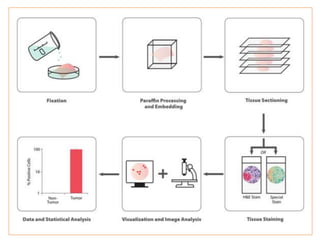
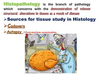
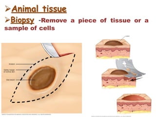
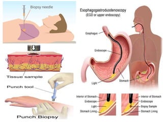
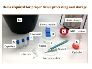
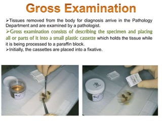
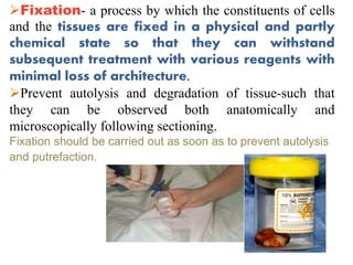
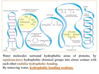
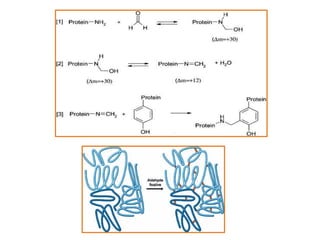
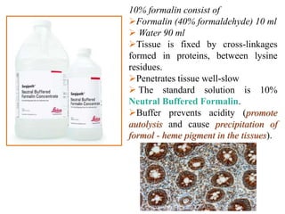
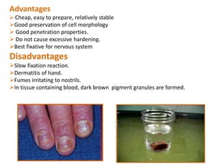
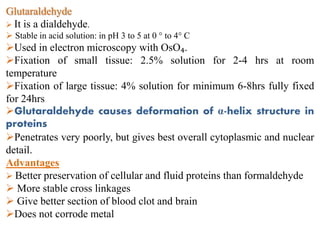
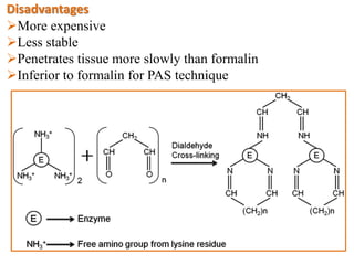
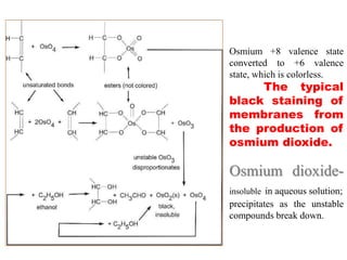
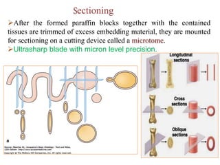
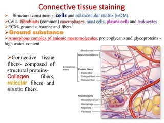
Histopathology is the branch of pathology
which concerns with the demonstration of minute
structural alterations in tissues as a result of disease
Sources for tissue study in Histology
Cadavers
Autopsy -Post-mortem examination
Animal tissue
Biopsy -Remove a piece of tissue or a
sample of cells
Scope
Useful in establishing the pathogenesis and
pathology of any disease caused by bacteria, virus,
chlamydia, rickettsia, mycoplasma, parasite, toxin,
poisons etc.
There are certain diseases in which histopathological examination of tissues
is the only alternative to diagnose the disease. e.g. Bovine
spongiform encephalopathy. The agent of this
disease takes a very long incubation period and very
difficult to isolate and there is no immune response and
inflammation in animal. Therefore, histopathology remains the only
alternative for confirmatory diagnosis.
In some cases, tissues from dead animals
are only available material for laboratory
diagnosis.
Histopathological examination
of tissues typically starts with
surgery or biopsy.
Collection of Samples:
Small piece of tissue (as early as possible)
Piece is removed with sharp knife
At the time of tissue collection, it should be kept in mind that the
representative tissue piece should include the part of lesion and a
part of normal tissue,
Tissues should be collected directly in the fixative and not in any
other pot or water.
The tissue pieces from hollow organs like intestines, oviduct etc.,
should be cut transversely.
Labelling:
Tissue is accompanied by a tag or label, bearing the
lab number given to specimen at start, through all
stages.
Sample collection and preservation
Items required for proper tissue processing and storage.
Tissues are saved in different cassettes having different color
Tissue specimens received
in the surgical pathology
laboratory -lists the patient
information and history
along with a description
of the site of origin.
The specimens are
accessioned by giving them
a number that will identify
each specimen for each
patient.
Tissues removed from the body for diagnosis arrive in the Pathology
Department and are examined by a pathologist.
Gross examination consists of describing the specimen and placing
all or parts of it into a small plastic cassette which holds the tissue while
it is being processed to a paraffin block.
Initially, the cassettes are placed into a fixative.
Fixation- a process by which the constituents of cells
and the tissues are fixed in a physical and partly
chemical state so that they can withstand
subsequent treatment with various reagents with
minimal loss of architecture.
Prevent autolysis and degradation of tissue-such that
they can be observed both anatomically and
microscopically following sectioning.
Fixation should be carried out as soon as to prevent autolysis
and putrefaction.
AIM AND EFFECTS OF FIXATION:
Rapid and even penetration.
To preserve cells and tissues in a life like manner
as possible.
Stabilize labile elements.
Must be rigid to allow sectioning.
Must allow staining.
Optical contrast must be induced
for morphological examination.
Allow long storage of tissues.
METHODS OF FIXATION
Heat fixation (physical fixation):
Dried smear- at room temperature,
Passed through flame of Bunsen burner- kills and adheres the
tissue to the slide.
Increasing temperature accelerates the process of fixation.
Excessive heat, particularly if -prolonged, can damage cells and
cause substantial shrinkage and hardening of the specimen.
Slide warmer Microincinerator Chemical fixation
The simplest form of fixation is heat.
Boiling an egg precipitates the proteins and on cutting, the yolk and egg white
can be identified separately.
Each component is less soluble in water after heat fixation than the same
component of a fresh egg.
In histopathology, heat is primarily used to accelerate other forms of
fixation as well as the steps of tissue processing.
Microwave fixation:
Principle
Exposure to electromagnetic fields (microwave)
Oscillation of water molecules and protein at 2450
times per second induces kinetic motion.
Instantaneous and uniform heat
Microwaves- little effect on tissue beyond a depth of 4 cm.
Control of the following parameters is important to the microwave
fixation method:
Specimens with one dimension less than 1 cm;
Irradiation temperatures lower than 50 degrees ⁰C;
Irradiation time less than 50 s;
Two ways of microwave technology is used for
tissue fixation-
Fresh tissue, placed in isotonic solution- irradiated to produce
primary fixation -“microwave fixation or microwave
stabilization. No chemical fixative is used.
Specimens can be placed in buffered formalin or some other
fixative and, at a later stage microwaved to assist the fixative action
of the fixing agent (referred to as ‘microwave-assisted fixation”).
Microwave-assisted fixation is much more commonly used than
primary microwave fixation.
Low toxicity fixatives containing glyoxal, have been
developed for use in microwave-assisted fixation.
Microwaving carried out while the tissue is
in fixative-hazard from toxic fumes!
Perfusion Fixation (chemical):
The fixative is injected into the heart with the injection volume. The
fixative spreads through the entire body.
This has the advantage of preserving perfect morphology, but the
disadvantages that the subject dies and the cost is high.
I
N
V
I
V
O
Immersion (chemical):
It is the most routinely used method, performed by placing
small pieces of tissue into a relatively large volume of
fixative (minimum 10 times of tissue volume).
Characteristics Of A
Fixative
Support high quality and consistent staining with (H&E) both
initially and after storage of the paraffin blocks.
Prevent short- and long-term destruction of the micro-architecture of
the tissue by stopping the activity of catabolic enzymes and hence
autolysis,.
Destruction of infectious agents, -maintain tissue and cellular
integrity.
Important to have good toxicological and flammability profiles
that permit the safe use of the fixative.
Versatile
Preserve small & large specimens and support histochemical,
immuno-histochemical, in situ hybridization and other specialized
procedures.
Penetrate and fix tissues rapidly, -shelf life of at least one year.
Readily disposable/recyclable and support long-term tissue storage
Cost effective.
Non-toxic and non-allergenic
Chemical fixation utilizes organic or non-organic solutions
to maintain adequate morphological preservation.
Cellular architecture maintained- lipoproteins and fibrous
proteins (collagen)
Coagulation of proteins-makes them
insoluble.
Coagulant fixatives -cytoplasmic flocculation
Not useful -ultrastructural analysis.
Commonly used (e.g. 50-60% ethanol, 80% methanol).
Remove and replace free water from tissue.
Coagulant fixatives
Molecules of water participate in hydrogen bonding
Removal of water destabilizes hydrogen
bonding.
Disruption the tertiary structure of proteins-
denaturation- changes their physical properties-insolubility and
loss of function
Water molecules surround hydrophobic areas of proteins, by
repulsion-force hydrophobic chemical groups into closer contact with
each other-stabilize hydrophobic bonding.
By removing water, hydrophobic bonding weakens.
Other types of coagulant fixative
Acidic coagulants such as picric acid and trichloroacetic acid
Change the charges on the ionizable side chains of protein- disruption
electrostatic and hydrogen bonding.
Trichloroacetic acid (Cl3CCOOH) can penetrate hydrophobic
domains of proteins and the anion produced reacts with charged
amine groups- precipitates proteins.
 Picric acid forms salts with basic groups of proteins-
proteins coagulate
Fixation by picric acid produces brighter staining.
Cross-linking fixatives
‘Covalent additive fixatives'.
Examples; Formaldehyde, Glutaraldehyde and other
aldehydes. e.g. Chloral hydrate and Glyoxal.
Metal salts such as Mercuric and Zinc
chloride. and other metallic compounds
such as Osmium tetroxide.
Formaldehyde
Powerful reducing agent.
 Most common fixative -fixation of biopsy specimen.
 Formalin: 40% formaldehyde in water.
Forms methylene bridges between protein molecules.
Method
4mm block - 8hrs at room temperature
4mm block - 2hrs at 45°C
In aqueous solution formaldehyde forms methylene hydrate
a methylene glycol as the first step in fixation.
Methylene hydrate reacts with several side chains of proteins
to form reactive hydroxymethyl side groups
10% formalin consist of
Formalin (40% formaldehyde) 10 ml
 Water 90 ml
Tissue is fixed by cross-linkages
formed in proteins, between lysine
residues.
Penetrates tissue well-slow
 The standard solution is 10%
Neutral Buffered Formalin.
Buffer prevents acidity (promote
autolysis and cause precipitation of
formol - heme pigment in the tissues).
Advantages
 Cheap, easy to prepare, relatively stable
Good preservation of cell morphology
 Good penetration properties.
 Do not cause excessive hardening.
Best fixative for nervous system
Disadvantages
Slow fixation reaction.
Dermatitis of hand.
Fumes irritating to nostrils.
In tissue containing blood, dark brown pigment granules are formed.
Glutaraldehyde
 It is a dialdehyde.
 Stable in acid solution: in pH 3 to 5 at 0 ° to 4° C
Used in electron microscopy with OsO₄.
Fixation of small tissue: 2.5% solution for 2-4 hrs at room
temperature
Fixation of large tissue: 4% solution for minimum 6-8hrs fully fixed
for 24hrs
Glutaraldehyde causes deformation of α-helix structure in
proteins
Penetrates very poorly, but gives best overall cytoplasmic and nuclear
detail.
Advantages
 Better preservation of cellular and fluid proteins than formaldehyde
 More stable cross linkages
 Give better section of blood clot and brain
Does not corrode metal
Disadvantages
More expensive
Less stable
Penetrates tissue more slowly than formalin
Inferior to formalin for PAS technique
Reacts with ammonium salts, amines, amides, amino acids, and
sulfydryl groups, by an additive reaction and hardens tissues.
It is especially reactive with cysteine forming a
dimercaptide.
Mercury-based fixatives -toxic -handle with care!.
penetrate slowly-specimens must be thin.
A potential replacement for mercuric chloride is zinc sulfate.
Advantages:
 Better staining of nuclei and connective tissue.
 Give best results with metachromatic staining
2(RSH) + HgCl₂ Dimercaptide-Hg²++2H++2Cl‾
Mercuric chloride
Osmium tetroxide (Os0₄)
Toxic volatile solid
Soluble in water as well as non-polar solvents
React with hydrophilic and hydrophobic sites -side chains of
proteins, causing cross-linking.
The reactive sites include sulfydryl, phenolic, hydroxyl, carboxyl,
amide, and heterocyclic groups.
OsO₄-interacts with nucleic acids,---- (the 2,3-glycol moiety in
terminal ribose groups and the 5-6 double bond of thymine).
Nuclei fixed in OsO₄ and dehydrated with alcohol-show clumping
of DNA.
Prevented by prefixation- potassium permanganate / postfixation
with uranyl acetate, or by adding calcium ions and tryptophan during
fixation.
Osmium tetroxide solution + buffers, -standard
fixative for electron microscopy.
Reaction of osmium with unsaturated bonds within lipids and
phospholipids.
Used as stain -blackens fat-various lipid-containing materials such
as the myelin sheaths (galactocerebroside) of nerve fiber.
Osmium +8 valence state
converted to +6 valence
state, which is colorless.
The typical
black staining of
membranes from
the production of
osmium dioxide.
Osmium dioxide-
insoluble in aqueous solution;
precipitates as the unstable
compounds break down.
TISSUE PROCESSING
It is impractical to place the entire organ under a
routine light microscope for study- Too large, but also
opaque-impossible to examine its micro-components.
A small portion -specific tissue or organ -excised
from a given organ and processed for microscopic
analysis.
Distortions, and loss of
components due to the preparation
process are almost always present.
Washing of the Tissue
Under running tap water overnight (12 hours) remove
excessive fixative solution.
Once fixed, the tissue must be treated to allow the cutting of the
thin sections required for viewing under the microscope.
The procedures designed to prepare the tissue for sectioning are
collectively known as tissue processing.
First, the sample is dehydrated by immersion in a series of aqueous
alcohol solutions gradually moving to pure alcohol.
The tissue is then soaked in an appropriate solvent to remove the
alcohol.
Finally the tissue is embedded in paraffin wax, which enables the
cutting of sections of between 3 and 10 microns thickness.
Tissues embedded in paraffin, can be sectioned at anywhere from 3
to 10 microns. The technique of getting fixed tissue into paraffin is
called tissue processing.
The main steps in this process are dehydration and clearing.
Most fixatives are aqueous,
Necessary to prepare the
tissue for embedding in non-
aqueous media like paraffin.
Large fraction of tissue–
water.
Collected tissues, once fixed,
are then dehydrated in
graded solutions
of alcohol or other
dehydrating agent.
Graded series of alcohol
baths, beginning with 50%
alcohol -progressing in graded
steps to 100% alcohol, are
used (dehydration),
Alcohol penetrates tissue
quickly and the water is
replaced with alcohol
,
Tissue dehydration
The next step is "clearing" -removal of the dehydrant
with a substance that will be miscible with the embedding
medium (paraffin).
Clearing agent is Xylene, Chloroform, Methyl salicylate
, Clearite (long chain aliphatic hydrocarbons)
Most tissue processing is done using automated machines that carry
out the steps automatically.
Tissues coming off a tissue processor are in a plastic box ready for
the embedding stage
It is the process by which tissues are
surrounded by a medium such as agar,
gelatin, or wax which when solidified will
provide sufficient external
support during sectioning.
Tissue is impregnated with melted wax.
the tissue is placed in a suitable container
of melted paraffin until it is completely
infiltrated,
Melted wax occupies spaces formerly
occupied by water.
Alternatives to paraffin include-Methyl
Methacrylate, Glycol Methacrylate,
Araldite, and Epon.
Impregnation/Infiltration/Embedding
After the formed paraffin blocks together with the contained
tissues are trimmed of excess embedding material, they are mounted
for sectioning on a cutting device called a microtome.
Ultrasharp blade with micron level precision.
Sectioning
Paraffin blocks can be sectioned with high-carbon steel blades.
Plastic blocks (methacrylate, araldite, or epon) are sectioned with glass or
diamond knives.
A glass knife can section down to about 0.1 micron.
Ultrathin sections for electron microscopy (below 100 nm) are best done with a
diamond knife
They are then picked up on a
glass slide.
Heated in oven-15 minutes to
help the section adhere to the
slide.
To preserve antigenicity
(immunostaining)-
adhesive-coated slides - Starch,
Albumin, Resins and
combinations thereof
 The adhered sections are then
ready for further processing.
Frozen Sections
The piece(s) of tissue-snap frozen in a cryogenic liquid or cold
environment (-20 to -70 Celsius).
Freezing -tissue solid -section with microtome.
Frozen sections -cryostat.
Cryostat -refrigerated box containing a microtome.
Temperature --20 to -30 Celsius.
Tissue sections are cut and picked up on a glass slide.
The sections are now ready for staining.
De-paraffinization
Embedding process - reversed to De-paraffinize wax out
of the tissue - allowing water soluble dyes to penetrate the
sections.
Before any staining, the slides are
"DE-PARAFFINIZED" by running them through
xylene (or substitutes) to alcohols
No staining can be done on
tissues containing paraffin
Sections prepared-colourless - different components cannot be visualised.
Staining- artificial coloration of a substance to facilitate examination by
the use of a colored organic molecule called dye.
The purpose of staining is that of
To outline the tissue and cellular components
To identify tissue
To establish the presence or absence of disease processes
 Staining provides visual contrast between the constituent parts of
a tissue section.
The depth of colouration is affected by chemical affinity, density, and
permeability.
Dyes -stain tissue components more or less selectively-form salt
linkages with oppositely charged tissues
BASIC PRINCIPLES OF STAINING
Dyes -colored organic compounds able to selectively bind to tissue
components.
CHROMOPHORE-a part of the molecule capable of
absorbing light strongly at certain wavelength and transmitting or
reflecting at others.
eg; C=C, C=O, C=N, N=N, N=O, NO₂.
AUXOCHROMES -ionizable groups -attached to the chromophore
modifying the ability of chromophore to absorb light, altering the
wavelength or the absorption intensity.
Acidic-COOH, -OH, -SO₃H, and
Basic- primary, secondary and tertiary amines (-NH2, -NHR,
-NR₂ ).
Histological staining -use of dyes to highlight specific intra- or
extracellular elements within tissue.
Human eye responds to wavelengths of light between 400 and 700
nanometers. (white light).
Absorption of light energy -promotes el‾ to a higher energy
level.
The energy absorbed can be re-emitted at a longer wavelength (fluorescence),
or dissipated as heat (simple absorbance).
Chemical interactions
Tissue-dye binding. Ionic bond or
electrostatic
interaction-attractions
between opposite charges of
dye and tissue.
Covalent bonds-
Eg; Schiff's reagent-
covalently attaches to
aldehydes in carbohydrate
identification by
Periodic Acid- Schiff staining
(PAS).
Hydrogen bonds -produced between hydrogen and a highly
electronegative atom, usually O₂ or N₂.
Hydrophobic interactions -maintain the dye in the tissue by means the
exclusion of water from hydrophobic regions, stabilizing the two groups
involved.
Van der Waals
Following are some terms related to dyes:
Mordants - A mordanting substance is considered part of the
stain, and in this way it may change the reaction of the stain. For
example, hematoxylin is an acid, but as it is almost always used in
conjunction with alum or iron (the mordant) it becomes a basic stain.
Amphophilic - It is a term used to indicate that the tissue stains
with both the basic and the acidic dyes.
Neutrophilic - No special affinity for either the basic or acidic
components of a dye.
Metachromasia - Production of a color during staining which is
different from the original color of the stain.
Toluidine Blue, Bismarck brown, Crystal violet, Cresyl Violet,
Safranin, Acid Fuschin
Basic and Acid Dyes
Basic dyes are cationic.
They form salts with tissue anions, -phosphate groups of
nucleic acids-sulfate groups of glycosaminoglycans.
Basophilic-designates the components of a cell or tissue, which
take up the basic stain rather than the acid stain of a
combination.
Nuclei are basophilic.
Acid dyes are anionic.
They form salts with cationic groups in cells and tissues -ionized
amino groups of proteins.
Eosinophilic components are cationic compounds that have an
affinity for that acid dye.
The cytoplasm is usually acidophilic.
H&E stain - preliminary stain applied to the tissue sections
Hematoxylin-Eosin (H and E) -widely used staining to identify nucleus
and cytoplasm in cells.
Hematoxylin (basic dye), -stains acidic structures –nucleus-(DNA), ribosomes and
rough endoplasmic reticulum( RNA).
Hematoxylin - natural compound –(logwood) Haematoxylum campechianum.
Extract-oxidized hematein, (active staining component)-Ripening
Hematoxylin staining requires-use of mordant (most commonly aluminum salts)
and stains the nuclear components of cells a dark blue.
Hematoxylin used in combination with eosin because eosin stains cytoplasmic
organelles -shades of pink, red or orange.
broad range of morphological information.
Classification
Stains for carbohydrates
Nucleic acid stains
Lipid stains
Connective tissue stains
Stains for microorganisms
Stains for pigments and minerals
Periodic Acid-Schiff (PAS) staining, one of the most
useful histological stains.
Presence of an adjacent pair of hydroxyl groups
in sugars, which can be oxidized with periodic acid
yielding two aldehyde groups.
In the next step these groups are detected with the chromogenic
Schiff’s reagent, which covalently combines with the aldehydes
to produce a red-purple compound
PAS strongly stains glycogen and certain types of
mucins, mucoprotein, glycoprotein, as well as fungi.
Identification -basement membranes due to the sugar moieties of its
proteoglycans.
Glycogen- normally found in liver, endocervix and muscle;
Pathologically present in carcinomas of liver,
kidney, pancreas and bladder, glycogen
storage disease
Glycogen and mucins: magenta
Nuclei: blue
The most common stains -lipids -Sudan dyes (Sudan
black, Sudan III, Sudan IV) and also Oil red O.
Oil red -identify the most hydrophobic lipids, while SUDAN BLACK is able to
stain these lipids in addition to other less hydrophobic such as phospholipids and
sphingomyelins.
Sudan dyes-study adipogenesis in tissues, Cardiovascular diseases
Lipid stains -useful to evaluate - effect of diets, clinical
drugs and herbal medicine in evolution of atherosclerotic
lesions.
DNA - Feulgen stain
Aldehyde groups react with Schiff’s reagent resulting in a purple staining.
Feulgen staining has been used in the past to demonstrate the banded karyotype
of mouse,
Feulgen to detect nuclear and cytogenetic abnormalities such as chromatin
condensation, karyorrhexis, presence of vacuoles
Combination Methyl green-Pyronin Y
Methyl green is specific for DNA, staining green the nuclei,
while Pyronin Y is specific for RNA staining red the
nucleoli.
Connective tissue staining
 Structural constituents; cells and extracellular matrix (ECM).
Cells- fibroblasts (common) macrophages, mast cells, plasma cells and leukocytes
ECM- ground substance and fibers.
Ground substance
Amorphous complex of anionic macromolecules, proteoglycans and glycoproteins -
high water content.
Connective tissue
fibers- composed of
structural proteins-
Collagen fibers,
reticular fibers and
elastic fibers.
STAINING
Multi-step (Masson's and Mallory's Trichrome)
One-step (Gomori’s Trichrome)
The trichrome stain uses two or more acid dyes (multi-step) in
conjunction with a polyacid (molybdophosphoric or tungstophosphoric
acids) to differentiate two basic components in contrasting colors,
commonly to demonstrate collagen fibers in contrast to smooth muscle .
Highlight the supporting collagen-sections from a variety of organs.
Helps to determine the pattern of tissue injury.
Trichrome will also aid in identifying normal structures, such as connective tissue
capsules of organs, the lamina propria of gastrointestinal tract, and the
bronchovascular structures in lungs.
Steps
Nuclear stain-Hematoxylin
Erythrocytes stain (optional)
Plasma stain with first acid dye-stain all the tissue
Displacement with poly-acid-displace the plasma stain from collagen
Fiber stain with second acid dye-displace the poly acid and deeply
stain collagen without replacing the red stain
Masson's
and
Mallory's
Trichrome
Collagen
fibres-
blue-green
Cytoplasm-
pale red
Nucleus-red
Glomerulus and
surrounding tubules.
An elastic tissue stain helps to outline arteries, because arteries, and the aorta,
contain elastic fibers.
The Van Gieson method for elastic fibers provides good contrast.
Verhoeff-Van Gieson staining
Weigert’s resorcin-Fuchsin staining
Orcein staining
The first step in both Verhoeff and Weigert’s stains -oxidative treatment with
iodine in Verhoeff stain and
Permanganate in Weigert’s stain -convert the disulfide bridges
into anionic sulfonic acid derivatives (basophilic) - rupture of disulfide bridges,
(disulfide bridges keep -elastic fibers highly cross-linked).
Basophilic- react with cationic dyes- Hematoxylin in Verhoeff stain and Basic
Fuchsin in Weigert’s stain
The final step is the counterstain, usually carried out with Van
Gieson (mixture of picric acid and acid fuchsin) or Iron
Hematoxylin.
Coarse fibers are intensely stained, but the staining of the fine fibers
may be less than satisfactory.
Asbestosis disease,
The study of granuloma annulare (benign skin lesion)
Elastic fibers—blue/black
Reticular fibers, -extremely thin, (0.5-2.0 µm)-
Cannot be visualized by regular staining
Silver impregnation.
Gordon and Sweet staining
Staining principle is based on formation of colloidal metallic silver.
After exposure to silver-external reducing agent needs is applied to complete the
reaction, (Argyrophilic reaction),
RETICULIN STAINING
Steps
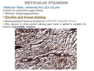
Oxidation (converts sugars -OH to –CHO), by phosphomolybdic acid or
KMnO₄.
Sensitization- treated with Ferric ammonium sulfate.
(Reticulin fibers- little affinity for silver)
Impregnation -with AgNO₃ or [Ag(NH₃)₂]+,
Sensitized sites accept silver.
Reduction- tissue sections-uncolored-reducing agent HCHO
HCHO + Ag -complex HCOOH + Ag (s) (brown-
black)
Contrasting
Ag deposits (brown-black) ----- AuCl₃ (purple-black)
Removal of by adding sodium thiosulfate.
Counterstain- nuclear fast red.
Study architecture of parenchymal organs
Study benign and malignant bone marrow disorders
Calcium
Bone mineralization,
Muscle and blood vessel contraction,
Nerve conduction,
Clot formation
Regulation of enzymes activity.
Bones and teeth (hydroxyapatite),
Abnormal deposition -lung or lymph nodes infections,
Necrotic tissue in lesion of atherosclerosis
Nephrocalcinosis among others.
Stains used Von Kossa and Alizarin red S.
Alizarin red S, detects small amounts of calcium present
(sensitive).
It is a chelating stain -anionic dye binds -calcium, -also able to bind to
other metals like Mn, Ba.
Incubation with Alizarin red solution, -removal of excess dye
Quick dehydration in acetone.
Birefringent bright orange-red precipitate is formed.
Recognition of calcification in coronary arteries and
aorta,
Field of osteoporosis, osteoblast mineralization has
demonstrate the effect of drugs and hormones on
bone metabolism regulation.
Study mineralization processes,
Identification of articular cartilage calcifications
Nephrocalcinosis
Identification and determination of osteogenic differentiation
Endogenous pigment -Melanin
In the skin & substantia nigra in the brain,
Masson-Fontana silver and Schmorl’s ferric ferricyanide.
Masson-Fontana reaction -melanin is
argentaffin (reduce silver).
Tissue treated-ammoniacal silver solution (reducing
groups of the tissue convert silver ions into colloidal metallic silver, colored
brown-black)
Color intensification –AuCl₃ solution,
Counterstain -neutral red or nuclear fast red
Not completely specific for melanin
Demonstrate substances -similar reducing properties such as argentaffin cell
granules or lipofuschins.
 Malignant melanomas -benign tumors derived from melanocytes
Cytoplasm of
skin
keratinocytes
Coverslipping
The stained section on the slide must be covered with a thin piece plastic or glass -
protect the tissue from being scratched- to provide better
optical quality for viewing under the microscope, and
to preserve the tissue section for
years to come.
Apply a single drop of
mounting medium upon
tissue section.
Hold coverslip at 45º
allowing the drop to spread
along the edge of the slip.
Let go of slip and allow
medium to spread slowly.
Decalcification
Firm -not section properly with paraffin embedding
owing to the difference in densities between calcium and paraffin
Carried out between fixation and processing steps.
Calcified tissues-Bone
Immersion in solutions containing mineral acids, organic
acids, or EDTA are the predominant methods used.
Electrolysis (slow- not suited for routine daily use)
Strong mineral acids HNO₃,HCl are used dense cortical
bone because they will remove large quantities of calcium at a rapid
rate- damage cellular morphology- not recommended for delicate
tissues such as bone marrow.
Organic acids – CH₃COOH, HCOOH are better -bone marrow
and other soft tissues.
Organic acids act more slowly than mineral
acids
10% HCOOH is the best all-around decalcifier.
a)tooth before decalcification b)tooth after
decalcification with nitric acid
Analysis
Microscopy- LM, SEM, TEM, FM, STEM .
Immuno-fluorecence imaging .
CCD camera (charge-coupled device).
Point spectroscopy -Raman spectroscopy, FT-IR
spectroscopy, (IR) absorption spectroscopy.
Spectral imaging- Multi-spectral (MS) and
Hyper-spectral (HS) Imaging
Spectroscopic imaging (MRS)
2-D, 3-D graphs.
References
nationaldiagnostics.com
Bancroft's Theory and Practice of Histological Techniques By Christopher
Layton, John D. Bancroft Elsevier Health Sciences
Dyes and stains: From molecular structure to histological application;
Frontiers in Bioscience
Chemical Principles in Tissue Clearing and Staining Protocols for Whole-
Body Cell Profiling ; Cell and Developmental Biology
Histological Techniques: An Introduction for Beginners in Toxicology; By
Robert Maynard, Noel Downes, Brenda Finney Royal Society of Chemistry
Histopathological Image Analysis: Annual Review of Biomedical Engineering
Slideshare
Histopathological techniques -sectioning, STAINING, EMBEDDING, fixaton, microtomy,

Histopathological techniques -sectioning, STAINING, EMBEDDING, fixaton, microtomy,

  • 4.
    Histopathology is thebranch of pathology which concerns with the demonstration of minute structural alterations in tissues as a result of disease Sources for tissue study in Histology Cadavers Autopsy -Post-mortem examination
  • 5.
    Animal tissue Biopsy -Removea piece of tissue or a sample of cells
  • 8.
    Scope Useful in establishingthe pathogenesis and pathology of any disease caused by bacteria, virus, chlamydia, rickettsia, mycoplasma, parasite, toxin, poisons etc. There are certain diseases in which histopathological examination of tissues is the only alternative to diagnose the disease. e.g. Bovine spongiform encephalopathy. The agent of this disease takes a very long incubation period and very difficult to isolate and there is no immune response and inflammation in animal. Therefore, histopathology remains the only alternative for confirmatory diagnosis. In some cases, tissues from dead animals are only available material for laboratory diagnosis.
  • 9.
    Histopathological examination of tissuestypically starts with surgery or biopsy.
  • 10.
    Collection of Samples: Smallpiece of tissue (as early as possible) Piece is removed with sharp knife At the time of tissue collection, it should be kept in mind that the representative tissue piece should include the part of lesion and a part of normal tissue, Tissues should be collected directly in the fixative and not in any other pot or water. The tissue pieces from hollow organs like intestines, oviduct etc., should be cut transversely. Labelling: Tissue is accompanied by a tag or label, bearing the lab number given to specimen at start, through all stages. Sample collection and preservation
  • 11.
    Items required forproper tissue processing and storage.
  • 12.
    Tissues are savedin different cassettes having different color
  • 14.
    Tissue specimens received inthe surgical pathology laboratory -lists the patient information and history along with a description of the site of origin. The specimens are accessioned by giving them a number that will identify each specimen for each patient.
  • 15.
    Tissues removed fromthe body for diagnosis arrive in the Pathology Department and are examined by a pathologist. Gross examination consists of describing the specimen and placing all or parts of it into a small plastic cassette which holds the tissue while it is being processed to a paraffin block. Initially, the cassettes are placed into a fixative.
  • 18.
    Fixation- a processby which the constituents of cells and the tissues are fixed in a physical and partly chemical state so that they can withstand subsequent treatment with various reagents with minimal loss of architecture. Prevent autolysis and degradation of tissue-such that they can be observed both anatomically and microscopically following sectioning. Fixation should be carried out as soon as to prevent autolysis and putrefaction.
  • 19.
    AIM AND EFFECTSOF FIXATION: Rapid and even penetration. To preserve cells and tissues in a life like manner as possible. Stabilize labile elements. Must be rigid to allow sectioning. Must allow staining. Optical contrast must be induced for morphological examination. Allow long storage of tissues.
  • 20.
    METHODS OF FIXATION Heatfixation (physical fixation): Dried smear- at room temperature, Passed through flame of Bunsen burner- kills and adheres the tissue to the slide. Increasing temperature accelerates the process of fixation. Excessive heat, particularly if -prolonged, can damage cells and cause substantial shrinkage and hardening of the specimen. Slide warmer Microincinerator Chemical fixation
  • 21.
    The simplest formof fixation is heat. Boiling an egg precipitates the proteins and on cutting, the yolk and egg white can be identified separately. Each component is less soluble in water after heat fixation than the same component of a fresh egg. In histopathology, heat is primarily used to accelerate other forms of fixation as well as the steps of tissue processing.
  • 22.
    Microwave fixation: Principle Exposure toelectromagnetic fields (microwave) Oscillation of water molecules and protein at 2450 times per second induces kinetic motion. Instantaneous and uniform heat Microwaves- little effect on tissue beyond a depth of 4 cm.
  • 23.
    Control of thefollowing parameters is important to the microwave fixation method: Specimens with one dimension less than 1 cm; Irradiation temperatures lower than 50 degrees ⁰C; Irradiation time less than 50 s;
  • 24.
    Two ways ofmicrowave technology is used for tissue fixation- Fresh tissue, placed in isotonic solution- irradiated to produce primary fixation -“microwave fixation or microwave stabilization. No chemical fixative is used. Specimens can be placed in buffered formalin or some other fixative and, at a later stage microwaved to assist the fixative action of the fixing agent (referred to as ‘microwave-assisted fixation”). Microwave-assisted fixation is much more commonly used than primary microwave fixation. Low toxicity fixatives containing glyoxal, have been developed for use in microwave-assisted fixation. Microwaving carried out while the tissue is in fixative-hazard from toxic fumes!
  • 25.
    Perfusion Fixation (chemical): Thefixative is injected into the heart with the injection volume. The fixative spreads through the entire body. This has the advantage of preserving perfect morphology, but the disadvantages that the subject dies and the cost is high. I N V I V O
  • 26.
    Immersion (chemical): It isthe most routinely used method, performed by placing small pieces of tissue into a relatively large volume of fixative (minimum 10 times of tissue volume).
  • 27.
  • 28.
    Support high qualityand consistent staining with (H&E) both initially and after storage of the paraffin blocks. Prevent short- and long-term destruction of the micro-architecture of the tissue by stopping the activity of catabolic enzymes and hence autolysis,. Destruction of infectious agents, -maintain tissue and cellular integrity. Important to have good toxicological and flammability profiles that permit the safe use of the fixative. Versatile Preserve small & large specimens and support histochemical, immuno-histochemical, in situ hybridization and other specialized procedures. Penetrate and fix tissues rapidly, -shelf life of at least one year. Readily disposable/recyclable and support long-term tissue storage Cost effective. Non-toxic and non-allergenic
  • 29.
    Chemical fixation utilizesorganic or non-organic solutions to maintain adequate morphological preservation.
  • 30.
    Cellular architecture maintained-lipoproteins and fibrous proteins (collagen) Coagulation of proteins-makes them insoluble. Coagulant fixatives -cytoplasmic flocculation Not useful -ultrastructural analysis. Commonly used (e.g. 50-60% ethanol, 80% methanol). Remove and replace free water from tissue. Coagulant fixatives Molecules of water participate in hydrogen bonding Removal of water destabilizes hydrogen bonding. Disruption the tertiary structure of proteins- denaturation- changes their physical properties-insolubility and loss of function
  • 31.
    Water molecules surroundhydrophobic areas of proteins, by repulsion-force hydrophobic chemical groups into closer contact with each other-stabilize hydrophobic bonding. By removing water, hydrophobic bonding weakens.
  • 32.
    Other types ofcoagulant fixative Acidic coagulants such as picric acid and trichloroacetic acid Change the charges on the ionizable side chains of protein- disruption electrostatic and hydrogen bonding. Trichloroacetic acid (Cl3CCOOH) can penetrate hydrophobic domains of proteins and the anion produced reacts with charged amine groups- precipitates proteins.  Picric acid forms salts with basic groups of proteins- proteins coagulate Fixation by picric acid produces brighter staining.
  • 33.
    Cross-linking fixatives ‘Covalent additivefixatives'. Examples; Formaldehyde, Glutaraldehyde and other aldehydes. e.g. Chloral hydrate and Glyoxal. Metal salts such as Mercuric and Zinc chloride. and other metallic compounds such as Osmium tetroxide.
  • 34.
    Formaldehyde Powerful reducing agent. Most common fixative -fixation of biopsy specimen.  Formalin: 40% formaldehyde in water. Forms methylene bridges between protein molecules. Method 4mm block - 8hrs at room temperature 4mm block - 2hrs at 45°C In aqueous solution formaldehyde forms methylene hydrate a methylene glycol as the first step in fixation. Methylene hydrate reacts with several side chains of proteins to form reactive hydroxymethyl side groups
  • 36.
    10% formalin consistof Formalin (40% formaldehyde) 10 ml  Water 90 ml Tissue is fixed by cross-linkages formed in proteins, between lysine residues. Penetrates tissue well-slow  The standard solution is 10% Neutral Buffered Formalin. Buffer prevents acidity (promote autolysis and cause precipitation of formol - heme pigment in the tissues).
  • 37.
    Advantages  Cheap, easyto prepare, relatively stable Good preservation of cell morphology  Good penetration properties.  Do not cause excessive hardening. Best fixative for nervous system Disadvantages Slow fixation reaction. Dermatitis of hand. Fumes irritating to nostrils. In tissue containing blood, dark brown pigment granules are formed.
  • 38.
    Glutaraldehyde  It isa dialdehyde.  Stable in acid solution: in pH 3 to 5 at 0 ° to 4° C Used in electron microscopy with OsO₄. Fixation of small tissue: 2.5% solution for 2-4 hrs at room temperature Fixation of large tissue: 4% solution for minimum 6-8hrs fully fixed for 24hrs Glutaraldehyde causes deformation of α-helix structure in proteins Penetrates very poorly, but gives best overall cytoplasmic and nuclear detail. Advantages  Better preservation of cellular and fluid proteins than formaldehyde  More stable cross linkages  Give better section of blood clot and brain Does not corrode metal
  • 39.
    Disadvantages More expensive Less stable Penetratestissue more slowly than formalin Inferior to formalin for PAS technique
  • 40.
    Reacts with ammoniumsalts, amines, amides, amino acids, and sulfydryl groups, by an additive reaction and hardens tissues. It is especially reactive with cysteine forming a dimercaptide. Mercury-based fixatives -toxic -handle with care!. penetrate slowly-specimens must be thin. A potential replacement for mercuric chloride is zinc sulfate. Advantages:  Better staining of nuclei and connective tissue.  Give best results with metachromatic staining 2(RSH) + HgCl₂ Dimercaptide-Hg²++2H++2Cl‾ Mercuric chloride
  • 41.
    Osmium tetroxide (Os0₄) Toxicvolatile solid Soluble in water as well as non-polar solvents React with hydrophilic and hydrophobic sites -side chains of proteins, causing cross-linking. The reactive sites include sulfydryl, phenolic, hydroxyl, carboxyl, amide, and heterocyclic groups. OsO₄-interacts with nucleic acids,---- (the 2,3-glycol moiety in terminal ribose groups and the 5-6 double bond of thymine). Nuclei fixed in OsO₄ and dehydrated with alcohol-show clumping of DNA. Prevented by prefixation- potassium permanganate / postfixation with uranyl acetate, or by adding calcium ions and tryptophan during fixation. Osmium tetroxide solution + buffers, -standard fixative for electron microscopy.
  • 42.
    Reaction of osmiumwith unsaturated bonds within lipids and phospholipids. Used as stain -blackens fat-various lipid-containing materials such as the myelin sheaths (galactocerebroside) of nerve fiber.
  • 43.
    Osmium +8 valencestate converted to +6 valence state, which is colorless. The typical black staining of membranes from the production of osmium dioxide. Osmium dioxide- insoluble in aqueous solution; precipitates as the unstable compounds break down.
  • 44.
    TISSUE PROCESSING It isimpractical to place the entire organ under a routine light microscope for study- Too large, but also opaque-impossible to examine its micro-components. A small portion -specific tissue or organ -excised from a given organ and processed for microscopic analysis. Distortions, and loss of components due to the preparation process are almost always present.
  • 45.
    Washing of theTissue Under running tap water overnight (12 hours) remove excessive fixative solution. Once fixed, the tissue must be treated to allow the cutting of the thin sections required for viewing under the microscope. The procedures designed to prepare the tissue for sectioning are collectively known as tissue processing. First, the sample is dehydrated by immersion in a series of aqueous alcohol solutions gradually moving to pure alcohol. The tissue is then soaked in an appropriate solvent to remove the alcohol. Finally the tissue is embedded in paraffin wax, which enables the cutting of sections of between 3 and 10 microns thickness. Tissues embedded in paraffin, can be sectioned at anywhere from 3 to 10 microns. The technique of getting fixed tissue into paraffin is called tissue processing. The main steps in this process are dehydration and clearing.
  • 46.
    Most fixatives areaqueous, Necessary to prepare the tissue for embedding in non- aqueous media like paraffin. Large fraction of tissue– water. Collected tissues, once fixed, are then dehydrated in graded solutions of alcohol or other dehydrating agent. Graded series of alcohol baths, beginning with 50% alcohol -progressing in graded steps to 100% alcohol, are used (dehydration), Alcohol penetrates tissue quickly and the water is replaced with alcohol , Tissue dehydration
  • 47.
    The next stepis "clearing" -removal of the dehydrant with a substance that will be miscible with the embedding medium (paraffin). Clearing agent is Xylene, Chloroform, Methyl salicylate , Clearite (long chain aliphatic hydrocarbons)
  • 48.
    Most tissue processingis done using automated machines that carry out the steps automatically. Tissues coming off a tissue processor are in a plastic box ready for the embedding stage
  • 50.
    It is theprocess by which tissues are surrounded by a medium such as agar, gelatin, or wax which when solidified will provide sufficient external support during sectioning. Tissue is impregnated with melted wax. the tissue is placed in a suitable container of melted paraffin until it is completely infiltrated, Melted wax occupies spaces formerly occupied by water. Alternatives to paraffin include-Methyl Methacrylate, Glycol Methacrylate, Araldite, and Epon. Impregnation/Infiltration/Embedding
  • 56.
    After the formedparaffin blocks together with the contained tissues are trimmed of excess embedding material, they are mounted for sectioning on a cutting device called a microtome. Ultrasharp blade with micron level precision. Sectioning
  • 57.
    Paraffin blocks canbe sectioned with high-carbon steel blades. Plastic blocks (methacrylate, araldite, or epon) are sectioned with glass or diamond knives. A glass knife can section down to about 0.1 micron. Ultrathin sections for electron microscopy (below 100 nm) are best done with a diamond knife
  • 59.
    They are thenpicked up on a glass slide. Heated in oven-15 minutes to help the section adhere to the slide. To preserve antigenicity (immunostaining)- adhesive-coated slides - Starch, Albumin, Resins and combinations thereof  The adhered sections are then ready for further processing.
  • 60.
    Frozen Sections The piece(s)of tissue-snap frozen in a cryogenic liquid or cold environment (-20 to -70 Celsius). Freezing -tissue solid -section with microtome. Frozen sections -cryostat. Cryostat -refrigerated box containing a microtome. Temperature --20 to -30 Celsius. Tissue sections are cut and picked up on a glass slide. The sections are now ready for staining.
  • 61.
    De-paraffinization Embedding process -reversed to De-paraffinize wax out of the tissue - allowing water soluble dyes to penetrate the sections. Before any staining, the slides are "DE-PARAFFINIZED" by running them through xylene (or substitutes) to alcohols No staining can be done on tissues containing paraffin
  • 62.
    Sections prepared-colourless -different components cannot be visualised. Staining- artificial coloration of a substance to facilitate examination by the use of a colored organic molecule called dye. The purpose of staining is that of To outline the tissue and cellular components To identify tissue To establish the presence or absence of disease processes  Staining provides visual contrast between the constituent parts of a tissue section. The depth of colouration is affected by chemical affinity, density, and permeability. Dyes -stain tissue components more or less selectively-form salt linkages with oppositely charged tissues
  • 64.
    BASIC PRINCIPLES OFSTAINING Dyes -colored organic compounds able to selectively bind to tissue components. CHROMOPHORE-a part of the molecule capable of absorbing light strongly at certain wavelength and transmitting or reflecting at others. eg; C=C, C=O, C=N, N=N, N=O, NO₂. AUXOCHROMES -ionizable groups -attached to the chromophore modifying the ability of chromophore to absorb light, altering the wavelength or the absorption intensity. Acidic-COOH, -OH, -SO₃H, and Basic- primary, secondary and tertiary amines (-NH2, -NHR, -NR₂ ).
  • 65.
    Histological staining -useof dyes to highlight specific intra- or extracellular elements within tissue. Human eye responds to wavelengths of light between 400 and 700 nanometers. (white light). Absorption of light energy -promotes el‾ to a higher energy level. The energy absorbed can be re-emitted at a longer wavelength (fluorescence), or dissipated as heat (simple absorbance).
  • 66.
    Chemical interactions Tissue-dye binding.Ionic bond or electrostatic interaction-attractions between opposite charges of dye and tissue. Covalent bonds- Eg; Schiff's reagent- covalently attaches to aldehydes in carbohydrate identification by Periodic Acid- Schiff staining (PAS). Hydrogen bonds -produced between hydrogen and a highly electronegative atom, usually O₂ or N₂. Hydrophobic interactions -maintain the dye in the tissue by means the exclusion of water from hydrophobic regions, stabilizing the two groups involved. Van der Waals
  • 67.
    Following are someterms related to dyes: Mordants - A mordanting substance is considered part of the stain, and in this way it may change the reaction of the stain. For example, hematoxylin is an acid, but as it is almost always used in conjunction with alum or iron (the mordant) it becomes a basic stain. Amphophilic - It is a term used to indicate that the tissue stains with both the basic and the acidic dyes. Neutrophilic - No special affinity for either the basic or acidic components of a dye. Metachromasia - Production of a color during staining which is different from the original color of the stain. Toluidine Blue, Bismarck brown, Crystal violet, Cresyl Violet, Safranin, Acid Fuschin
  • 69.
    Basic and AcidDyes Basic dyes are cationic. They form salts with tissue anions, -phosphate groups of nucleic acids-sulfate groups of glycosaminoglycans. Basophilic-designates the components of a cell or tissue, which take up the basic stain rather than the acid stain of a combination. Nuclei are basophilic. Acid dyes are anionic. They form salts with cationic groups in cells and tissues -ionized amino groups of proteins. Eosinophilic components are cationic compounds that have an affinity for that acid dye. The cytoplasm is usually acidophilic.
  • 70.
    H&E stain -preliminary stain applied to the tissue sections Hematoxylin-Eosin (H and E) -widely used staining to identify nucleus and cytoplasm in cells. Hematoxylin (basic dye), -stains acidic structures –nucleus-(DNA), ribosomes and rough endoplasmic reticulum( RNA). Hematoxylin - natural compound –(logwood) Haematoxylum campechianum. Extract-oxidized hematein, (active staining component)-Ripening Hematoxylin staining requires-use of mordant (most commonly aluminum salts) and stains the nuclear components of cells a dark blue. Hematoxylin used in combination with eosin because eosin stains cytoplasmic organelles -shades of pink, red or orange. broad range of morphological information.
  • 71.
    Classification Stains for carbohydrates Nucleicacid stains Lipid stains Connective tissue stains Stains for microorganisms Stains for pigments and minerals
  • 73.
    Periodic Acid-Schiff (PAS)staining, one of the most useful histological stains. Presence of an adjacent pair of hydroxyl groups in sugars, which can be oxidized with periodic acid yielding two aldehyde groups. In the next step these groups are detected with the chromogenic Schiff’s reagent, which covalently combines with the aldehydes to produce a red-purple compound PAS strongly stains glycogen and certain types of mucins, mucoprotein, glycoprotein, as well as fungi. Identification -basement membranes due to the sugar moieties of its proteoglycans. Glycogen- normally found in liver, endocervix and muscle; Pathologically present in carcinomas of liver, kidney, pancreas and bladder, glycogen storage disease
  • 74.
    Glycogen and mucins:magenta Nuclei: blue
  • 75.
    The most commonstains -lipids -Sudan dyes (Sudan black, Sudan III, Sudan IV) and also Oil red O. Oil red -identify the most hydrophobic lipids, while SUDAN BLACK is able to stain these lipids in addition to other less hydrophobic such as phospholipids and sphingomyelins. Sudan dyes-study adipogenesis in tissues, Cardiovascular diseases Lipid stains -useful to evaluate - effect of diets, clinical drugs and herbal medicine in evolution of atherosclerotic lesions.
  • 76.
    DNA - Feulgenstain Aldehyde groups react with Schiff’s reagent resulting in a purple staining. Feulgen staining has been used in the past to demonstrate the banded karyotype of mouse, Feulgen to detect nuclear and cytogenetic abnormalities such as chromatin condensation, karyorrhexis, presence of vacuoles Combination Methyl green-Pyronin Y Methyl green is specific for DNA, staining green the nuclei, while Pyronin Y is specific for RNA staining red the nucleoli.
  • 77.
    Connective tissue staining Structural constituents; cells and extracellular matrix (ECM). Cells- fibroblasts (common) macrophages, mast cells, plasma cells and leukocytes ECM- ground substance and fibers. Ground substance Amorphous complex of anionic macromolecules, proteoglycans and glycoproteins - high water content. Connective tissue fibers- composed of structural proteins- Collagen fibers, reticular fibers and elastic fibers.
  • 78.
    STAINING Multi-step (Masson's andMallory's Trichrome) One-step (Gomori’s Trichrome) The trichrome stain uses two or more acid dyes (multi-step) in conjunction with a polyacid (molybdophosphoric or tungstophosphoric acids) to differentiate two basic components in contrasting colors, commonly to demonstrate collagen fibers in contrast to smooth muscle . Highlight the supporting collagen-sections from a variety of organs. Helps to determine the pattern of tissue injury. Trichrome will also aid in identifying normal structures, such as connective tissue capsules of organs, the lamina propria of gastrointestinal tract, and the bronchovascular structures in lungs. Steps Nuclear stain-Hematoxylin Erythrocytes stain (optional) Plasma stain with first acid dye-stain all the tissue Displacement with poly-acid-displace the plasma stain from collagen Fiber stain with second acid dye-displace the poly acid and deeply stain collagen without replacing the red stain
  • 79.
  • 81.
    An elastic tissuestain helps to outline arteries, because arteries, and the aorta, contain elastic fibers. The Van Gieson method for elastic fibers provides good contrast. Verhoeff-Van Gieson staining Weigert’s resorcin-Fuchsin staining Orcein staining
  • 82.
    The first stepin both Verhoeff and Weigert’s stains -oxidative treatment with iodine in Verhoeff stain and Permanganate in Weigert’s stain -convert the disulfide bridges into anionic sulfonic acid derivatives (basophilic) - rupture of disulfide bridges, (disulfide bridges keep -elastic fibers highly cross-linked). Basophilic- react with cationic dyes- Hematoxylin in Verhoeff stain and Basic Fuchsin in Weigert’s stain The final step is the counterstain, usually carried out with Van Gieson (mixture of picric acid and acid fuchsin) or Iron Hematoxylin. Coarse fibers are intensely stained, but the staining of the fine fibers may be less than satisfactory. Asbestosis disease, The study of granuloma annulare (benign skin lesion)
  • 83.
  • 84.
    Reticular fibers, -extremelythin, (0.5-2.0 µm)- Cannot be visualized by regular staining Silver impregnation. Gordon and Sweet staining Staining principle is based on formation of colloidal metallic silver. After exposure to silver-external reducing agent needs is applied to complete the reaction, (Argyrophilic reaction), RETICULIN STAINING
  • 85.
    Steps Oxidation (converts sugars-OH to –CHO), by phosphomolybdic acid or KMnO₄. Sensitization- treated with Ferric ammonium sulfate. (Reticulin fibers- little affinity for silver) Impregnation -with AgNO₃ or [Ag(NH₃)₂]+, Sensitized sites accept silver. Reduction- tissue sections-uncolored-reducing agent HCHO HCHO + Ag -complex HCOOH + Ag (s) (brown- black) Contrasting Ag deposits (brown-black) ----- AuCl₃ (purple-black) Removal of by adding sodium thiosulfate. Counterstain- nuclear fast red. Study architecture of parenchymal organs Study benign and malignant bone marrow disorders
  • 86.
    Calcium Bone mineralization, Muscle andblood vessel contraction, Nerve conduction, Clot formation Regulation of enzymes activity. Bones and teeth (hydroxyapatite), Abnormal deposition -lung or lymph nodes infections, Necrotic tissue in lesion of atherosclerosis Nephrocalcinosis among others. Stains used Von Kossa and Alizarin red S.
  • 87.
    Alizarin red S,detects small amounts of calcium present (sensitive). It is a chelating stain -anionic dye binds -calcium, -also able to bind to other metals like Mn, Ba. Incubation with Alizarin red solution, -removal of excess dye Quick dehydration in acetone. Birefringent bright orange-red precipitate is formed. Recognition of calcification in coronary arteries and aorta, Field of osteoporosis, osteoblast mineralization has demonstrate the effect of drugs and hormones on bone metabolism regulation. Study mineralization processes, Identification of articular cartilage calcifications Nephrocalcinosis Identification and determination of osteogenic differentiation
  • 88.
    Endogenous pigment -Melanin Inthe skin & substantia nigra in the brain, Masson-Fontana silver and Schmorl’s ferric ferricyanide. Masson-Fontana reaction -melanin is argentaffin (reduce silver). Tissue treated-ammoniacal silver solution (reducing groups of the tissue convert silver ions into colloidal metallic silver, colored brown-black) Color intensification –AuCl₃ solution, Counterstain -neutral red or nuclear fast red Not completely specific for melanin Demonstrate substances -similar reducing properties such as argentaffin cell granules or lipofuschins.  Malignant melanomas -benign tumors derived from melanocytes
  • 89.
  • 90.
    Coverslipping The stained sectionon the slide must be covered with a thin piece plastic or glass - protect the tissue from being scratched- to provide better optical quality for viewing under the microscope, and to preserve the tissue section for years to come. Apply a single drop of mounting medium upon tissue section. Hold coverslip at 45º allowing the drop to spread along the edge of the slip. Let go of slip and allow medium to spread slowly.
  • 91.
  • 92.
    Firm -not sectionproperly with paraffin embedding owing to the difference in densities between calcium and paraffin Carried out between fixation and processing steps. Calcified tissues-Bone Immersion in solutions containing mineral acids, organic acids, or EDTA are the predominant methods used. Electrolysis (slow- not suited for routine daily use) Strong mineral acids HNO₃,HCl are used dense cortical bone because they will remove large quantities of calcium at a rapid rate- damage cellular morphology- not recommended for delicate tissues such as bone marrow. Organic acids – CH₃COOH, HCOOH are better -bone marrow and other soft tissues. Organic acids act more slowly than mineral acids 10% HCOOH is the best all-around decalcifier.
  • 93.
    a)tooth before decalcificationb)tooth after decalcification with nitric acid
  • 94.
    Analysis Microscopy- LM, SEM,TEM, FM, STEM . Immuno-fluorecence imaging . CCD camera (charge-coupled device). Point spectroscopy -Raman spectroscopy, FT-IR spectroscopy, (IR) absorption spectroscopy. Spectral imaging- Multi-spectral (MS) and Hyper-spectral (HS) Imaging Spectroscopic imaging (MRS) 2-D, 3-D graphs.
  • 96.
    References nationaldiagnostics.com Bancroft's Theory andPractice of Histological Techniques By Christopher Layton, John D. Bancroft Elsevier Health Sciences Dyes and stains: From molecular structure to histological application; Frontiers in Bioscience Chemical Principles in Tissue Clearing and Staining Protocols for Whole- Body Cell Profiling ; Cell and Developmental Biology Histological Techniques: An Introduction for Beginners in Toxicology; By Robert Maynard, Noel Downes, Brenda Finney Royal Society of Chemistry Histopathological Image Analysis: Annual Review of Biomedical Engineering Slideshare