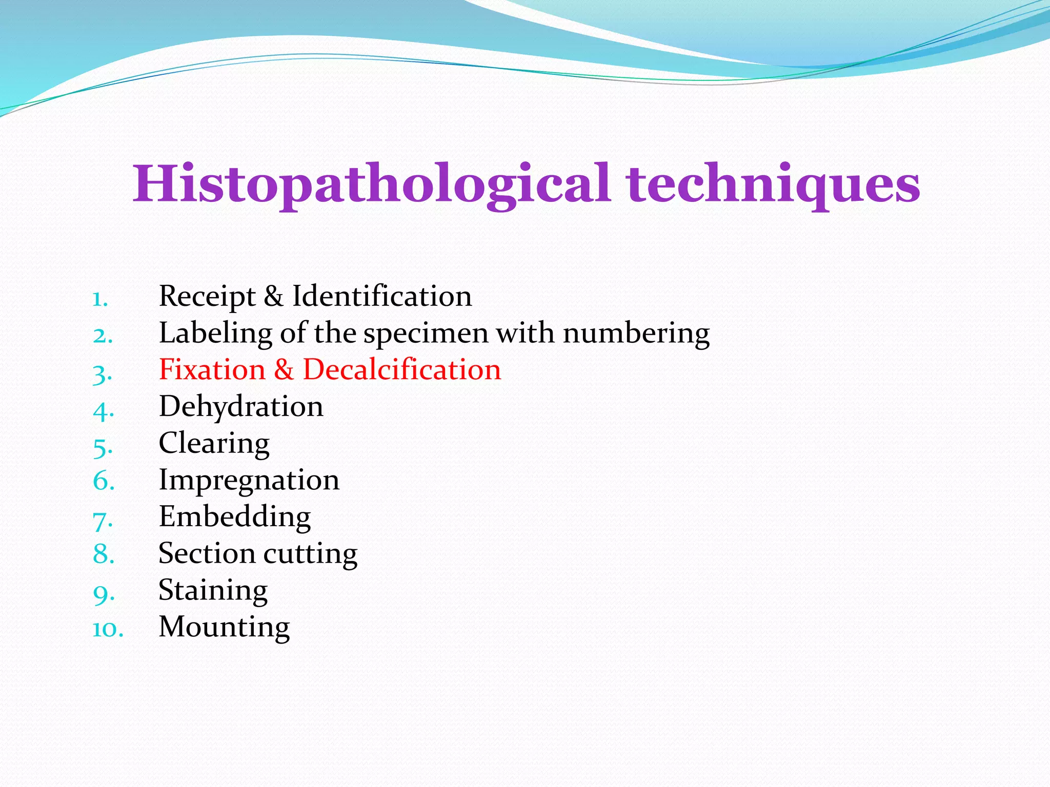
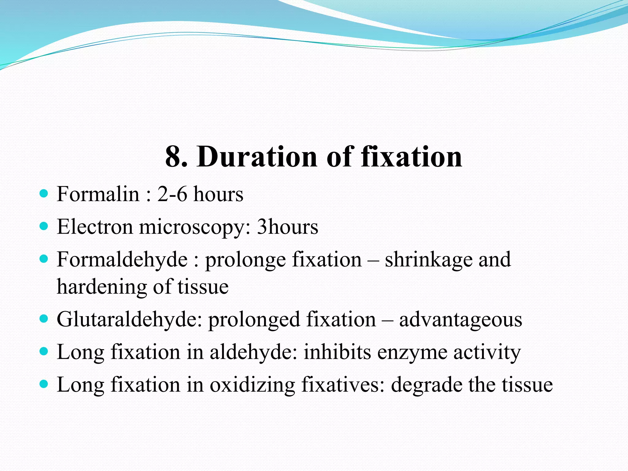
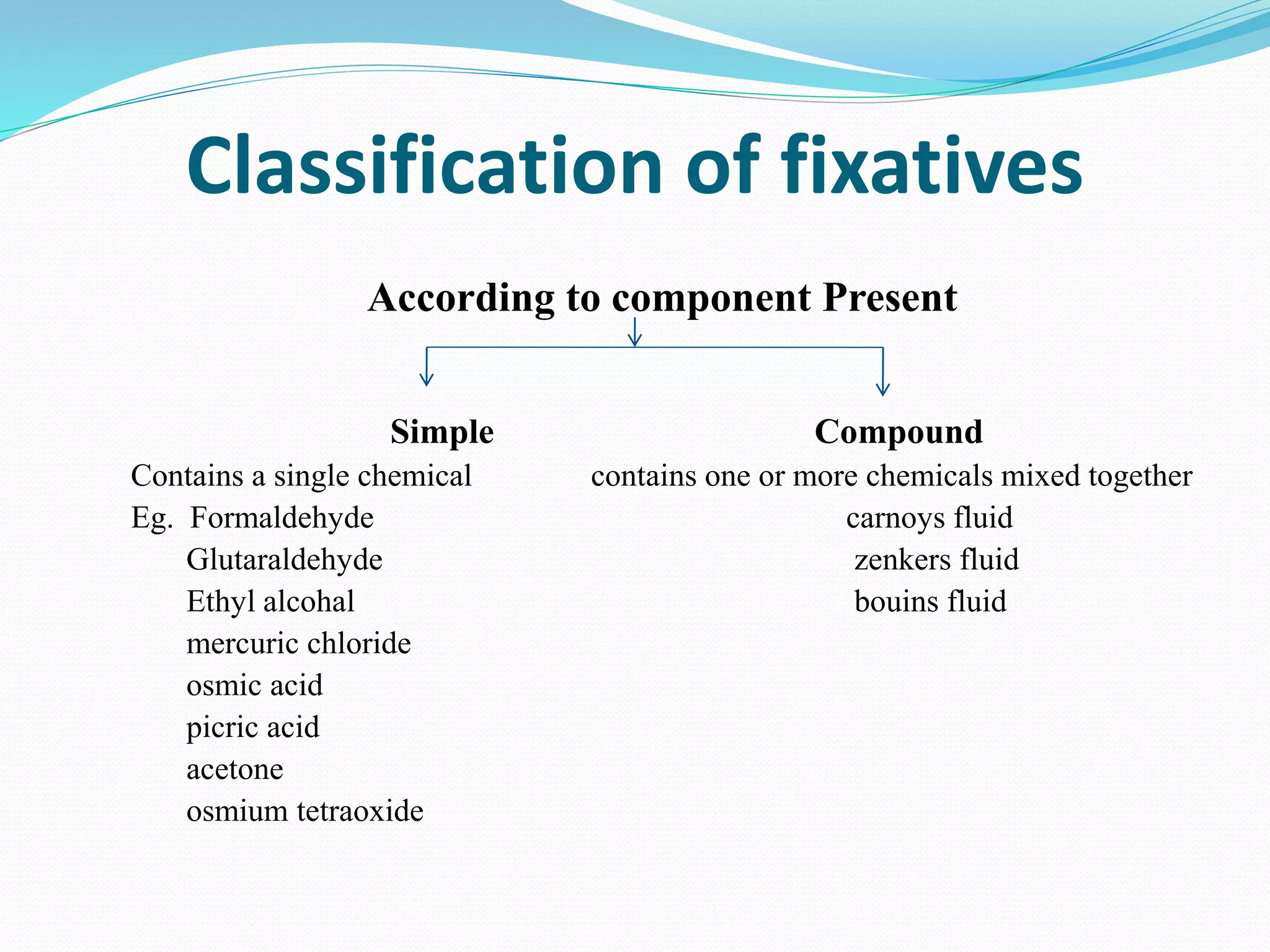
Dr. Vasim Akram Ansari
 HISTOLOGY :
It is the branch of science which deals with the gross &
microscopic study of normal tissue .
 HISTOPATHOLOGY :
It is the branch of pathology which deals with the gross &
microscopic study of tissue affected by disease.
Tissue for study can be obtained from:
 Biopsies
 Autopsies/Postmortem Specimens
Histopathological techniques
1. Receipt & Identification
2. Labeling of the specimen with numbering
3. Fixation & Decalcification
4. Dehydration
5. Clearing
6. Impregnation
7. Embedding
8. Section cutting
9. Staining
10. Mounting
Purpose of fixation
Fixation preserves a sample of biological material
(tissue or cells) as close to its natural state as possible in the
process of preparing tissue for examination.
i) They disable intrinsic biomolecules—
particularly proteolytic enzymes—which otherwise
digests or damages the sample.
ii) Fixatives are toxic to most common microorganisms
(bacteria in particular) that might cause extrinsic
damage to tissue sample.
iii) At molecular level they increase the mechanical strength or
stability of cells which help to preserve
the morphology (shape and structure) of the sample.
iv) Alteration of refractive indices of cell components so that
unstained components are easily visualized.
v) Preservation of chemical compounds so that further
histochemistry is possible.
vi) Certain fixatives also intensify the staining character of tissue
esp. with haematoxylin.
Methods of fixation
 Immersion
 Perfusion
 Vapour fixation
 Coating/spray
 Freeze drying
 Microwave fixation/stabilization
Important Factor in Fixation
Factor Affecting Fixation
1. Buffer and PH
Fixation is best carried out close to neutral pH, in the range of 6-
8. Hypoxia of tissues lowers the pH, so there must be buffering
capacity in the fixative to prevent excessive acidity.. Common
buffers include phosphate, bicarbonate, cacodylate, and veronal.
Commercial formalin is buffered with phosphate at a pH of 7.
2. Temperature
Most tissue fixed at room temperature.
Electron microscopy and histochemistry - 0-4○c
Increasing the temperature,, will increase the speed of fixation.
3. Penetration Capacity
Depends upon the diffusability of each individual fixative, which
is a constant. Formalin and alcohol penetrate the best, and
glutaraldehyde the worst. Tissues should be sectioned thinly (3 to
4 mm) for rapid penetration.
4. VOLUME
There should be a 20:1 ratio of fixative to tissue. Therefore,
the fixative should be changed at intervals to avoid
exhaustion of the fixative.
5. Agitation
Agitation increase the speed of penetration of fluid into the tissue
Speed of agitation is important.
6. Osmolality of fixative
 Hypertonic solution : cell shrinkage
 Hypotonic solution: swelling of cells and poor fixation
 Best : slightly hypertonic solution
7. CONCENTRATION
 of fixative should be adjusted down to the lowest
level possible, too high a concentration may adversely
affect the tissues and produce artefact similar to
excessive heat.
8. Duration of fixation
 Formalin : 2-6 hours
 Electron microscopy: 3hours
 Formaldehyde : prolonge fixation – shrinkage and
hardening of tissue
 Glutaraldehyde: prolonged fixation – advantageous
 Long fixation in aldehyde: inhibits enzyme activity
 Long fixation in oxidizing fixatives: degrade the tissue
Classification of fixatives
According to component Present
Simple Compound
Contains a single chemical contains one or more chemicals mixed together
Eg. Formaldehyde carnoys fluid
Glutaraldehyde zenkers fluid
Ethyl alcohal bouins fluid
mercuric chloride
osmic acid
picric acid
acetone
osmium tetraoxide
According to action upon cells & tissue
Micro Anatomical Cytological Histochemical
They preserve microscopic preserve intracellular used to demonstrate enzyme
structure of tissue structure of tissue & other substance of cells
eg. Formal saline eg. Buffered neutral formaline
formal calcium cold acetone
Buffered neutral formaline absolute alcohal
Zenkers fluid
Bouins fluid
Nuclear Cytoplasmic
Carneys fluid champys fluid
Clarks fluid formal saline
Flemings fluid formal calcium
According to action
physical methods Chemical methods
eg. Heating
microwaving
freeze drying
Agents that form covelent Agents that remove free Agents which denature protiens
Cross links water from the tissue and nucleic acid through
And hence precipitate change in PH and salt formation
And coagulate protiens
eg. Formaldehyde eg. Methanol eg. Acetic acid
gluteraldehyde ethanol trichloroacetic acid
acetone zinc acetate
According to tolerance action
tolerant fluid non tolerant fluid
Eg. Formaline eg. carnoys fluid
Commonly use fixatives & their
mechenism of action
Time required for fixation
 At room temperature – 12 hours
 For small biopsies – 4 - 6 hours
 At 650 c fixation occurs in – 2 hours
ETHYLALCOHOL
colorless, powerful dehydrating and Precipitating agent which acts by
reducing the solubility of protein molecules and (often) by disrupting
the hydrophobic interactions of proteins.
Coagulate protein but not nucleioprotien
Precipitate glycogen
Slow to penetrate, hardens and shrinks tissue.
But penetrates rapidly in combination with Carony’s fixative.
Can be used for immunoflorescence to detect certain enzymes.
METHYL ALCOHOL is used for fixing blood and bone marrow
smears.
Most commonly used fixatives in
laboratories
FORMALDEHYDE CONTAINING FIXATIVE
A.10% FORMALINE
Formaldehyde (40%)- 10 ml
Distilled water – 90 ml
B .10% FORMAL SALINE
Formaldehyde (40%)- 100 ml
Sodium chloride - 9 gm
Distilled water – 900 ml
It is cheap &penetrates rapidly
Does not over hardens the tissues
C. BUFFERED FORMALIN :(preferred fixative)
Formalin -10 ml
sodium dihydrogen phosphate - 0.4 gm
Anhydrous disodium hydrogen phosphate - 0.65 gm
distilled water - 90 ml
Advantage – prevent the formation of formaline pigments
 D. FORMOL CALCIUM
 i) Formalin-10 ml
 ii) Ca Acetate-2.0 gm
 iii)Water to make upto 100ml
 Has neutral pH
 Formalin pigment not formed.
 E. FORMALIN AMMONIUM BROMIDE
 i) Formalin-15 ml
 ii) Distilled WATER – 85ml
 iii) Ammonium bromide -2.0 gm
 Preserves neurological tissues specially when
 gold & silver impregnation is employed.
F. ALCOHOLIC FORMALIN
i)40% Formalin - 10ml
ii) 70-95 alcohol - 90 ml
G. ACETIC ALCOHOLIC FORMALIN
i) Formalin - 5.0ml
ii) Glacial acetic acid - 5.0 ml
iii) 70% alcohol - 90 ml
i) Formalin -10 ml
ii) Sucrose -7.5 gm
iii) M/15 phosphate to make 100 ml
 Used for combined cytochemistry & electron
microscopy.
 Excellent preservative for fine structure of
phospholopids & some enzymes.
 Should be used cold (40 C) on fresh tissue.
H. BUFFERED FORMOL SUCROSE:
i) Saturated picric acid(1.2 gm/100 ml) - 75 ml
ii) Formaldehyde - 25 ml
iii) Glacial acetic acid - 5 ml
 Excellent fixative for testicular & intestinal
biopsy as it gives good nuclear details.
 Good fixative for glycogen.
 Excess picric acid is removed by alcohol
treatment.
I. BOUIN’S FLUID:
ALCOHOL CONTAINING FIXATIVE
A) CARNOY’S FLUID:
i) Absolute alcohol -60 ml
ii) Chloroform -30 ml
iii) Glacial acetic acid - 10 ml
 Excellent nuclear fixation
 Good fixative for carbohydrate.
 Nissel’s substance & glycogen are preserved
 Fixation complete in 1-2 hrs
 It dissolves cytoplasmic elements
B. METHCARN
i) Absolute alcohol -60 ml
ii) Chloroform -30 ml
iii) Glacial acetic acid - 10 ml
C. WOLMANS SOLUTION
i) Absolute alcohol -60 ml
ii) Glacial acetic acid - 10 ml
D. ACETIC ALCOHOLIC FORMALIN
i) Formaldehyde - 5.0ml
ii) acetic acid - 5.0 ml
iii) alcohol - 90 ml
i) Absolute alcohol -75 ml
ii) Glacial acetic acid -25 ml
 Good nuclear as well as cytoplasmic element.
 Excellent for chromosomal analysis.
E. CLARK’S FLUID:
Picric acid containing fixatives
A. BOUIN’S FLUID
i) Saturated aqueous picric acid - 75 ml
ii) Formalin(40%) - 25 ml
iii) Glacial acetic acid - 5 ml
 Excellent fixative for testicular & intestinal biopsy
as it gives good nuclear details.
 Good fixative for glycogen.
 Excess picric acid is removed by alcohol treatment.
B. ROSSMANS FLUID
100% ethanol saturated with picric acid – 90 ml
Neutralized commercial formaline – 10 ml
C. GENDERS FLUID
90% ethanol saturated with picric acid – 80 ml
40% formaldehyde – 15 ml
Glacial acetic acid – 5ml
MERCURY SALT CONTAINING FIXATIVES
A. ZENKERS FLUID
i) Mercuric chloride - 5.0 gm
ii) Potassium dichromate - 2.5 gm
iii) Sodium sulphate - 1.0 gm
iv) Distilled H2O to make 100 ml
v) Add immediately before use glacial acetic acid -5 ml
 Good routine fixative
 Gives rapid & even penetration
Note:- Washing of tissue in running H2O is done to
remove excess dichromate.
B. ZENKER-FORMOL(HELLY’S FLUID)
Solution A
Distilled water – 100 ml
potassium dichromate – 2.5 gm
Mercuric chloride – 5 gm
Sodium sulfate – 1 gm
Solution B
40% formaldehyde – 5 ml
Add solution A to solution B immediately before use
 Used as both cyptoplasmic fixative & a microanatomical
fixative, particularly for bone narrow & blood forming
organs.
C. B5 FIXATIVE
STOCK REAGENT A
i) Mercuric chloride -12 gm
ii) Sodium acetate -2.5 gm
iii) Distilled H2O - 200 ml
STOCK REAGENT B
10% buffered neutral formaline
To prepare a working solution mix 90 ml stock reagent A with
10 ml stock reagent B
Fixation time 5-8 hours
 Used in fixation of Lymph Node biopsies to improve cytological
details and enhance immunoreactivity with anti-immunoglobulin
antiserum used in phenotyping of B cell neoplas
D. SUSA FLUID
i) Mercuric chloride - 4.5 gm
ii) NaCl - 0.5 gm
iii) Trichloracetic acid - 2.0 gm
iv) Acetic acid - 4.0 gm
v) 40% formaldehyde – 20 ml
Distilled water to make 100 ml
 Excellent fixative for routine biopsy.
 Allows brilliant staining with good cytological
details.
 Gives rapid & even penetration.
OTHERS FIXATIVES
NEW COMER’S FLUID:
i) Isopropranolol - 60 ml
ii) Propionic acid - 40 ml
iii) Petroleum ether -10 ml
iv) Acetone -10 ml
v) Dioxane -10 ml
 For fixation of chromosomes
 Fixes and preserves mucopolysaccharides
 Fixation is complete in 12-18 hrs.
Soln A - i) Formalin – 128 ml
ii) Acetic acid - 16 ml
Soln B - i) 1% Chromic acid
Mix 9 ml of soln A + 16 ml of Soln B
immediately before use.
 Useful in demonstrating mitotic figures &
chromosomes.
 Tissue should be washed well in running water before
use.
SANFELICE FLUID:
CHAMPY’S FLUID
i) 3% Na dichromate - 7 ml
ii) 17% Chromic acid - 7 ml
iii) 2% Osmium tera oxide - 4 ml
 Preserves the mitochondrial fat & lipids.
 Tissue must be washed overnight after fixation.
HOPE FIXATIVES
 Hepes Glutamic Acid Buffer Mediated Organic Solvent Protection Effect
 HOPE® - Formalin-free fixation technique for molecular pathology and
research
 The HOPE®-Fixation technique requires HOPE® Fixative System I
and HOPE® Fixative System II to complete the protocol.
 Applications:
 PCR
 RT-PCR
 Immunohistochemistry
 In situ hybridization targeting RNA and DNA
 Benefits:
 Achieve IHC on paraffin tissue without antigen unmasking
 IHC using antibodies which do not work on formalin fixed parafffin
embedded tissue
 No more overfixed formalin tissue samples
 Unmatched results in RNA and DNA in situ hybridization
 Excellent conservation of nucleic acids for PCR and RT-PCR
SECONDARY FIXATION
 A second fixative is used e.g. Mercuric chloride for 4 hrs after
the tissue is being fixed by Formalin.
 It provide firmer texture to tissues.
 It also accentuates the staining.
TISSUE FIXATIVE OF CHOICE TIME FOR FIXATION
Routine Formalin 10-12 hrs
GIT Biopsies Buffered formaldehyde 4-6 hrs
Testicular Biopsy Bouin’s fluid 4-6 hrs
Liver biopsy Buffered formaldehyde 4-12 hrs
Bone marrow biopsy Bouin’s fluid 2 ½ hrs followed by washing in
running water overnight
Spleen & blood filled
cavities
Zenker’s fluid 1-6 hrs
Lymph node B5 Working Solution 12-18 hrs
Mitochondria & Nissl body Carnoy’s fluid 1-2 hrs
Chromosome/Cell culture Clark’s fluid 1-2 hrs
DECALCIFICATON
Process of removing the calcium salt from bone, teeth
and other calcified tissues and making them amenable
for sectioning is known as DECALCIFICATION.
CRITERIA- Good Decalcificant
1. Complete removal of calcium.
2. Absence of damage to tissue cells or fibers.
3. Subsequent staining not altered.
4. Short time required for decalcification.
Purpose of decalcification
 To make bone flexible and easy for pathological
investigation. This is necessary in order to obtain soft
section of bone using the microtome.
 Faliure of decalcification results in torn ragged section
and damage to cutting edge of microtome knife.
Principle of decalcification
 Insoluble salt converted to soluble salt by the action
of decalcifying agent so that tissue become soft.
 Chelating agent binds to calcium ions present in the
bone and decalcification is carried out.
Method of decalcification
 Acid decalcifying agents
 Ion exchange resins with acid and decalcifying fluids
 Electrolyte decalcification
 Chelating agents
ACID DECALCIFYING AGENTS
 Acids are further divided into -weak acid and strong acid.
 weak acid are formic acid and trichloroacetic acid.
 Strong acid are nitric acid and hydrochloric acid.
AQUEOUS NITRIC ACID(5-10%)
 Nitric acid – 5ml
 Distilled water – 95ml
 Procedure:
Place specimen in solution till decalcification is
complete.
 Wash in running water for 30 mins.
 Neutralize for at least 5 hrs. in 10% formalin
 Wash in running water overnight.
Overexposure impairs nuclear staining.
FORMIC ACID( 5-10%)
 Brings out fairly rapid decalcification.
 Nuclear staining is better.
 But requires neutralization and thorough
washing prior to dehydration.
PERENYL’S FLUID
i)10% Nitric acid - 40 ml
ii)Absolute alcohol - 30 ml.
iii)0.5% Chromic acid - 30 ml.
It is slow for decalcifying hard bone but excellent fluid for
small deposits of calcium eg. calcified arteries, coin
lesions and calcified glands.
FORMALIN NITRIC ACID
i)Formalin -10 ml
ii)Distilled water -80 ml
iii)Nitric acid -10ml
 Nitric acid causes serious deterioration of nuclear
stainability which is partially inhibited by formaldehyde
GOODING AND STELWART’S FLUID
i)90% formic acid 5-10ml.
ii)Formalin 5ml
iii)Distilled water to 100 ml.
EVAN’S AND KRAJIAN FLUID
i)20% aqueous trisodium citrate 65 ml
ii)90% formic acid 35 ml
ION EXCHANGE RESIN
Remove calcium ion from fluid .
These are Ammoniated salt of sulfonated resin along with
various concentrations of formic acid.
Resin is layered on the bottom of container to a depth of ½
inch or 1.25cm
Advantages include-Faster decalcification,Tissue
preservation and better cellular details.
CHELATING AGENTS
 Organic compounds which have the power of binding
certain metals.
Ethylene-diamene-tetra-acetic acid, disodium salt called
Versenate is used.
 Slow process but has little or no effect on other tissue
elements
i)Versenate 10 gm.
ii)Distilled water 100 ml
 Time 7-21 days
Advantages of EDTA
 Deposits of iron and other metals may also be
removed.
 Tissue not hardened after decalcification.
 Its can be good for bone,teeth and any calcified tissue.
 This is also preffered solution for decalcifying bone
material for transmission electron microscopy.
END POINT OF DECALCIFICATION
1. Flexibility method
Unreliable, causes damage to the tissue.
2. X-ray method
Best method but not cost effective.
3. Chemical Method
Take 5 ml of decalcifying fluid from the bottom of container
which has been in contact with the tissue for 6-12 hrs.
Add 5 ml each of 5% ammonium oxalate and 5% ammonium
hydroxide. Mix and wait for 15-30 min.
Presence of a “Cloudy solution” caused by Calcium oxalate
indicates that specimen is not thoroughly decalcified
TREATMENT OF HARD TISSUES
Concentrated Sulphuric acid causes keratin to completely
dissolve from the tissue sections but much tissue distortion
occurs.
For softening of keratin following solutions used:-
i) Mercuric chloride - 4 gm
ii) Chromic acid - 0.5 gm
iii) Nitric acid (Conc.) - 10 ml
iv) Ethyl alcohol 95% - 50 ml
v) Distilled water - 200 ml
LENDRUM’S TECHNIQUE
 Very useful for tissues which became hard at the time of
fixation.
 Following washing out of the fixative, tissue is
immersed in a 4% aqueous solution of phenol for 1-3
days.
SURFACE DECALCIFICATION
The surface of the block to be decalcified is
trimmed with scalpel.
 The block is then placed in solution of 1% HCL face
downwards so that acid bathes the cut surface for 15-60
min.
Here penetration and decalcification is sufficient for few
sections only.
Protocol for small sized bony tissue
(mild Decalcification protocol)
 bone marrow biopsies
 J needle bone biopsies
 Curettage and small bone biopsies
 Procedure:
 overnight fixation in acetic acid-zinc-formalin (AZF)
fixative.
 Decalcification in a mixture of formic acid and formaline
(10% formic acid -10 ml 40% formaldehyde -5 ml
distilled water -85 ml ) for 5-6 hours
 Advantage- antigenicity is largely preserved
Protocol for large sized and harder bony bits
 For large mendibulectomies and limb specimen
 Procedure:
 Large specimen cut with bone saw into 3-5 mm thick
section and Put in cassette.
 These put in mixture of 10% buffered formalin(90ml) and
nitric acid (10ml) overnight.
 Now decalcified using 10% nitric acid .
Protocol for firm tissue which are less calcified
(Light Decalcification protocol)
 tracheal margins, pinna of ear
 Procedure:
 These put in mixture of 10% buffered formalin(95ml) and
nitric acid (5ml) overnight.
 Now decalcified using 5% nitric acid 5-6 hours.
THANK YOU

fixation and decalcification

  • 1.
  • 2.
     HISTOLOGY : Itis the branch of science which deals with the gross & microscopic study of normal tissue .  HISTOPATHOLOGY : It is the branch of pathology which deals with the gross & microscopic study of tissue affected by disease. Tissue for study can be obtained from:  Biopsies  Autopsies/Postmortem Specimens
  • 3.
    Histopathological techniques 1. Receipt& Identification 2. Labeling of the specimen with numbering 3. Fixation & Decalcification 4. Dehydration 5. Clearing 6. Impregnation 7. Embedding 8. Section cutting 9. Staining 10. Mounting
  • 5.
    Purpose of fixation Fixationpreserves a sample of biological material (tissue or cells) as close to its natural state as possible in the process of preparing tissue for examination. i) They disable intrinsic biomolecules— particularly proteolytic enzymes—which otherwise digests or damages the sample. ii) Fixatives are toxic to most common microorganisms (bacteria in particular) that might cause extrinsic damage to tissue sample.
  • 6.
    iii) At molecularlevel they increase the mechanical strength or stability of cells which help to preserve the morphology (shape and structure) of the sample. iv) Alteration of refractive indices of cell components so that unstained components are easily visualized. v) Preservation of chemical compounds so that further histochemistry is possible. vi) Certain fixatives also intensify the staining character of tissue esp. with haematoxylin.
  • 7.
    Methods of fixation Immersion  Perfusion  Vapour fixation  Coating/spray  Freeze drying  Microwave fixation/stabilization
  • 8.
  • 19.
    Factor Affecting Fixation 1.Buffer and PH Fixation is best carried out close to neutral pH, in the range of 6- 8. Hypoxia of tissues lowers the pH, so there must be buffering capacity in the fixative to prevent excessive acidity.. Common buffers include phosphate, bicarbonate, cacodylate, and veronal. Commercial formalin is buffered with phosphate at a pH of 7.
  • 20.
    2. Temperature Most tissuefixed at room temperature. Electron microscopy and histochemistry - 0-4○c Increasing the temperature,, will increase the speed of fixation.
  • 21.
    3. Penetration Capacity Dependsupon the diffusability of each individual fixative, which is a constant. Formalin and alcohol penetrate the best, and glutaraldehyde the worst. Tissues should be sectioned thinly (3 to 4 mm) for rapid penetration.
  • 22.
    4. VOLUME There shouldbe a 20:1 ratio of fixative to tissue. Therefore, the fixative should be changed at intervals to avoid exhaustion of the fixative.
  • 23.
    5. Agitation Agitation increasethe speed of penetration of fluid into the tissue Speed of agitation is important.
  • 24.
    6. Osmolality offixative  Hypertonic solution : cell shrinkage  Hypotonic solution: swelling of cells and poor fixation  Best : slightly hypertonic solution
  • 25.
    7. CONCENTRATION  offixative should be adjusted down to the lowest level possible, too high a concentration may adversely affect the tissues and produce artefact similar to excessive heat.
  • 26.
    8. Duration offixation  Formalin : 2-6 hours  Electron microscopy: 3hours  Formaldehyde : prolonge fixation – shrinkage and hardening of tissue  Glutaraldehyde: prolonged fixation – advantageous  Long fixation in aldehyde: inhibits enzyme activity  Long fixation in oxidizing fixatives: degrade the tissue
  • 27.
    Classification of fixatives Accordingto component Present Simple Compound Contains a single chemical contains one or more chemicals mixed together Eg. Formaldehyde carnoys fluid Glutaraldehyde zenkers fluid Ethyl alcohal bouins fluid mercuric chloride osmic acid picric acid acetone osmium tetraoxide
  • 28.
    According to actionupon cells & tissue Micro Anatomical Cytological Histochemical They preserve microscopic preserve intracellular used to demonstrate enzyme structure of tissue structure of tissue & other substance of cells eg. Formal saline eg. Buffered neutral formaline formal calcium cold acetone Buffered neutral formaline absolute alcohal Zenkers fluid Bouins fluid Nuclear Cytoplasmic Carneys fluid champys fluid Clarks fluid formal saline Flemings fluid formal calcium
  • 29.
    According to action physicalmethods Chemical methods eg. Heating microwaving freeze drying Agents that form covelent Agents that remove free Agents which denature protiens Cross links water from the tissue and nucleic acid through And hence precipitate change in PH and salt formation And coagulate protiens eg. Formaldehyde eg. Methanol eg. Acetic acid gluteraldehyde ethanol trichloroacetic acid acetone zinc acetate
  • 30.
    According to toleranceaction tolerant fluid non tolerant fluid Eg. Formaline eg. carnoys fluid
  • 32.
    Commonly use fixatives& their mechenism of action
  • 35.
    Time required forfixation  At room temperature – 12 hours  For small biopsies – 4 - 6 hours  At 650 c fixation occurs in – 2 hours
  • 41.
    ETHYLALCOHOL colorless, powerful dehydratingand Precipitating agent which acts by reducing the solubility of protein molecules and (often) by disrupting the hydrophobic interactions of proteins. Coagulate protein but not nucleioprotien Precipitate glycogen Slow to penetrate, hardens and shrinks tissue. But penetrates rapidly in combination with Carony’s fixative. Can be used for immunoflorescence to detect certain enzymes. METHYL ALCOHOL is used for fixing blood and bone marrow smears.
  • 49.
    Most commonly usedfixatives in laboratories FORMALDEHYDE CONTAINING FIXATIVE A.10% FORMALINE Formaldehyde (40%)- 10 ml Distilled water – 90 ml B .10% FORMAL SALINE Formaldehyde (40%)- 100 ml Sodium chloride - 9 gm Distilled water – 900 ml It is cheap &penetrates rapidly Does not over hardens the tissues
  • 50.
    C. BUFFERED FORMALIN:(preferred fixative) Formalin -10 ml sodium dihydrogen phosphate - 0.4 gm Anhydrous disodium hydrogen phosphate - 0.65 gm distilled water - 90 ml Advantage – prevent the formation of formaline pigments
  • 51.
     D. FORMOLCALCIUM  i) Formalin-10 ml  ii) Ca Acetate-2.0 gm  iii)Water to make upto 100ml  Has neutral pH  Formalin pigment not formed.  E. FORMALIN AMMONIUM BROMIDE  i) Formalin-15 ml  ii) Distilled WATER – 85ml  iii) Ammonium bromide -2.0 gm  Preserves neurological tissues specially when  gold & silver impregnation is employed.
  • 52.
    F. ALCOHOLIC FORMALIN i)40%Formalin - 10ml ii) 70-95 alcohol - 90 ml G. ACETIC ALCOHOLIC FORMALIN i) Formalin - 5.0ml ii) Glacial acetic acid - 5.0 ml iii) 70% alcohol - 90 ml
  • 53.
    i) Formalin -10ml ii) Sucrose -7.5 gm iii) M/15 phosphate to make 100 ml  Used for combined cytochemistry & electron microscopy.  Excellent preservative for fine structure of phospholopids & some enzymes.  Should be used cold (40 C) on fresh tissue. H. BUFFERED FORMOL SUCROSE:
  • 54.
    i) Saturated picricacid(1.2 gm/100 ml) - 75 ml ii) Formaldehyde - 25 ml iii) Glacial acetic acid - 5 ml  Excellent fixative for testicular & intestinal biopsy as it gives good nuclear details.  Good fixative for glycogen.  Excess picric acid is removed by alcohol treatment. I. BOUIN’S FLUID:
  • 55.
    ALCOHOL CONTAINING FIXATIVE A)CARNOY’S FLUID: i) Absolute alcohol -60 ml ii) Chloroform -30 ml iii) Glacial acetic acid - 10 ml  Excellent nuclear fixation  Good fixative for carbohydrate.  Nissel’s substance & glycogen are preserved  Fixation complete in 1-2 hrs  It dissolves cytoplasmic elements
  • 56.
    B. METHCARN i) Absolutealcohol -60 ml ii) Chloroform -30 ml iii) Glacial acetic acid - 10 ml C. WOLMANS SOLUTION i) Absolute alcohol -60 ml ii) Glacial acetic acid - 10 ml
  • 57.
    D. ACETIC ALCOHOLICFORMALIN i) Formaldehyde - 5.0ml ii) acetic acid - 5.0 ml iii) alcohol - 90 ml
  • 58.
    i) Absolute alcohol-75 ml ii) Glacial acetic acid -25 ml  Good nuclear as well as cytoplasmic element.  Excellent for chromosomal analysis. E. CLARK’S FLUID:
  • 59.
    Picric acid containingfixatives A. BOUIN’S FLUID i) Saturated aqueous picric acid - 75 ml ii) Formalin(40%) - 25 ml iii) Glacial acetic acid - 5 ml  Excellent fixative for testicular & intestinal biopsy as it gives good nuclear details.  Good fixative for glycogen.  Excess picric acid is removed by alcohol treatment.
  • 60.
    B. ROSSMANS FLUID 100%ethanol saturated with picric acid – 90 ml Neutralized commercial formaline – 10 ml C. GENDERS FLUID 90% ethanol saturated with picric acid – 80 ml 40% formaldehyde – 15 ml Glacial acetic acid – 5ml
  • 61.
    MERCURY SALT CONTAININGFIXATIVES A. ZENKERS FLUID i) Mercuric chloride - 5.0 gm ii) Potassium dichromate - 2.5 gm iii) Sodium sulphate - 1.0 gm iv) Distilled H2O to make 100 ml v) Add immediately before use glacial acetic acid -5 ml  Good routine fixative  Gives rapid & even penetration Note:- Washing of tissue in running H2O is done to remove excess dichromate.
  • 62.
    B. ZENKER-FORMOL(HELLY’S FLUID) SolutionA Distilled water – 100 ml potassium dichromate – 2.5 gm Mercuric chloride – 5 gm Sodium sulfate – 1 gm Solution B 40% formaldehyde – 5 ml Add solution A to solution B immediately before use  Used as both cyptoplasmic fixative & a microanatomical fixative, particularly for bone narrow & blood forming organs.
  • 63.
    C. B5 FIXATIVE STOCKREAGENT A i) Mercuric chloride -12 gm ii) Sodium acetate -2.5 gm iii) Distilled H2O - 200 ml STOCK REAGENT B 10% buffered neutral formaline To prepare a working solution mix 90 ml stock reagent A with 10 ml stock reagent B Fixation time 5-8 hours  Used in fixation of Lymph Node biopsies to improve cytological details and enhance immunoreactivity with anti-immunoglobulin antiserum used in phenotyping of B cell neoplas
  • 64.
    D. SUSA FLUID i)Mercuric chloride - 4.5 gm ii) NaCl - 0.5 gm iii) Trichloracetic acid - 2.0 gm iv) Acetic acid - 4.0 gm v) 40% formaldehyde – 20 ml Distilled water to make 100 ml  Excellent fixative for routine biopsy.  Allows brilliant staining with good cytological details.  Gives rapid & even penetration.
  • 65.
    OTHERS FIXATIVES NEW COMER’SFLUID: i) Isopropranolol - 60 ml ii) Propionic acid - 40 ml iii) Petroleum ether -10 ml iv) Acetone -10 ml v) Dioxane -10 ml  For fixation of chromosomes  Fixes and preserves mucopolysaccharides  Fixation is complete in 12-18 hrs.
  • 66.
    Soln A -i) Formalin – 128 ml ii) Acetic acid - 16 ml Soln B - i) 1% Chromic acid Mix 9 ml of soln A + 16 ml of Soln B immediately before use.  Useful in demonstrating mitotic figures & chromosomes.  Tissue should be washed well in running water before use. SANFELICE FLUID:
  • 67.
    CHAMPY’S FLUID i) 3%Na dichromate - 7 ml ii) 17% Chromic acid - 7 ml iii) 2% Osmium tera oxide - 4 ml  Preserves the mitochondrial fat & lipids.  Tissue must be washed overnight after fixation.
  • 68.
    HOPE FIXATIVES  HepesGlutamic Acid Buffer Mediated Organic Solvent Protection Effect  HOPE® - Formalin-free fixation technique for molecular pathology and research  The HOPE®-Fixation technique requires HOPE® Fixative System I and HOPE® Fixative System II to complete the protocol.  Applications:  PCR  RT-PCR  Immunohistochemistry  In situ hybridization targeting RNA and DNA  Benefits:  Achieve IHC on paraffin tissue without antigen unmasking  IHC using antibodies which do not work on formalin fixed parafffin embedded tissue  No more overfixed formalin tissue samples  Unmatched results in RNA and DNA in situ hybridization  Excellent conservation of nucleic acids for PCR and RT-PCR
  • 70.
    SECONDARY FIXATION  Asecond fixative is used e.g. Mercuric chloride for 4 hrs after the tissue is being fixed by Formalin.  It provide firmer texture to tissues.  It also accentuates the staining.
  • 71.
    TISSUE FIXATIVE OFCHOICE TIME FOR FIXATION Routine Formalin 10-12 hrs GIT Biopsies Buffered formaldehyde 4-6 hrs Testicular Biopsy Bouin’s fluid 4-6 hrs Liver biopsy Buffered formaldehyde 4-12 hrs Bone marrow biopsy Bouin’s fluid 2 ½ hrs followed by washing in running water overnight Spleen & blood filled cavities Zenker’s fluid 1-6 hrs Lymph node B5 Working Solution 12-18 hrs Mitochondria & Nissl body Carnoy’s fluid 1-2 hrs Chromosome/Cell culture Clark’s fluid 1-2 hrs
  • 76.
    DECALCIFICATON Process of removingthe calcium salt from bone, teeth and other calcified tissues and making them amenable for sectioning is known as DECALCIFICATION.
  • 77.
    CRITERIA- Good Decalcificant 1.Complete removal of calcium. 2. Absence of damage to tissue cells or fibers. 3. Subsequent staining not altered. 4. Short time required for decalcification.
  • 78.
    Purpose of decalcification To make bone flexible and easy for pathological investigation. This is necessary in order to obtain soft section of bone using the microtome.  Faliure of decalcification results in torn ragged section and damage to cutting edge of microtome knife.
  • 79.
    Principle of decalcification Insoluble salt converted to soluble salt by the action of decalcifying agent so that tissue become soft.  Chelating agent binds to calcium ions present in the bone and decalcification is carried out.
  • 80.
    Method of decalcification Acid decalcifying agents  Ion exchange resins with acid and decalcifying fluids  Electrolyte decalcification  Chelating agents
  • 81.
    ACID DECALCIFYING AGENTS Acids are further divided into -weak acid and strong acid.  weak acid are formic acid and trichloroacetic acid.  Strong acid are nitric acid and hydrochloric acid.
  • 82.
    AQUEOUS NITRIC ACID(5-10%) Nitric acid – 5ml  Distilled water – 95ml  Procedure: Place specimen in solution till decalcification is complete.  Wash in running water for 30 mins.  Neutralize for at least 5 hrs. in 10% formalin  Wash in running water overnight. Overexposure impairs nuclear staining.
  • 83.
    FORMIC ACID( 5-10%) Brings out fairly rapid decalcification.  Nuclear staining is better.  But requires neutralization and thorough washing prior to dehydration.
  • 84.
    PERENYL’S FLUID i)10% Nitricacid - 40 ml ii)Absolute alcohol - 30 ml. iii)0.5% Chromic acid - 30 ml. It is slow for decalcifying hard bone but excellent fluid for small deposits of calcium eg. calcified arteries, coin lesions and calcified glands.
  • 85.
    FORMALIN NITRIC ACID i)Formalin-10 ml ii)Distilled water -80 ml iii)Nitric acid -10ml  Nitric acid causes serious deterioration of nuclear stainability which is partially inhibited by formaldehyde
  • 86.
    GOODING AND STELWART’SFLUID i)90% formic acid 5-10ml. ii)Formalin 5ml iii)Distilled water to 100 ml. EVAN’S AND KRAJIAN FLUID i)20% aqueous trisodium citrate 65 ml ii)90% formic acid 35 ml
  • 87.
    ION EXCHANGE RESIN Removecalcium ion from fluid . These are Ammoniated salt of sulfonated resin along with various concentrations of formic acid. Resin is layered on the bottom of container to a depth of ½ inch or 1.25cm Advantages include-Faster decalcification,Tissue preservation and better cellular details.
  • 88.
    CHELATING AGENTS  Organiccompounds which have the power of binding certain metals. Ethylene-diamene-tetra-acetic acid, disodium salt called Versenate is used.  Slow process but has little or no effect on other tissue elements i)Versenate 10 gm. ii)Distilled water 100 ml  Time 7-21 days
  • 89.
    Advantages of EDTA Deposits of iron and other metals may also be removed.  Tissue not hardened after decalcification.  Its can be good for bone,teeth and any calcified tissue.  This is also preffered solution for decalcifying bone material for transmission electron microscopy.
  • 90.
    END POINT OFDECALCIFICATION 1. Flexibility method Unreliable, causes damage to the tissue. 2. X-ray method Best method but not cost effective. 3. Chemical Method Take 5 ml of decalcifying fluid from the bottom of container which has been in contact with the tissue for 6-12 hrs. Add 5 ml each of 5% ammonium oxalate and 5% ammonium hydroxide. Mix and wait for 15-30 min. Presence of a “Cloudy solution” caused by Calcium oxalate indicates that specimen is not thoroughly decalcified
  • 91.
    TREATMENT OF HARDTISSUES Concentrated Sulphuric acid causes keratin to completely dissolve from the tissue sections but much tissue distortion occurs. For softening of keratin following solutions used:- i) Mercuric chloride - 4 gm ii) Chromic acid - 0.5 gm iii) Nitric acid (Conc.) - 10 ml iv) Ethyl alcohol 95% - 50 ml v) Distilled water - 200 ml
  • 92.
    LENDRUM’S TECHNIQUE  Veryuseful for tissues which became hard at the time of fixation.  Following washing out of the fixative, tissue is immersed in a 4% aqueous solution of phenol for 1-3 days.
  • 93.
    SURFACE DECALCIFICATION The surfaceof the block to be decalcified is trimmed with scalpel.  The block is then placed in solution of 1% HCL face downwards so that acid bathes the cut surface for 15-60 min. Here penetration and decalcification is sufficient for few sections only.
  • 94.
    Protocol for smallsized bony tissue (mild Decalcification protocol)  bone marrow biopsies  J needle bone biopsies  Curettage and small bone biopsies  Procedure:  overnight fixation in acetic acid-zinc-formalin (AZF) fixative.  Decalcification in a mixture of formic acid and formaline (10% formic acid -10 ml 40% formaldehyde -5 ml distilled water -85 ml ) for 5-6 hours  Advantage- antigenicity is largely preserved
  • 95.
    Protocol for largesized and harder bony bits  For large mendibulectomies and limb specimen  Procedure:  Large specimen cut with bone saw into 3-5 mm thick section and Put in cassette.  These put in mixture of 10% buffered formalin(90ml) and nitric acid (10ml) overnight.  Now decalcified using 10% nitric acid .
  • 96.
    Protocol for firmtissue which are less calcified (Light Decalcification protocol)  tracheal margins, pinna of ear  Procedure:  These put in mixture of 10% buffered formalin(95ml) and nitric acid (5ml) overnight.  Now decalcified using 5% nitric acid 5-6 hours.
  • 97.

Editor's Notes