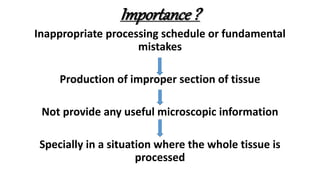
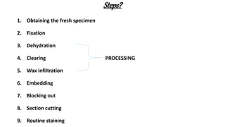
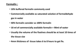
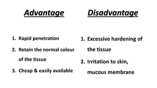
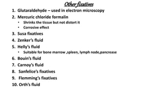
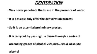
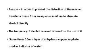
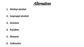
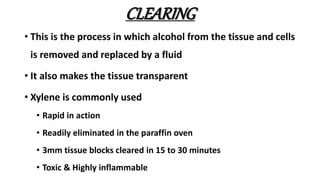
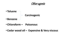
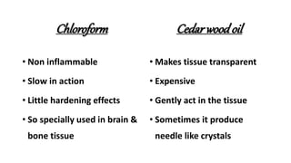
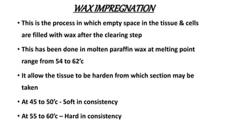
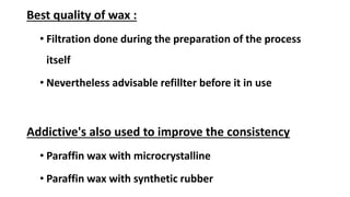
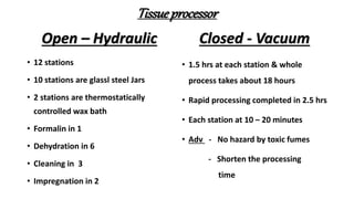
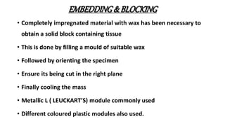
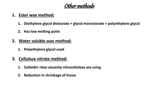
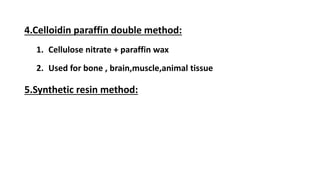
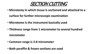
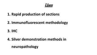
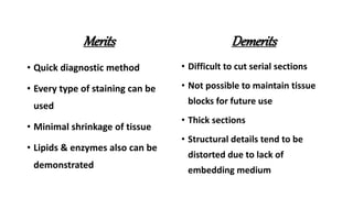
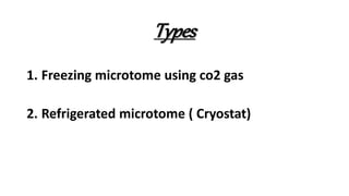
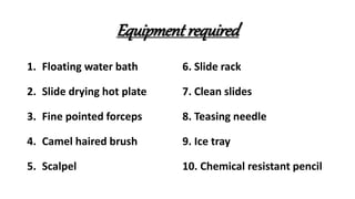
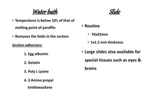
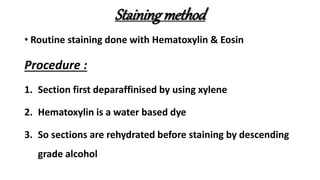
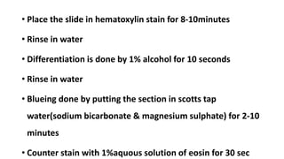
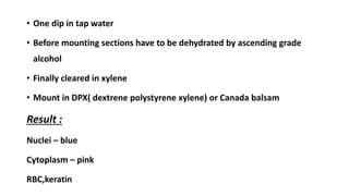
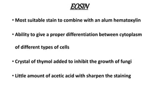
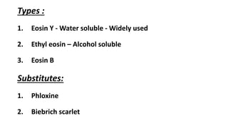
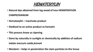
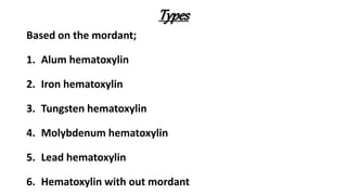
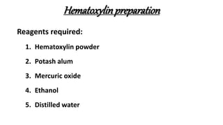
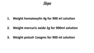
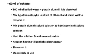
PROCESSING OF
TISSUE
Dr.T.Arivazhagan
Post Graduate
Department Of Pathology
INTRODUCTION
• It describes the steps required to take animal or
human tissue from the fixation to the state where it
completely infiltrated with a suitable histological
wax and can be embedded and ready for section ,
cutting on the microtome.
Types
• Manual – Hand processing
• Automated – Machine processing
• Tissue transfer – Specimens are transferred
• Fluid transfer – Specimen held in a single container and the
fluids are pumped in & out
Importance ?
Inappropriate processing schedule or fundamental
mistakes
Production of improper section of tissue
Not provide any useful microscopic information
Specially in a situation where the whole tissue is
processed
Steps?
1. Obtaining the fresh specimen
2. Fixation
3. Dehydration
4. Clearing PROCESSING
5. Wax infiltration
6. Embedding
7. Blocking out
8. Section cutting
9. Routine staining
Obtainingspecimen
• Separate room is required for specimen reception
• Dissection area must have
• Good lighting
• Good ventilation
• Non absorbent wipe clean surface
• Other equipment's
• Prior to fixation some tissue from the specimen need to
reserve for other departmental assessment
Specimendissectionplan
1.Small samples:
• Rarely need dissection
• It may be placed in a nylon bag in order to prevent them of falling
• Eosin used as a marker
• Recommended to count the small tissue at the time dissection to
verify prior to section cutting
2.Core biopsies:
• Tissue laid out in a longitudinal fashion so that majority of the
tissue can be cut
3.Skin biopsies:
• Sections are best managed in sequential / serial transverse section
4.Bowel specimens:
• Large specimens are sampled at multiple blocks
• Resected margins also included as a part of dissection
• Particular attention is given for lymph node
5.Gynecological specimen:
• Commonly cervical biopsy obtained
• Uterine specimens sampled in terms of
cervix,endometrium,myometrium along with common benign lesions
6.Breast specimens:
• Usually need for inked margins for orientation purpose
• Lymph node status also assessed
FIXATION
• Main aim of this step is preserving the cells & tissue
components with minimal distortion of the tissue.
Effects:
• Prevent the putrification & autolysis
• Hardness the tissue
• Make cells to insensitive to hyper & hypotonic solutions
• Act as a mordant
• Induce optical contrast for good morphologic examination
Ideal fixatives:
1. Should be cheap & easily available
2. Should be stable & safe to handle
3. Should have rapid on of action
4. Should cause minimal loss of tissue
5. Should give even penetration
6. Should retain the normal colour of the tissue
7. Should not bind to the reactive groups
Types
1.Simple – Contain one substance
2.Compound – Contain 2 or more substance
Also divided into;
1.Micro anatomical - Preserve the anatomy of the tissue
2.Cytological – Preserve the intra cellular constituents
3.Histochemical – Demonstration of histochemical
constituents & enzymes
Formalin :
• 10% buffered formalin commonly used
• Commercially available as saturated solution of formaldehyde
gas in water
• 40% formalin consider as 100% formalin
• 10 ml of commercially available formalin + 90ml of water
• Usually the volume of the fixatives should be at least 10 times of
the tissue size
• 4mm thickness of tissue takes 6 to 8 hours to get fix.
Advantage
1. Rapid penetration
2. Retain the normal colour
of the tissue
3. Cheap & easily available
Disadvantage
1. Excessive hardening of
the tissue
2. Irritation to skin,
mucous membrane
Otherfixatives
1. Glutaraldehyde – used in electron microscopy
2. Mercuric chloride formalin
• Shrinks the tissue but not distort it
• Corrosive effect
3. Susa fixatives
4. Zenker’s fluid
5. Helly’s fluid
• Suitable for bone marrow ,spleen, lymph node,pancrease
6. Bouin’s fluid
7. Carnoy’s fluid
8. Sanfelice’s fixatives
9. Flemming’s fixatives
10. Orth’s fluid
DEHYDRATION
• Wax never penetrate the tissue in the presence of water
• It is possible only after the dehydration process
• So it is an essential preliminary process
• It is carryout by passing the tissue through a series of
ascending grades of alcohol 70%,80%,90% & absolute
alcohol
• Reason – in order to prevent the distortion of tissue when
transfer a tissue from an aqueous medium to absolute
alcohol directly
• The frequency of alcohol renewal is based on the use of it
• Some times 10mm layer of anhydrous copper sulphate
used as indicator of water.
Alternatives
1. Methyl alcohol
2. Isopropyl alcohol
3. Acetone
4. Pyridine
5. Dioxane
6. Cellosolve
CLEARING
• This is the process in which alcohol from the tissue and cells
is removed and replaced by a fluid
• It also makes the tissue transparent
• Xylene is commonly used
• Rapid in action
• Readily eliminated in the paraffin oven
• 3mm tissue blocks cleared in 15 to 30 minutes
• Toxic & Highly inflammable
Otheragents
• Toluene
Carcinogenic
• Benzene
• Chloroform – Poisonous
• Cedar wood oil – Expensive & Very viscous
Chloroform
• Non inflammable
• Slow in action
• Little hardening effects
• So specially used in brain &
bone tissue
Cedarwoodoil
• Makes tissue transparent
• Expensive
• Gently act in the tissue
• Sometimes it produce
needle like crystals
WAXIMPREGNATION
• This is the process in which empty space in the tissue & cells
are filled with wax after the clearing step
• This has been done in molten paraffin wax at melting point
range from 54 to 62’c
• It allow the tissue to be harden from which section may be
taken
• At 45 to 50’c - Soft in consistency
• At 55 to 60’c – Hard in consistency
Best quality of wax :
• Filtration done during the preparation of the process
itself
• Nevertheless advisable refillter before it in use
Addictive's also used to improve the consistency
• Paraffin wax with microcrystalline
• Paraffin wax with synthetic rubber
Tissueprocessor
Open – Hydraulic
• 12 stations
• 10 stations are glassl steel Jars
• 2 stations are thermostatically
controlled wax bath
• Formalin in 1
• Dehydration in 6
• Cleaning in 3
• Impregnation in 2
Closed - Vacuum
• 1.5 hrs at each station & whole
process takes about 18 hours
• Rapid processing completed in 2.5 hrs
• Each station at 10 – 20 minutes
• Adv - No hazard by toxic fumes
- Shorten the processing
time
EMBEDDING&BLOCKING
• Completely impregnated material with wax has been necessary to
obtain a solid block containing tissue
• This is done by filling a mould of suitable wax
• Followed by orienting the specimen
• Ensure its being cut in the right plane
• Finally cooling the mass
• Metallic L ( LEUCKART’S) module commonly used
• Different coloured plastic modules also used.
Othermethods
1. Ester wax method:
1. Diethylene glycol distearate + glycol monosterate + polyethylene glycol
2. Has low melting point
2. Water soluble wax method:
1. Polyethylene glycol used
3. Cellulose nitrate method:
1. Celloidin +low viscosity nitrocellulose are using
2. Reduction in shrinkage of tissue
4.Celloidin paraffin double method:
1. Cellulose nitrate + paraffin wax
2. Used for bone , brain,muscle,animal tissue
5.Synthetic resin method:
SECTIONCUTTING
• Microtomy in which tissue is sectioned and attached to a
surface for further microscopic examination
• Microtome is the instrument basically used
• Thickness range from 1 micrometer to several hundred
micrometer
• Common range is 3-8 micrometer
• Both paraffin & frozen sections are used
Types
1. Rotary microtome
2. Base sledge microtome
3. Rotary rocking microtome
4. Sliding microtome
5. Ultra microtome
RotaryMicrotome
• Most commonly used
• Basic mechanism is rotation of a fine advanced hand wheel by
360 degree
• Knife is fixed
• Tissue block is movable
• It may be manual, semi automated, fully automated
• Ability to cut thin 2 – 3 micrometer sections
• Honing & Stropping- Process of sharpened the knife
Sliding microtome
• Used in the cutting of cellulose nitrate embedded
tissue
• Tissue block is fixed
• Knife is movable
Base sledgemicrotome
• Used for very hard tissue or large blocks
• Ex: brain & heart tissue
• Base is moved to and fro run against the knife to
produce section
Rotary rockingmicrotome
• Commonly used in cryostats
• Knife is immovable
• Tissue blocks is held in a spring bearing rocking arm
ULTRAMICROTOME:
• Used exclusively for electron microscope
Typesof knives
1. A- Biconcave
2. B- Planoconcave
3. C- Wedge shaped
4. D-Tool edge
Freezingmicrotome
• Designed for the preparation of frozen section of
fixed or unfixed tissue
• It does not need preliminary embedding step
• The stage is connected to an cylinder which has co2
for rapid cooling
FROZENSECTION
•Tissue become firm
•Tissue is rapidly frozen
•Ice acting as embedding medium
•Sections are produced with out using of
dehydration,clearing,wax embedding steps
Uses
1. Rapid production of sections
2. Immunofluorescent methodology
3. IHC
4. Silver demonstration methods in
neuropathology
Merits
• Quick diagnostic method
• Every type of staining can be
used
• Minimal shrinkage of tissue
• Lipids & enzymes also can be
demonstrated
Demerits
• Difficult to cut serial sections
• Not possible to maintain tissue
blocks for future use
• Thick sections
• Structural details tend to be
distorted due to lack of
embedding medium
Types
1. Freezing microtome using co2 gas
2. Refrigerated microtome ( Cryostat)
Equipment required
1. Floating water bath
2. Slide drying hot plate
3. Fine pointed forceps
4. Camel haired brush
5. Scalpel
6. Slide rack
7. Clean slides
8. Teasing needle
9. Ice tray
10. Chemical resistant pencil
Waterbath
• Temperature is below 10’c of that of
melting point of paraffin
• Removes the folds in the section
Section adhesives:
1. Egg albumin
2. Gelatin
3. Poly L Lysine
4. 3 Amino propyl
triethoxysilane
Slide
• Routine
• 76x25mm
• 1x1.2 mm thickness
• Large slides also available for
special tissues such as eyes &
brains
Stainingmethod
• Routine staining done with Hematoxylin & Eosin
Procedure :
1. Section first deparaffinised by using xylene
2. Hematoxylin is a water based dye
3. So sections are rehydrated before staining by descending
grade alcohol
• Place the slide in hematoxylin stain for 8-10minutes
• Rinse in water
• Differentiation is done by 1% alcohol for 10 seconds
• Rinse in water
• Blueing done by putting the section in scotts tap
water(sodium bicarbonate & magnesium sulphate) for 2-10
minutes
• Counter stain with 1%aquous solution of eosin for 30 sec
• One dip in tap water
• Before mounting sections have to be dehydrated by ascending grade
alcohol
• Finally cleared in xylene
• Mount in DPX( dextrene polystyrene xylene) or Canada balsam
Result :
Nuclei – blue
Cytoplasm – pink
RBC,keratin
EOSIN
• Most suitable stain to combine with an alum hematoxylin
• Ability to give a proper differentiation between cytoplasm
of different types of cells
• Crystal of thymol added to inhibit the growth of fungi
• Little amount of acetic acid with sharpen the staining
Types :
1. Eosin Y - Water soluble - Widely used
2. Ethyl eosin – Alcohol soluble
3. Eosin B
Substitutes:
1. Phloxine
2. Biebrich scarlet
HEMATOXYLIN
• Natural dye obtained from log wood of tree HEMATOXYLON
CAMPECHIANUM
• Hematoxylin – inactivate product
• Oxidized to an active product as hematin
• This process know as ripening
• Done by naturally in sunlight or chemically by addition of sodium
iodate mercuric oxide,kmno4
• Mordant – helps in penetration the stain particles to the tissue
Types
Based on the mordant;
1. Alum hematoxylin
2. Iron hematoxylin
3. Tungsten hematoxylin
4. Molybdenum hematoxylin
5. Lead hematoxylin
6. Hematoxylin with out mordant
Hematoxylinpreparation
Reagents required:
1. Hematoxylin powder
2. Potash alum
3. Mercuric oxide
4. Ethanol
5. Distilled water
Steps
1. Weight hematoxylin 4g for 900 ml solution
2. Weight mercuric oxide 2g for 900ml solution
3. Weight potash 1oogms for 900 ml solution
• 60ml of ethanol
• 800 ml of boiled water + potash alum till it is dissolved
• Mix 4g of hematoxylin in 60 ml of ethanol and shake well to
dissolve it
• Mix potash alum dissolved solution to hematoxylin dissolved
solution
• Heat the solution & add mercuric oxide
• Keep on heating till pinkish colour appear
• Then cool it
• Stain ready to use

Processing of tissue

  • 1.
  • 2.
    INTRODUCTION • It describesthe steps required to take animal or human tissue from the fixation to the state where it completely infiltrated with a suitable histological wax and can be embedded and ready for section , cutting on the microtome.
  • 3.
    Types • Manual –Hand processing • Automated – Machine processing • Tissue transfer – Specimens are transferred • Fluid transfer – Specimen held in a single container and the fluids are pumped in & out
  • 4.
    Importance ? Inappropriate processingschedule or fundamental mistakes Production of improper section of tissue Not provide any useful microscopic information Specially in a situation where the whole tissue is processed
  • 5.
    Steps? 1. Obtaining thefresh specimen 2. Fixation 3. Dehydration 4. Clearing PROCESSING 5. Wax infiltration 6. Embedding 7. Blocking out 8. Section cutting 9. Routine staining
  • 6.
    Obtainingspecimen • Separate roomis required for specimen reception • Dissection area must have • Good lighting • Good ventilation • Non absorbent wipe clean surface • Other equipment's • Prior to fixation some tissue from the specimen need to reserve for other departmental assessment
  • 7.
    Specimendissectionplan 1.Small samples: • Rarelyneed dissection • It may be placed in a nylon bag in order to prevent them of falling • Eosin used as a marker • Recommended to count the small tissue at the time dissection to verify prior to section cutting 2.Core biopsies: • Tissue laid out in a longitudinal fashion so that majority of the tissue can be cut
  • 8.
    3.Skin biopsies: • Sectionsare best managed in sequential / serial transverse section 4.Bowel specimens: • Large specimens are sampled at multiple blocks • Resected margins also included as a part of dissection • Particular attention is given for lymph node 5.Gynecological specimen: • Commonly cervical biopsy obtained • Uterine specimens sampled in terms of cervix,endometrium,myometrium along with common benign lesions
  • 9.
    6.Breast specimens: • Usuallyneed for inked margins for orientation purpose • Lymph node status also assessed
  • 10.
    FIXATION • Main aimof this step is preserving the cells & tissue components with minimal distortion of the tissue. Effects: • Prevent the putrification & autolysis • Hardness the tissue • Make cells to insensitive to hyper & hypotonic solutions • Act as a mordant • Induce optical contrast for good morphologic examination
  • 11.
    Ideal fixatives: 1. Shouldbe cheap & easily available 2. Should be stable & safe to handle 3. Should have rapid on of action 4. Should cause minimal loss of tissue 5. Should give even penetration 6. Should retain the normal colour of the tissue 7. Should not bind to the reactive groups
  • 12.
    Types 1.Simple – Containone substance 2.Compound – Contain 2 or more substance Also divided into; 1.Micro anatomical - Preserve the anatomy of the tissue 2.Cytological – Preserve the intra cellular constituents 3.Histochemical – Demonstration of histochemical constituents & enzymes
  • 13.
    Formalin : • 10%buffered formalin commonly used • Commercially available as saturated solution of formaldehyde gas in water • 40% formalin consider as 100% formalin • 10 ml of commercially available formalin + 90ml of water • Usually the volume of the fixatives should be at least 10 times of the tissue size • 4mm thickness of tissue takes 6 to 8 hours to get fix.
  • 14.
    Advantage 1. Rapid penetration 2.Retain the normal colour of the tissue 3. Cheap & easily available Disadvantage 1. Excessive hardening of the tissue 2. Irritation to skin, mucous membrane
  • 15.
    Otherfixatives 1. Glutaraldehyde –used in electron microscopy 2. Mercuric chloride formalin • Shrinks the tissue but not distort it • Corrosive effect 3. Susa fixatives 4. Zenker’s fluid 5. Helly’s fluid • Suitable for bone marrow ,spleen, lymph node,pancrease 6. Bouin’s fluid 7. Carnoy’s fluid 8. Sanfelice’s fixatives 9. Flemming’s fixatives 10. Orth’s fluid
  • 16.
    DEHYDRATION • Wax neverpenetrate the tissue in the presence of water • It is possible only after the dehydration process • So it is an essential preliminary process • It is carryout by passing the tissue through a series of ascending grades of alcohol 70%,80%,90% & absolute alcohol
  • 17.
    • Reason –in order to prevent the distortion of tissue when transfer a tissue from an aqueous medium to absolute alcohol directly • The frequency of alcohol renewal is based on the use of it • Some times 10mm layer of anhydrous copper sulphate used as indicator of water.
  • 18.
    Alternatives 1. Methyl alcohol 2.Isopropyl alcohol 3. Acetone 4. Pyridine 5. Dioxane 6. Cellosolve
  • 19.
    CLEARING • This isthe process in which alcohol from the tissue and cells is removed and replaced by a fluid • It also makes the tissue transparent • Xylene is commonly used • Rapid in action • Readily eliminated in the paraffin oven • 3mm tissue blocks cleared in 15 to 30 minutes • Toxic & Highly inflammable
  • 20.
    Otheragents • Toluene Carcinogenic • Benzene •Chloroform – Poisonous • Cedar wood oil – Expensive & Very viscous
  • 21.
    Chloroform • Non inflammable •Slow in action • Little hardening effects • So specially used in brain & bone tissue Cedarwoodoil • Makes tissue transparent • Expensive • Gently act in the tissue • Sometimes it produce needle like crystals
  • 22.
    WAXIMPREGNATION • This isthe process in which empty space in the tissue & cells are filled with wax after the clearing step • This has been done in molten paraffin wax at melting point range from 54 to 62’c • It allow the tissue to be harden from which section may be taken • At 45 to 50’c - Soft in consistency • At 55 to 60’c – Hard in consistency
  • 23.
    Best quality ofwax : • Filtration done during the preparation of the process itself • Nevertheless advisable refillter before it in use Addictive's also used to improve the consistency • Paraffin wax with microcrystalline • Paraffin wax with synthetic rubber
  • 24.
    Tissueprocessor Open – Hydraulic •12 stations • 10 stations are glassl steel Jars • 2 stations are thermostatically controlled wax bath • Formalin in 1 • Dehydration in 6 • Cleaning in 3 • Impregnation in 2 Closed - Vacuum • 1.5 hrs at each station & whole process takes about 18 hours • Rapid processing completed in 2.5 hrs • Each station at 10 – 20 minutes • Adv - No hazard by toxic fumes - Shorten the processing time
  • 25.
    EMBEDDING&BLOCKING • Completely impregnatedmaterial with wax has been necessary to obtain a solid block containing tissue • This is done by filling a mould of suitable wax • Followed by orienting the specimen • Ensure its being cut in the right plane • Finally cooling the mass • Metallic L ( LEUCKART’S) module commonly used • Different coloured plastic modules also used.
  • 26.
    Othermethods 1. Ester waxmethod: 1. Diethylene glycol distearate + glycol monosterate + polyethylene glycol 2. Has low melting point 2. Water soluble wax method: 1. Polyethylene glycol used 3. Cellulose nitrate method: 1. Celloidin +low viscosity nitrocellulose are using 2. Reduction in shrinkage of tissue
  • 27.
    4.Celloidin paraffin doublemethod: 1. Cellulose nitrate + paraffin wax 2. Used for bone , brain,muscle,animal tissue 5.Synthetic resin method:
  • 28.
    SECTIONCUTTING • Microtomy inwhich tissue is sectioned and attached to a surface for further microscopic examination • Microtome is the instrument basically used • Thickness range from 1 micrometer to several hundred micrometer • Common range is 3-8 micrometer • Both paraffin & frozen sections are used
  • 29.
    Types 1. Rotary microtome 2.Base sledge microtome 3. Rotary rocking microtome 4. Sliding microtome 5. Ultra microtome
  • 30.
    RotaryMicrotome • Most commonlyused • Basic mechanism is rotation of a fine advanced hand wheel by 360 degree • Knife is fixed • Tissue block is movable • It may be manual, semi automated, fully automated • Ability to cut thin 2 – 3 micrometer sections • Honing & Stropping- Process of sharpened the knife
  • 31.
    Sliding microtome • Usedin the cutting of cellulose nitrate embedded tissue • Tissue block is fixed • Knife is movable
  • 32.
    Base sledgemicrotome • Usedfor very hard tissue or large blocks • Ex: brain & heart tissue • Base is moved to and fro run against the knife to produce section
  • 33.
    Rotary rockingmicrotome • Commonlyused in cryostats • Knife is immovable • Tissue blocks is held in a spring bearing rocking arm ULTRAMICROTOME: • Used exclusively for electron microscope
  • 34.
    Typesof knives 1. A-Biconcave 2. B- Planoconcave 3. C- Wedge shaped 4. D-Tool edge
  • 35.
    Freezingmicrotome • Designed forthe preparation of frozen section of fixed or unfixed tissue • It does not need preliminary embedding step • The stage is connected to an cylinder which has co2 for rapid cooling
  • 36.
    FROZENSECTION •Tissue become firm •Tissueis rapidly frozen •Ice acting as embedding medium •Sections are produced with out using of dehydration,clearing,wax embedding steps
  • 37.
    Uses 1. Rapid productionof sections 2. Immunofluorescent methodology 3. IHC 4. Silver demonstration methods in neuropathology
  • 38.
    Merits • Quick diagnosticmethod • Every type of staining can be used • Minimal shrinkage of tissue • Lipids & enzymes also can be demonstrated Demerits • Difficult to cut serial sections • Not possible to maintain tissue blocks for future use • Thick sections • Structural details tend to be distorted due to lack of embedding medium
  • 39.
    Types 1. Freezing microtomeusing co2 gas 2. Refrigerated microtome ( Cryostat)
  • 40.
    Equipment required 1. Floatingwater bath 2. Slide drying hot plate 3. Fine pointed forceps 4. Camel haired brush 5. Scalpel 6. Slide rack 7. Clean slides 8. Teasing needle 9. Ice tray 10. Chemical resistant pencil
  • 41.
    Waterbath • Temperature isbelow 10’c of that of melting point of paraffin • Removes the folds in the section Section adhesives: 1. Egg albumin 2. Gelatin 3. Poly L Lysine 4. 3 Amino propyl triethoxysilane Slide • Routine • 76x25mm • 1x1.2 mm thickness • Large slides also available for special tissues such as eyes & brains
  • 42.
    Stainingmethod • Routine stainingdone with Hematoxylin & Eosin Procedure : 1. Section first deparaffinised by using xylene 2. Hematoxylin is a water based dye 3. So sections are rehydrated before staining by descending grade alcohol
  • 43.
    • Place theslide in hematoxylin stain for 8-10minutes • Rinse in water • Differentiation is done by 1% alcohol for 10 seconds • Rinse in water • Blueing done by putting the section in scotts tap water(sodium bicarbonate & magnesium sulphate) for 2-10 minutes • Counter stain with 1%aquous solution of eosin for 30 sec
  • 44.
    • One dipin tap water • Before mounting sections have to be dehydrated by ascending grade alcohol • Finally cleared in xylene • Mount in DPX( dextrene polystyrene xylene) or Canada balsam Result : Nuclei – blue Cytoplasm – pink RBC,keratin
  • 45.
    EOSIN • Most suitablestain to combine with an alum hematoxylin • Ability to give a proper differentiation between cytoplasm of different types of cells • Crystal of thymol added to inhibit the growth of fungi • Little amount of acetic acid with sharpen the staining
  • 46.
    Types : 1. EosinY - Water soluble - Widely used 2. Ethyl eosin – Alcohol soluble 3. Eosin B Substitutes: 1. Phloxine 2. Biebrich scarlet
  • 47.
    HEMATOXYLIN • Natural dyeobtained from log wood of tree HEMATOXYLON CAMPECHIANUM • Hematoxylin – inactivate product • Oxidized to an active product as hematin • This process know as ripening • Done by naturally in sunlight or chemically by addition of sodium iodate mercuric oxide,kmno4 • Mordant – helps in penetration the stain particles to the tissue
  • 48.
    Types Based on themordant; 1. Alum hematoxylin 2. Iron hematoxylin 3. Tungsten hematoxylin 4. Molybdenum hematoxylin 5. Lead hematoxylin 6. Hematoxylin with out mordant
  • 49.
    Hematoxylinpreparation Reagents required: 1. Hematoxylinpowder 2. Potash alum 3. Mercuric oxide 4. Ethanol 5. Distilled water
  • 50.
    Steps 1. Weight hematoxylin4g for 900 ml solution 2. Weight mercuric oxide 2g for 900ml solution 3. Weight potash 1oogms for 900 ml solution
  • 51.
    • 60ml ofethanol • 800 ml of boiled water + potash alum till it is dissolved • Mix 4g of hematoxylin in 60 ml of ethanol and shake well to dissolve it • Mix potash alum dissolved solution to hematoxylin dissolved solution • Heat the solution & add mercuric oxide • Keep on heating till pinkish colour appear • Then cool it • Stain ready to use