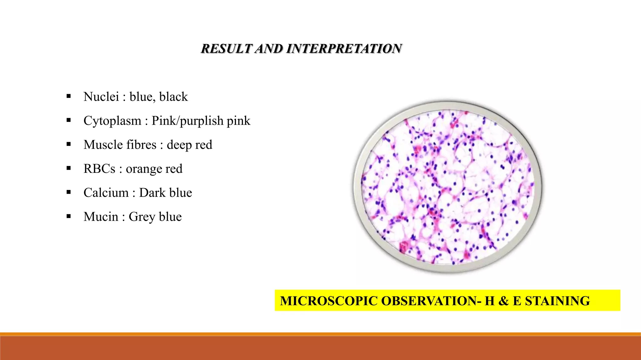
The document discusses H&E (hematoxylin and eosin) staining, which is the most widely used staining technique in histopathology. H&E staining differentially colors tissue components, with hematoxylin staining nuclei blue and eosin staining cytoplasm and other tissues shades of pink. The process involves deparaffinizing tissue sections, staining with hematoxylin, differentiating with acid, counterstaining with eosin, and mounting for examination under a microscope. H&E staining allows clear visualization and analysis of cells and structures to enable histopathological diagnosis.