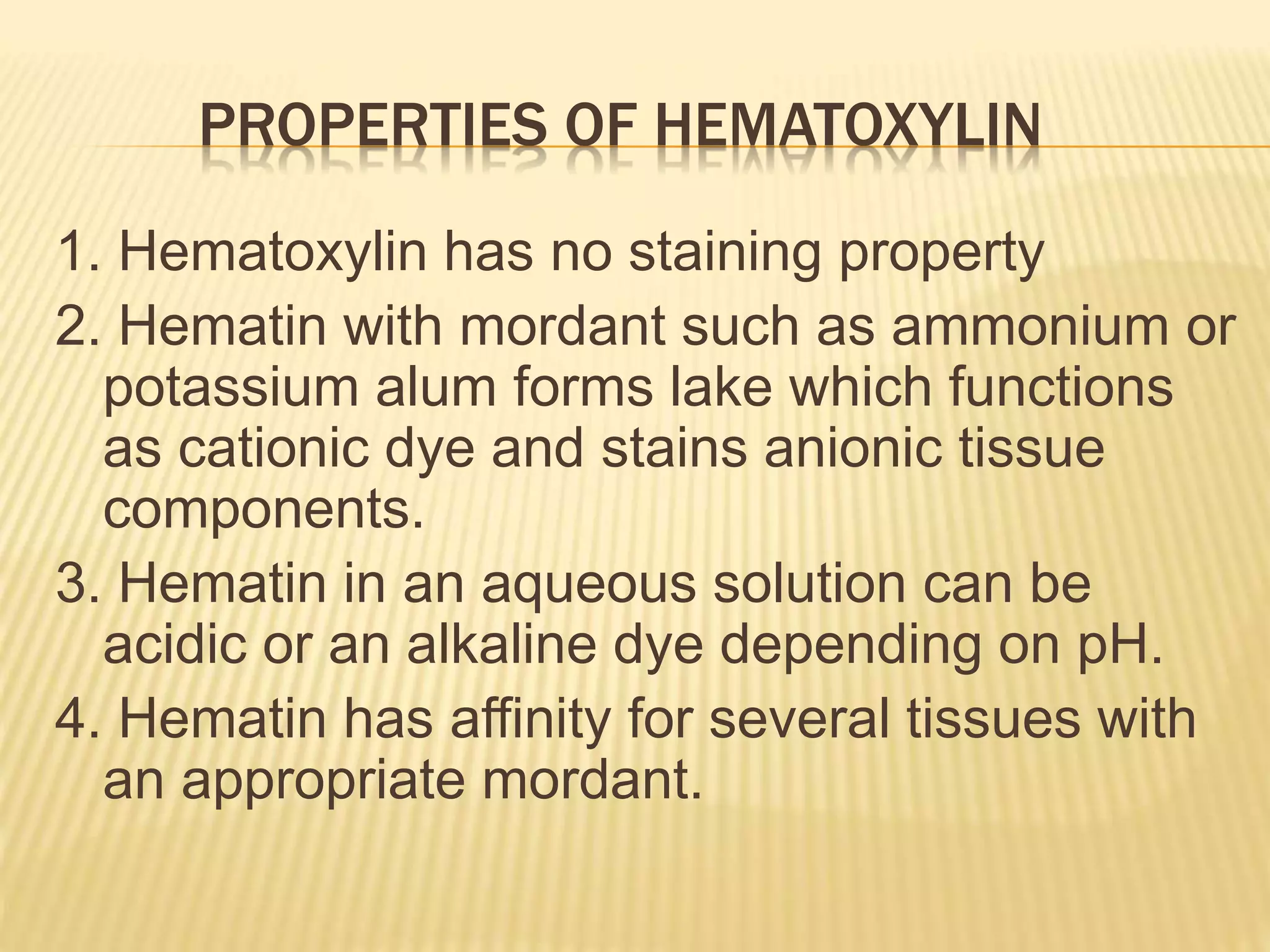
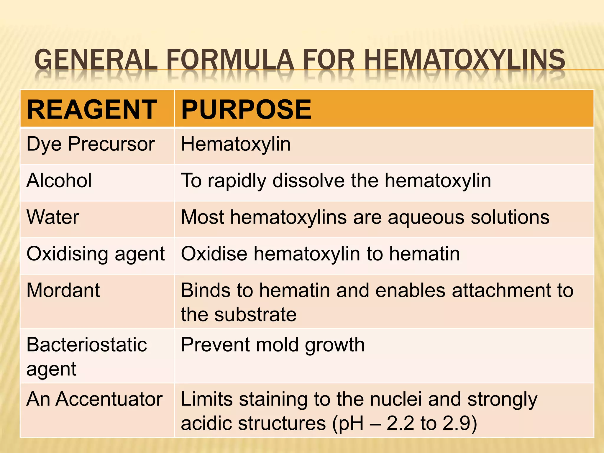
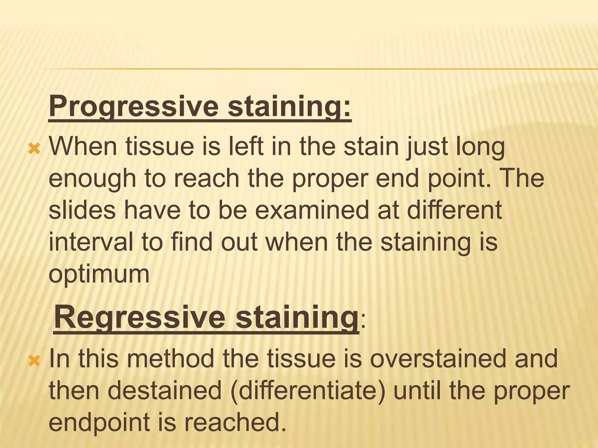
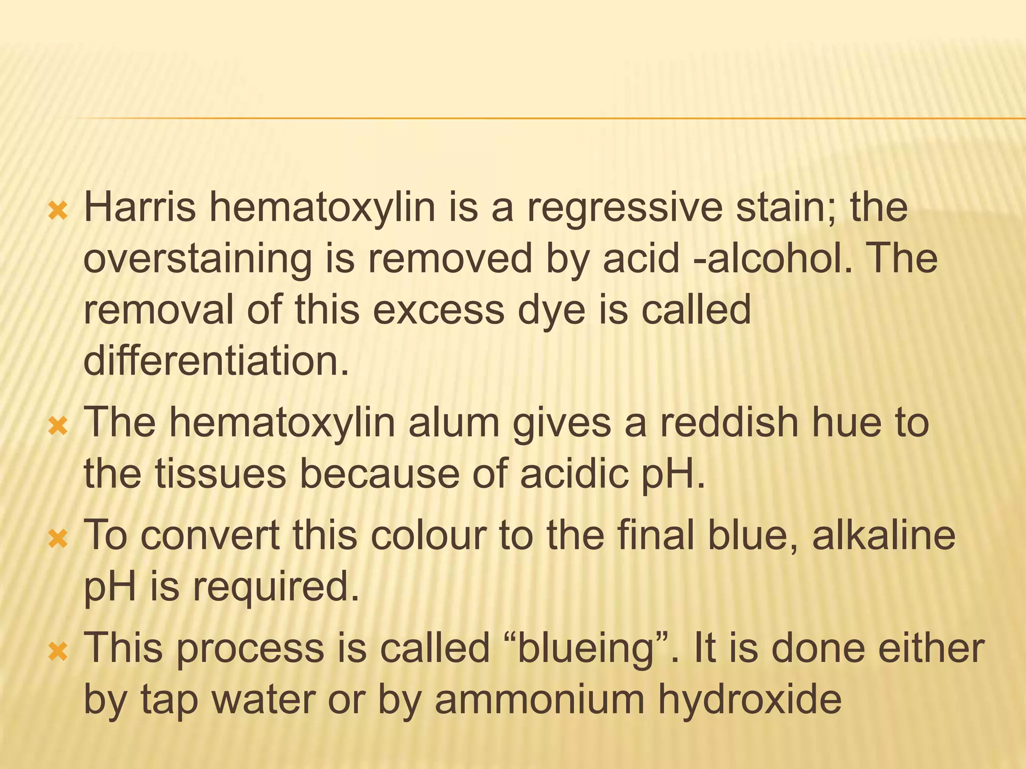
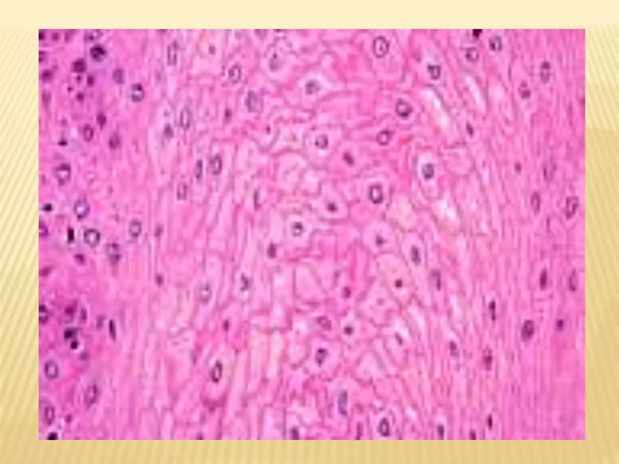
Hematoxylin and eosin (H&E) staining is the most common histology stain. Hematoxylin stains cell nuclei blue by binding to DNA and RNA, while eosin stains cytoplasm and extracellular components pink. The staining process involves deparaffinizing tissue sections, staining with hematoxylin, differentiating with acid to remove excess stain, staining with eosin, and mounting for examination. Hematoxylin is extracted from logwood and oxidized to hematin, which binds tissue as a cationic dye with a mordant like alum. Eosin Y is the typical counterstain used to visualize cytoplasm. Together, H&E staining provides excellent contrast to study cell and