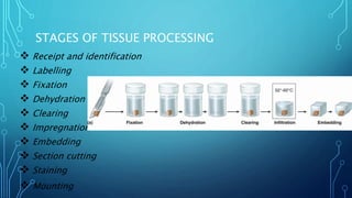
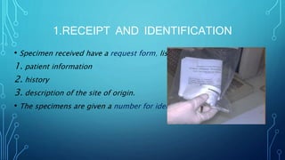
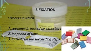
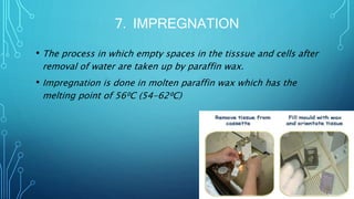
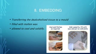
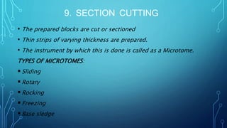
TISSUE PROCESSING
PRESENTED BY,
GOPIKA SUKUMARAN
CRI
CONTENT
• INTRODUCTION
• STAGES OF TISSUE PROCESSING
 RECEIPT AND IDENTIFICTION
 LABELLING
 FIXATION
 DECALCIFICATION
 DEHYDRATION
CONTENT (CONT)
 CLEARING
 IMPREGNATION
 EMBEDDING
 SECTION CUTTING
 STAINING
 MOUNTING
• CONCLUSION
• REFERENCE
INTRODUCTION
• Tissue processing is defined as the process of
1. preparing the tissue
2. by embedding it in a solid medium
3. that is firm enough to support it
4. give sufficient rigidity to enable thin sections
5. soft enough to enable the knife to cut the sections
with little damage to the knife or the tissue.
STAGES OF TISSUE PROCESSING
 Receipt and identification
 Labelling
 Fixation
 Dehydration
 Clearing
 Impregnation
 Embedding
 Section cutting
 Staining
 Mounting
1.RECEIPT AND IDENTIFICATION
• Specimen received have a request form, lists
1. patient information
2. history
3. description of the site of origin.
• The specimens are given a number for identification
2. LABELLING
Should be insoluble
Should not contaminate
Should not penetrate
Should not react
Clearly identifiable
Eg: India ink, silver nitrate
3.FIXATION
•Process in which
1.specimen is treated by exposing to fixative
2.for period of time
3.to facilitate the succeeding steps.
AIMS OF FIXATION:
Prevents putrification and autolysis.
Hardens the tissue which helps in section cutting
Acts as a mordant.
Increase the optical density.
Stable
and safe
to handle
Cheap &
easily
available
Should
cause
fixation
quickly
PROPERTIES
OF FIXATION
Should
cause
minimal
loss of
tissue
Retain
the color
of tissue
Should
give even
penetratio
n
TYPES OF FIXATION
Simple fixation
Compound fixation
SIMPLE FIXATIVES
 FORMALIN
 Commonly used fixative.
 Prepared by mixing 40% Formaldehyde gas in 100w/v of distilled water.
 GLUTARALDEHYDE
 OSMIUM TETRAOXIDE
 POTTASIUM DICHROMATE
 MERCURIC CHLORIDE
COMPOUND FIXATIVES
• MICROANATOMICAL FIXATIVES:
Used to preserve the anatomy of the tissues.
Eg. 10% Formal saline
Buffered formalin
• CYTOLOGICAL FIXATIVES:
Used to fix intracellular structures.
• HISTOCHEMICAL FIXATIVES:
Used to demonstrate the chemical constituents of the cell.
Eg. Formal saline
Cold acetone
Absolute alcohol
4.DECALCIFICATION
• Process of removal of the calcium salts from the specimen.
• The various agents used are:
 Nitric acid
 Hydrochloric acid
 Formic acid
 Pictric acid
 Acetic acid
 Citric acid
5.DEHYDRATION
• Process in which
1. The water content in tissue is completely reduced
2. By passing the tissue through increasing concentrations of
dehydrating agents.
• Various dehydrating agents:
 Ethyl alcohol
 Acetone
 Isopropyl alcohol
 Dioxane
6.CLEARING( DEALCOHOLIZATION)
• It is the procedure where the
1. alcohol in the tissue is replaced
2. by a fluid which will dissolve the wax
• The various clearing agents used are;
 Cedar wood oil : The best agent but is expensive.
 Benzene : It is carcinogenic.
 Xylene : It is most commonly used.
 Chloroform : Toxic and expensive.
7. IMPREGNATION
• The process in which empty spaces in the tisssue and cells after
removal of water are taken up by paraffin wax.
• Impregnation is done in molten paraffin wax which has the
melting point of 56⁰C (54-62⁰C)
8. EMBEDDING
• Transferring the dealcoholised tissue to a mould
• filled with molten wax
• allowed to cool and solidify.
9. SECTION CUTTING
• The prepared blocks are cut or sectioned
• Thin strips of varying thickness are prepared.
• The instrument by which this is done is called as a Microtome.
TYPES OF MICROTOMES:
Sliding
Rotary
Rocking
Freezing
Base sledge
10. STAINING
• Staining of the section is done to bring out the particular
details in the tissue under study.
• Commonly used stain
1. Haematoxylin
2. Eosin
11. MOUNTING
• Adhesives used for fixing the sections on the slides:
 Albumin solution
 Starch paste
 Gelatin
CONCLUSION
It is a critical step that needs to be monitored with utmost care.
Due to longer processing time, any mistakes alters the tissue, requiring
repetition.
They should not be under processed or over processed, as it may
hamper the tissue details.
To get sections of diagnostic value, processing of tissue should be done
with approppriate reagents and embedded in suitable embedding
medium depending upon type of tissue.
REFERENCES
• Textbook of Oral Histology & Embryology- ORBANS
• Textbook of Pathology- HARSH MOHAN- Second edition
• Wikipedia
Tissue processing

Tissue processing

  • 1.
  • 2.
    CONTENT • INTRODUCTION • STAGESOF TISSUE PROCESSING  RECEIPT AND IDENTIFICTION  LABELLING  FIXATION  DECALCIFICATION  DEHYDRATION
  • 3.
    CONTENT (CONT)  CLEARING IMPREGNATION  EMBEDDING  SECTION CUTTING  STAINING  MOUNTING • CONCLUSION • REFERENCE
  • 4.
    INTRODUCTION • Tissue processingis defined as the process of 1. preparing the tissue 2. by embedding it in a solid medium 3. that is firm enough to support it 4. give sufficient rigidity to enable thin sections 5. soft enough to enable the knife to cut the sections with little damage to the knife or the tissue.
  • 5.
    STAGES OF TISSUEPROCESSING  Receipt and identification  Labelling  Fixation  Dehydration  Clearing  Impregnation  Embedding  Section cutting  Staining  Mounting
  • 6.
    1.RECEIPT AND IDENTIFICATION •Specimen received have a request form, lists 1. patient information 2. history 3. description of the site of origin. • The specimens are given a number for identification
  • 7.
    2. LABELLING Should beinsoluble Should not contaminate Should not penetrate Should not react Clearly identifiable Eg: India ink, silver nitrate
  • 8.
    3.FIXATION •Process in which 1.specimenis treated by exposing to fixative 2.for period of time 3.to facilitate the succeeding steps.
  • 9.
    AIMS OF FIXATION: Preventsputrification and autolysis. Hardens the tissue which helps in section cutting Acts as a mordant. Increase the optical density.
  • 10.
    Stable and safe to handle Cheap& easily available Should cause fixation quickly PROPERTIES OF FIXATION Should cause minimal loss of tissue Retain the color of tissue Should give even penetratio n
  • 11.
    TYPES OF FIXATION Simplefixation Compound fixation
  • 12.
    SIMPLE FIXATIVES  FORMALIN Commonly used fixative.  Prepared by mixing 40% Formaldehyde gas in 100w/v of distilled water.  GLUTARALDEHYDE  OSMIUM TETRAOXIDE  POTTASIUM DICHROMATE  MERCURIC CHLORIDE
  • 13.
    COMPOUND FIXATIVES • MICROANATOMICALFIXATIVES: Used to preserve the anatomy of the tissues. Eg. 10% Formal saline Buffered formalin • CYTOLOGICAL FIXATIVES: Used to fix intracellular structures. • HISTOCHEMICAL FIXATIVES: Used to demonstrate the chemical constituents of the cell. Eg. Formal saline Cold acetone Absolute alcohol
  • 14.
    4.DECALCIFICATION • Process ofremoval of the calcium salts from the specimen. • The various agents used are:  Nitric acid  Hydrochloric acid  Formic acid  Pictric acid  Acetic acid  Citric acid
  • 15.
    5.DEHYDRATION • Process inwhich 1. The water content in tissue is completely reduced 2. By passing the tissue through increasing concentrations of dehydrating agents. • Various dehydrating agents:  Ethyl alcohol  Acetone  Isopropyl alcohol  Dioxane
  • 16.
    6.CLEARING( DEALCOHOLIZATION) • Itis the procedure where the 1. alcohol in the tissue is replaced 2. by a fluid which will dissolve the wax • The various clearing agents used are;  Cedar wood oil : The best agent but is expensive.  Benzene : It is carcinogenic.  Xylene : It is most commonly used.  Chloroform : Toxic and expensive.
  • 17.
    7. IMPREGNATION • Theprocess in which empty spaces in the tisssue and cells after removal of water are taken up by paraffin wax. • Impregnation is done in molten paraffin wax which has the melting point of 56⁰C (54-62⁰C)
  • 18.
    8. EMBEDDING • Transferringthe dealcoholised tissue to a mould • filled with molten wax • allowed to cool and solidify.
  • 19.
    9. SECTION CUTTING •The prepared blocks are cut or sectioned • Thin strips of varying thickness are prepared. • The instrument by which this is done is called as a Microtome. TYPES OF MICROTOMES: Sliding Rotary Rocking Freezing Base sledge
  • 20.
    10. STAINING • Stainingof the section is done to bring out the particular details in the tissue under study. • Commonly used stain 1. Haematoxylin 2. Eosin
  • 21.
    11. MOUNTING • Adhesivesused for fixing the sections on the slides:  Albumin solution  Starch paste  Gelatin
  • 22.
    CONCLUSION It is acritical step that needs to be monitored with utmost care. Due to longer processing time, any mistakes alters the tissue, requiring repetition. They should not be under processed or over processed, as it may hamper the tissue details. To get sections of diagnostic value, processing of tissue should be done with approppriate reagents and embedded in suitable embedding medium depending upon type of tissue.
  • 23.
    REFERENCES • Textbook ofOral Histology & Embryology- ORBANS • Textbook of Pathology- HARSH MOHAN- Second edition • Wikipedia