Angle Closure Glaucoma
•
8 likes•2,147 views

This document provides information about angle closure glaucoma from Amity Medical School. It defines glaucoma and discusses the anatomy of the anterior chamber and drainage angle. It describes the differences between open-angle glaucoma and angle closure glaucoma, including causes and risk factors. Treatment options are outlined, including medical management with eye drops and surgical options like laser trabeculoplasty and laser iridotomy.

Recommended
More Related Content
What's hot
What's hot (20)
Viewers also liked
Viewers also liked (19)
Similar to Angle Closure Glaucoma
Similar to Angle Closure Glaucoma (20)
Recently uploaded
Recently uploaded (20)
Angle Closure Glaucoma
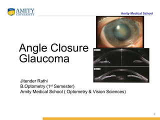
- 1. Amity Medical School Angle Closure Glaucoma 1 Jitender Rathi B.Optometry (1st Semester) Amity Medical School ( Optometry & Vision Sciences)
- 2. Amity Medical School • Anatomy • Aqueous Physiology and IOP • Definition of Glaucoma • Classification • Etiology • Treatment • Medical Management • Surgical Management 2 Outlines
- 4. Amity Medical School Anatomy of the drainage angle The anterior chamber(AC) • is that space, containing aqueous humor, which is bounded in front by the cornea and part of the sclera, and behind by the iris and part of the ciliary body. • Its normal depth in adults varies from 2.5-3.5mm. The angle of the anterior chamber.(iridocorneal angles) • Between the iris and the cornea at the periphery of the anterior chamber of the eye. • Refers to that peripheral recess bounded posteriorly by the root of the iris and the ciliary body and anteriorly by the corneo-scleral junction or the limbus. • There is an annular channel, called the canal of Schlemm. • The canal is separated from the aqueous in the anterior chamber by the trabecular meshwork. 4
- 5. Amity Medical School Aqueous Physiology and IOP • Fluid circulation within the eye.(Aqueous humor) • This creates the relatively constant and healthy pressure within the eye.(IOP) • Pressure is needed the eye Inflated, Nourished and helps in functioning properly. • Rate:2.0 µl/min (Micro Liter Per Minute) • Over production or inadequate drainage leads to elevate of IOP. 5
- 6. Amity Medical School Route of Aqueous Flow 6
- 8. Amity Medical School The trabecular meshwork is made up of circumferentially disposed flattened collagenous bands which criss-cross, leaving numerous tortuous passages through which the aqueous humor drains from the anterior chamber to the canal of Schlemm. The aqueous humor is a transparent colorless fluid which fills the anterior and posterior chambers of the eye. Its chief site of formation is the processes of the ciliary body. The volume of aqueous in the anterior chamber of the human eye is 0.25 ml. 8
- 9. Amity Medical School Glaucoma Introduction A common eye condition in which the fluid pressure inside the eye rises to a level higher than healthy for that eye. If untreated, it may damage the optic nerve, causing the loss of vision or even blindness. Glaucoma describes a number of ocular conditions characterized by: - 1-Raised intraocular pressure (IOP). 2-Optic nerve head damage(cupping). 3-Corresponding loss of visual field (VF). • IOP depends on the relationship between aqueous production and outflow. • The normal ocular tension is between 10-21mm.Hg.(Millimeters of mercury). • > 22mm Hg is considered abnormal. • Glaucoma remains one of the principal causes of blindness throughout the world. 9
- 10. Amity Medical School • It is a silent thief. • Scary part about this disease for both Doctor and patient is that It is a very silent disease of the eye, it is so silent that does not announce itself . • Patient must get it diagnosed as early as possible ,before it can drop vision. 10
- 11. Amity Medical School Currently, the World Health Organization ranks glaucoma as the second largest cause of blindness worldwide, behind cataract 11
- 12. Amity Medical School Classification of glaucoma. There are several types of glaucoma, however, the two most common are :- 1. open angle glaucoma (OAG), having a slow and insidious onset. 2. And angle closure glaucoma (ACG), which is less common and tends to be more acute. 12
- 13. Amity Medical School Open angle glaucoma (OAG). 13
- 14. Amity Medical School Open-angle glaucoma, the most common form of glaucoma, accounting for at least 90% of all glaucoma cases: • Is caused by the slow clogging of the drainage canals, resulting in increased eye pressure. • Has a wide and open angle between the iris and cornea. • Develops slowly and is a lifelong condition. • Has symptoms and damage that are not noticed. “Open-angle” means that the angle where the iris meets the cornea is as wide and open as it should be. Open-angle glaucoma is also called primary or chronic glaucoma. 14 OAG
- 16. Amity Medical School Angle closure glaucoma (ACG) 16
- 17. Amity Medical School Angle-closure glaucoma, a less common form of glaucoma: -Is caused by blocked drainage canals, resulting in a sudden rise in intraocular pressure. -Has a closed or narrow angle between the iris and cornea. -Develops very quickly. -Has symptoms and damage that are usually very noticeable. -Demands immediate medical attention. “It is also called acute glaucoma or narrow-angle glaucoma. Unlike open-angle glaucoma, angle-closure glaucoma is a result of the angle between the iris and cornea closing” Angle Closure glaucoma 17
- 18. Amity Medical School Causes of Narrow-Angle Glaucoma • Pupillary block: if the back of the iris adheres to the lens inside the eye, the pupillary channel becomes blocked. Then fluid backs up behind the iris, pushing the iris forward until it closes the drainage angle in the anterior chamber. • Iris plateau. In this condition, the iris is attached to the ciliary body too close to the trabecular meshwork, where drainage occurs. When the pupil dilates, the peripheral iris tissue bunches up in the drainage angle and can cover up the trabecular meshwork, causing IOP to rise quickly. • Hyperopia. People who are farsighted are more likely to have eyes with shallow anterior chambers and narrow angles, increasing their risk for angle-closure glaucoma from pupil dilation or aging changes in the eye. • Tumors and other causes. A tumor behind the iris, swelling associated with inflammation of the ciliary body (intermediate uveitis) and alteration of the shape of the eye after surgery for a detached retina also can cause angle-closure glaucoma. 18
- 19. Amity Medical School Risk factors for acute angle-closure glaucoma • Age. Being over age 60, the lens inside our eyes gets larger, increasing the risk for pupil block. Also, the anterior chamber tends to become increasingly shallow, and the drainage angle may narrow as we age. • Having a family history of the condition • Having certain medical conditions, such as diabetes, heart disease, high blood pressure. • Having certain eye conditions, such as smaller eyes Far- sightedness ( Hypropia). • Females-their eye ball is bit smaller as compare male. • Taking corticosteroid medications, especially eyedrops, for a long time. 19
- 20. Amity Medical School Angle closure in the plateau iris syndrome 20
- 21. Amity Medical SchoolSymptoms • Severe eye pain • Nausea and vomiting • Headache • Blurred vision and/or seeing haloes around lights (Haloes and blurred vision occur because the cornea is swollen.) • Profuse tearing 21
- 22. Amity Medical School Management Aims The aim of management is to lower IOP sufficiently to arrest progressive VF loss. 1/Medical treatment:Usually eye dropes. 2/Surgical treatment: For Open angle glaucoma (OAG) Trabeculoplasty provides a definitive reduction of IOP to within safe limits in the majority of cases. 22
- 23. Amity Medical School Laser Trabeculoplasty 23
- 24. Amity Medical School For Narrow Angle Glaucoma ;Leaser Iridology Is used 24
- 25. Amity Medical SchoolReferences . • 1-Book Clinical Ophthalmology (Glaucoma) Wills eye Institute Douglas J.Rhee • 2-Lacture Notes from Dr Najeeb -Youtube • Various discussions videos on Glaucoma 25