Download as PPS, PPTX
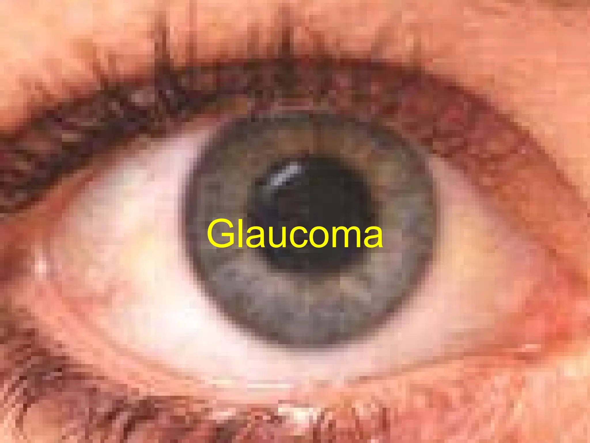
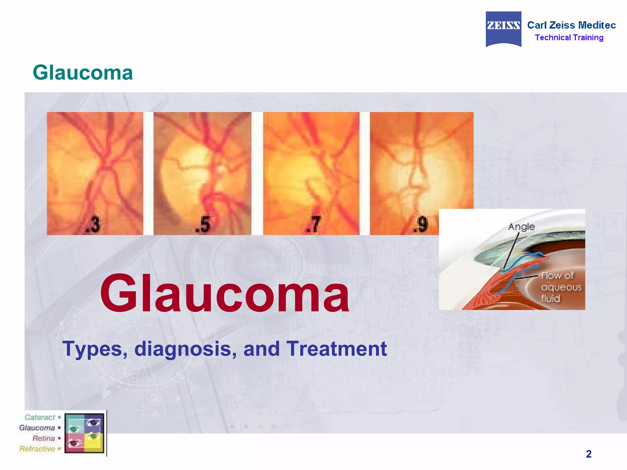
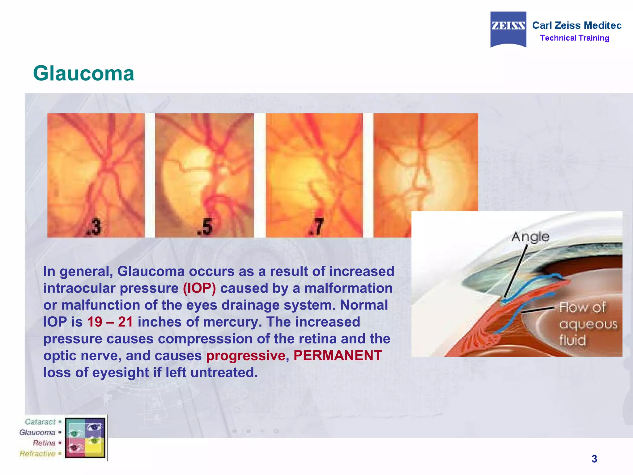


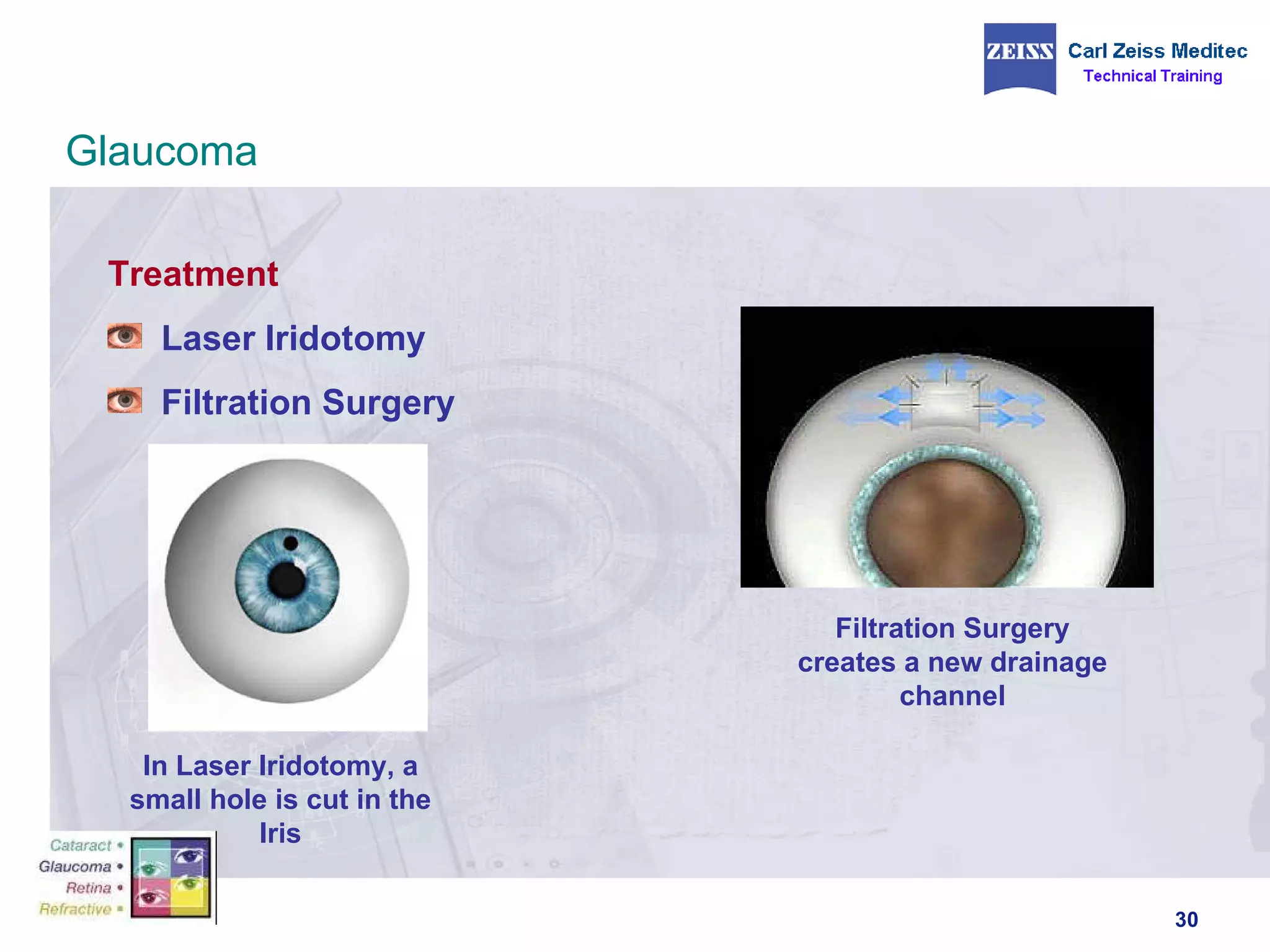

Glaucoma is caused by increased pressure in the eye (intraocular pressure) that damages the optic nerve and causes vision loss. It can be primary open-angle glaucoma, angle-closure glaucoma, congenital glaucoma, secondary glaucoma, pigmentary glaucoma, or normal tension glaucoma. Diagnosis involves tests of eye pressure, optic nerve examination, and visual field tests. Treatment options include eye medications, laser surgery, and filtration surgery to manage pressure and slow vision loss.


