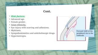
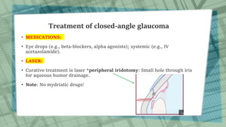
GLAUCOMA
Done by: Noor al-soub
Supervised by: Khalid al Zubi M.D
Definition
• Glaucoma is a heterogenous group of eye diseases causing optic neuropathy,
associated mostly with increased intraocular pressure (IOP). It has a characteristic
optic disc appearance (cupping) and leads to visual field defects.
• The result is damage to the optic nerve.
• Affects eye structures  atrophy of outer rim of optic nerve  peripheral vision
loss .
• Further IOP increase  loss of central vision.
• The two main types are open-angle glaucoma and angle-closure glaucoma.
Epidemiology
• Second leading cause of blindness in adults in the US following age-related
macular degeneration (AMD).
• Vision impairment in ∼ 10% of patients.
• Blindness in ∼ 5% of patients.
• Open-angle glaucoma accounts for 90% of all cases of glaucoma.
Pathophysiology
• Aqueous humor is produced by the ciliary body on the iris, flows from
the posterior chamber into the anterior chamber, and then drains back into
the venous system via the trabecular meshwork in the angle of the anterior
chamber.
• Average intraocular pressure (IOP) is between 10–21 mm Hg.
• Any process that disrupts the flow of aqueous humor may increase
intraocular pressure and lead to optic nerve damage and visual impairment.
Mechanism of Optic nerve fiber damage
• Mechanical damage by the raised IOP.
• Ischemia of nerve fibers caused by impaired perfusion pressure.
• The optic disc has a center
portion called the "cup" which
is normally quite small in
comparison to the entire optic
disc.
• In people with glaucoma,
increased pressure in the eye
and/or loss of blood flow to the
optic nerve causes the nerve
fibers to die.
• This causes the cup to become
larger in comparison to the
optic disc (increased cup: disc
ratio).
Cont.
• Normal cup: disc ratio is
0.30.4 or less.
• It varies between individuals.
• What is important in glaucoma
is the amount of increase in
cup: disc ratio, not the cup:
disc ratio itself!
Progression of glaucomatous cupping
Open angle glaucoma
primary glaucoma
• The angle is between cornea and the iris.
• The trabecular meshwork appears normal but there is an increased resistance to
the outflow due to :
• Thickening of the trabecular lamellae.
• Reduction in the number of lining trabecular cells.
• Increased extracellular materials in the trabecular meshwork.
• Risk factors:
• Age > 40 years.
• African descent.
• Diabetes mellitus.
• Family history of glaucoma.
Clinical features of open angle
glaucoma
• Initially asymptomatic.
• Bilateral, progressive visual field
loss (from peripheral to central).
• Over time, mild headaches,
impaired adaptation to darkness.
Diagnosis
• Slit-lamp examination of the
anterior segment.
• Tonometry: to measure increased
intraocular pressure.
• Gonioscopy: Special lens to
visualize angle.
• Fundoscopy: cupping and pallor of
optic disc, diffuse or focal
narrowing of the optic disc rim.
• Pachymetry: to measure the
thickness of the eye's cornea.
• Visual acuity and visual field
testing.
Slit lamp
examination
Fundoscopy
Tonometry
Gonioscopy
Treatment of open-angle glaucoma
Goal of therapy (target IOP): ≥ 25% decrease in pretreatment IOP
• MEDICATIONS:
• *Prostaglandin analogs (eye drops) most effective, alpha agonists  increase
outflow of aqueous humor.
• Beta-blockers, carbonic anhydrase inhibitors, alpha agonists  decrease
production of aqueous humor.
• LASER:
• Trabeculoplasty: opens trabecular meshwork, increase aqueous outflow.
• SURGERY:
• Trabeculectomy: creates an alternate drainage pathway.
Trabeculoplasty Trabeculectomy
Closed angle glaucoma
primary glaucoma
• It occurs through a process termed pupillary block. Normally, aqueous humor
is flows through the pupil into the anterior chamber and drains into the
trabecular meshwork to exit the eye. When the pupil is mid-dilated, the
distance between the iris and the lens is the shortest, and the two
structures can come into contact with each other. When this occurs,
aqueous humor cannot flow through the pupil into the anterior chamber, it
builds up and pushes the iris forward. When the iris is pushed against the
trabecular meshwork, aqueous humor cannot flow out of the eye (angle
closure), increasing IOP.
• Rapid buildup of pressure.
Cont.
• Risk factors:
• Advanced age.
• Female gender.
• Asian ethnicity.
• Eye injury with scarring and adhesions.
• Mydriasis.
• Sympathomimetics and anticholinergic drugs.
• Hypermetropia.
Clinical features of acute
closed angle glaucoma
• Sudden onset.
• Unilateral red, hard, and
severely painful eye.
• Frontal headaches.
• Vomiting, nausea.
• Corneal edema.
• blurred vision, halos around lights.
• Dilated, nonreactive pupil.
Clinical features of chronic
closed angle glaucoma
• Asymptomatic in early stages.
• Progressive vision loss beginning
with peripheral fields of vision.
Diagnosis:
• Slit-lamp examination, Tonometry and
Gonioscopy, Fundoscopy.
• Visual acuity: in acute angle-closure
glaucoma Corneal edema may
decrease visual acuity.
• Visual field testing.
Treatment of closed-angle glaucoma
• MEDICATIONS:
• Eye drops (e.g., beta-blockers, alpha agonists); systemic (e.g., IV
acetazolamide).
• LASER:
• Curative treatment is laser *peripheral iridotomy: Small hole through iris
for aqueous humor drainage.
• Note: No mydriatic drugs!
Normal Tension Glaucoma
• A form of glaucoma where glaucomatous changes are seen with normal
IOP (<21 mmHg).
• Here the optic nerve is unusually susceptible to the increased IOP or
has reduced blood flow.
• Presentation: young females, migraine, use of antihypertensive drugs.
• Ischemic theory is the prominent mechanism in this case.
Secondary glaucoma
• Develop as a complication from other conditions including :
• Eye injuries.
• Diabetes (diabetic retinopathy  blocks the drainage system).
• Inflammatory cells ( Uveitis ).
• Pigment from iris ( Pigment dispersion).
• Pseudoexfoliative glaucoma: deposition of amyloid produced by the
epithelium of the lens ,iris and cilliary body in the trabecular meshwork. Most
common.
• Drugs (topical or systemic steroids).
Congenital Glaucoma
• Congenital defect in the angle of the anterior chamber that obstructs the outflow
of the aqueous humour.
• Present at birth or within the first year of life.
• If <3 years old congenital glaucoma. If 3-16 years old  juvenile glaucoma.
• Presentation: enlarged globe (buphthalmos), enlarged cornea (megalocornea),
greyish-bluish discoloration of the cornea due to corneal edema, wide palpebral
fissure.
• Family history is important!
• Treatment:
• Surgical: *Goniotomy, Trabeculectomy and trabeculotomy.
• Medical and laser treatment may be needed later.

Glaucoma

  • 1.
    GLAUCOMA Done by: Nooral-soub Supervised by: Khalid al Zubi M.D
  • 2.
    Definition • Glaucoma isa heterogenous group of eye diseases causing optic neuropathy, associated mostly with increased intraocular pressure (IOP). It has a characteristic optic disc appearance (cupping) and leads to visual field defects. • The result is damage to the optic nerve. • Affects eye structures  atrophy of outer rim of optic nerve  peripheral vision loss . • Further IOP increase  loss of central vision. • The two main types are open-angle glaucoma and angle-closure glaucoma.
  • 3.
    Epidemiology • Second leadingcause of blindness in adults in the US following age-related macular degeneration (AMD). • Vision impairment in ∼ 10% of patients. • Blindness in ∼ 5% of patients. • Open-angle glaucoma accounts for 90% of all cases of glaucoma.
  • 4.
    Pathophysiology • Aqueous humoris produced by the ciliary body on the iris, flows from the posterior chamber into the anterior chamber, and then drains back into the venous system via the trabecular meshwork in the angle of the anterior chamber. • Average intraocular pressure (IOP) is between 10–21 mm Hg. • Any process that disrupts the flow of aqueous humor may increase intraocular pressure and lead to optic nerve damage and visual impairment.
  • 6.
    Mechanism of Opticnerve fiber damage • Mechanical damage by the raised IOP. • Ischemia of nerve fibers caused by impaired perfusion pressure.
  • 7.
    • The opticdisc has a center portion called the "cup" which is normally quite small in comparison to the entire optic disc. • In people with glaucoma, increased pressure in the eye and/or loss of blood flow to the optic nerve causes the nerve fibers to die. • This causes the cup to become larger in comparison to the optic disc (increased cup: disc ratio).
  • 8.
    Cont. • Normal cup:disc ratio is 0.30.4 or less. • It varies between individuals. • What is important in glaucoma is the amount of increase in cup: disc ratio, not the cup: disc ratio itself! Progression of glaucomatous cupping
  • 9.
    Open angle glaucoma primaryglaucoma • The angle is between cornea and the iris. • The trabecular meshwork appears normal but there is an increased resistance to the outflow due to : • Thickening of the trabecular lamellae. • Reduction in the number of lining trabecular cells. • Increased extracellular materials in the trabecular meshwork. • Risk factors: • Age > 40 years. • African descent. • Diabetes mellitus. • Family history of glaucoma.
  • 10.
    Clinical features ofopen angle glaucoma • Initially asymptomatic. • Bilateral, progressive visual field loss (from peripheral to central). • Over time, mild headaches, impaired adaptation to darkness. Diagnosis • Slit-lamp examination of the anterior segment. • Tonometry: to measure increased intraocular pressure. • Gonioscopy: Special lens to visualize angle. • Fundoscopy: cupping and pallor of optic disc, diffuse or focal narrowing of the optic disc rim. • Pachymetry: to measure the thickness of the eye's cornea. • Visual acuity and visual field testing.
  • 11.
  • 12.
    Treatment of open-angleglaucoma Goal of therapy (target IOP): ≥ 25% decrease in pretreatment IOP • MEDICATIONS: • *Prostaglandin analogs (eye drops) most effective, alpha agonists  increase outflow of aqueous humor. • Beta-blockers, carbonic anhydrase inhibitors, alpha agonists  decrease production of aqueous humor. • LASER: • Trabeculoplasty: opens trabecular meshwork, increase aqueous outflow. • SURGERY: • Trabeculectomy: creates an alternate drainage pathway.
  • 13.
  • 14.
    Closed angle glaucoma primaryglaucoma • It occurs through a process termed pupillary block. Normally, aqueous humor is flows through the pupil into the anterior chamber and drains into the trabecular meshwork to exit the eye. When the pupil is mid-dilated, the distance between the iris and the lens is the shortest, and the two structures can come into contact with each other. When this occurs, aqueous humor cannot flow through the pupil into the anterior chamber, it builds up and pushes the iris forward. When the iris is pushed against the trabecular meshwork, aqueous humor cannot flow out of the eye (angle closure), increasing IOP. • Rapid buildup of pressure.
  • 15.
    Cont. • Risk factors: •Advanced age. • Female gender. • Asian ethnicity. • Eye injury with scarring and adhesions. • Mydriasis. • Sympathomimetics and anticholinergic drugs. • Hypermetropia.
  • 16.
    Clinical features ofacute closed angle glaucoma • Sudden onset. • Unilateral red, hard, and severely painful eye. • Frontal headaches. • Vomiting, nausea. • Corneal edema. • blurred vision, halos around lights. • Dilated, nonreactive pupil. Clinical features of chronic closed angle glaucoma • Asymptomatic in early stages. • Progressive vision loss beginning with peripheral fields of vision. Diagnosis: • Slit-lamp examination, Tonometry and Gonioscopy, Fundoscopy. • Visual acuity: in acute angle-closure glaucoma Corneal edema may decrease visual acuity. • Visual field testing.
  • 17.
    Treatment of closed-angleglaucoma • MEDICATIONS: • Eye drops (e.g., beta-blockers, alpha agonists); systemic (e.g., IV acetazolamide). • LASER: • Curative treatment is laser *peripheral iridotomy: Small hole through iris for aqueous humor drainage. • Note: No mydriatic drugs!
  • 19.
    Normal Tension Glaucoma •A form of glaucoma where glaucomatous changes are seen with normal IOP (<21 mmHg). • Here the optic nerve is unusually susceptible to the increased IOP or has reduced blood flow. • Presentation: young females, migraine, use of antihypertensive drugs. • Ischemic theory is the prominent mechanism in this case.
  • 20.
    Secondary glaucoma • Developas a complication from other conditions including : • Eye injuries. • Diabetes (diabetic retinopathy  blocks the drainage system). • Inflammatory cells ( Uveitis ). • Pigment from iris ( Pigment dispersion). • Pseudoexfoliative glaucoma: deposition of amyloid produced by the epithelium of the lens ,iris and cilliary body in the trabecular meshwork. Most common. • Drugs (topical or systemic steroids).
  • 21.
    Congenital Glaucoma • Congenitaldefect in the angle of the anterior chamber that obstructs the outflow of the aqueous humour. • Present at birth or within the first year of life. • If <3 years old congenital glaucoma. If 3-16 years old  juvenile glaucoma. • Presentation: enlarged globe (buphthalmos), enlarged cornea (megalocornea), greyish-bluish discoloration of the cornea due to corneal edema, wide palpebral fissure. • Family history is important! • Treatment: • Surgical: *Goniotomy, Trabeculectomy and trabeculotomy. • Medical and laser treatment may be needed later.

Editor's Notes

  • #13 *Latanoprost , Travoprost , Bimatoprost .
  • #17 Acute angle-closure glaucoma is a medical emergency, as it can cause permanent vision loss if left untreated!
  • #18 peripheral iridotomy is first line for chronic closed angle glaucoma.
  • #22 *Goniotomy is a surgical procedure in which the doctor uses a lens called a goniolens to see the structures of the front part of the eye (anterior chamber). An opening is made in the trabecular meshwork, the group of tiny canals located in the drainage angle, where fluid leaves the eye.