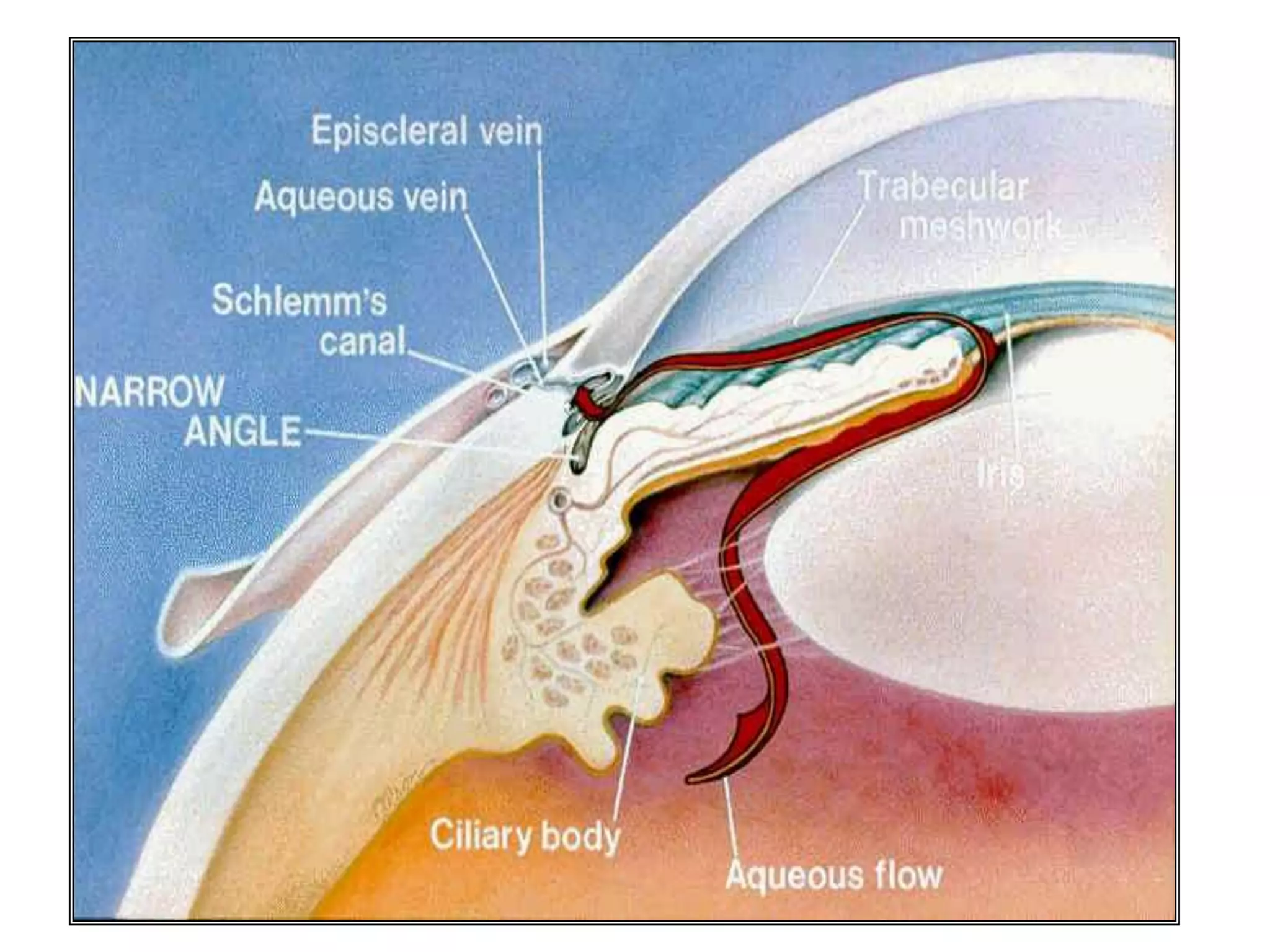
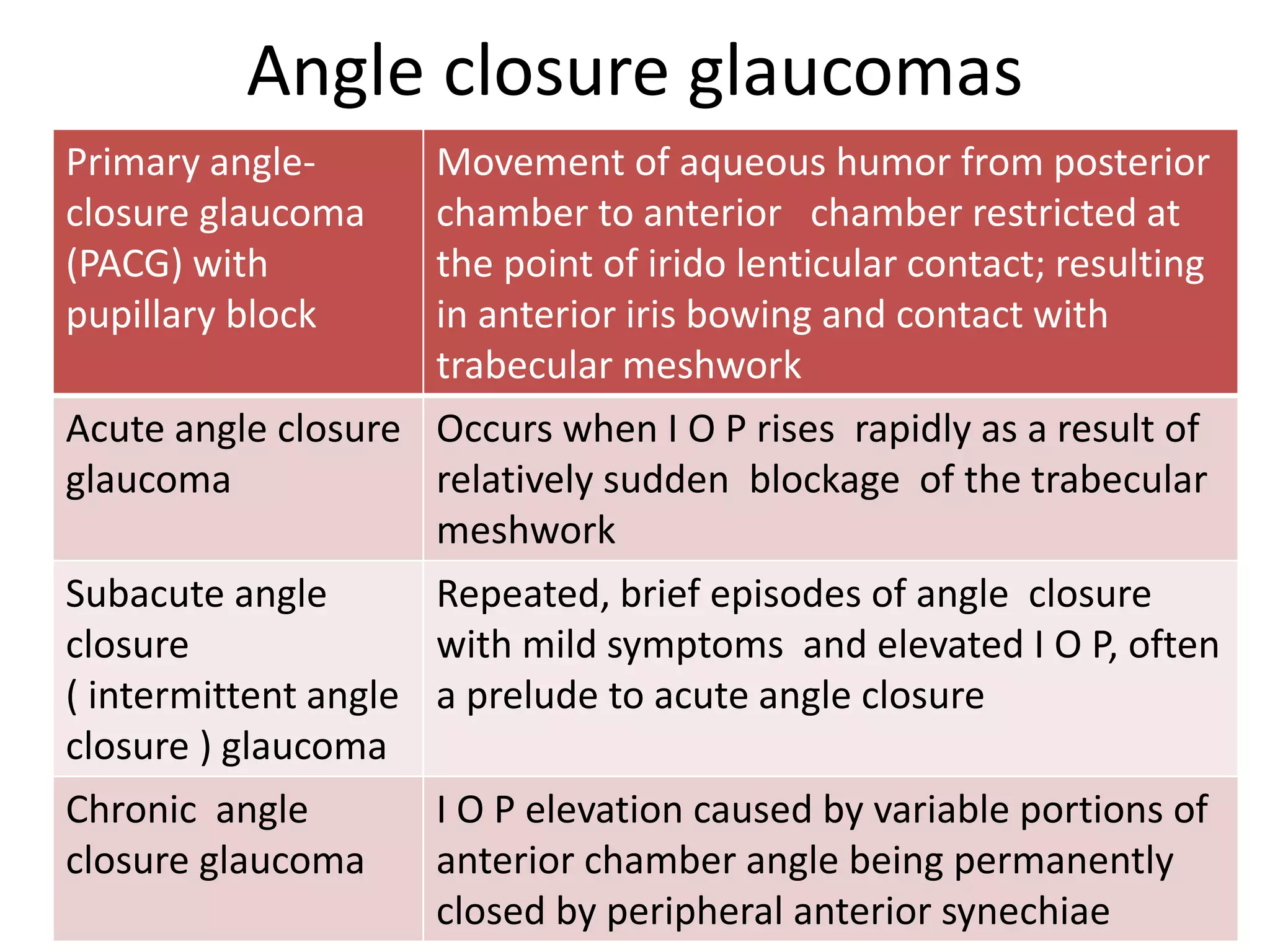
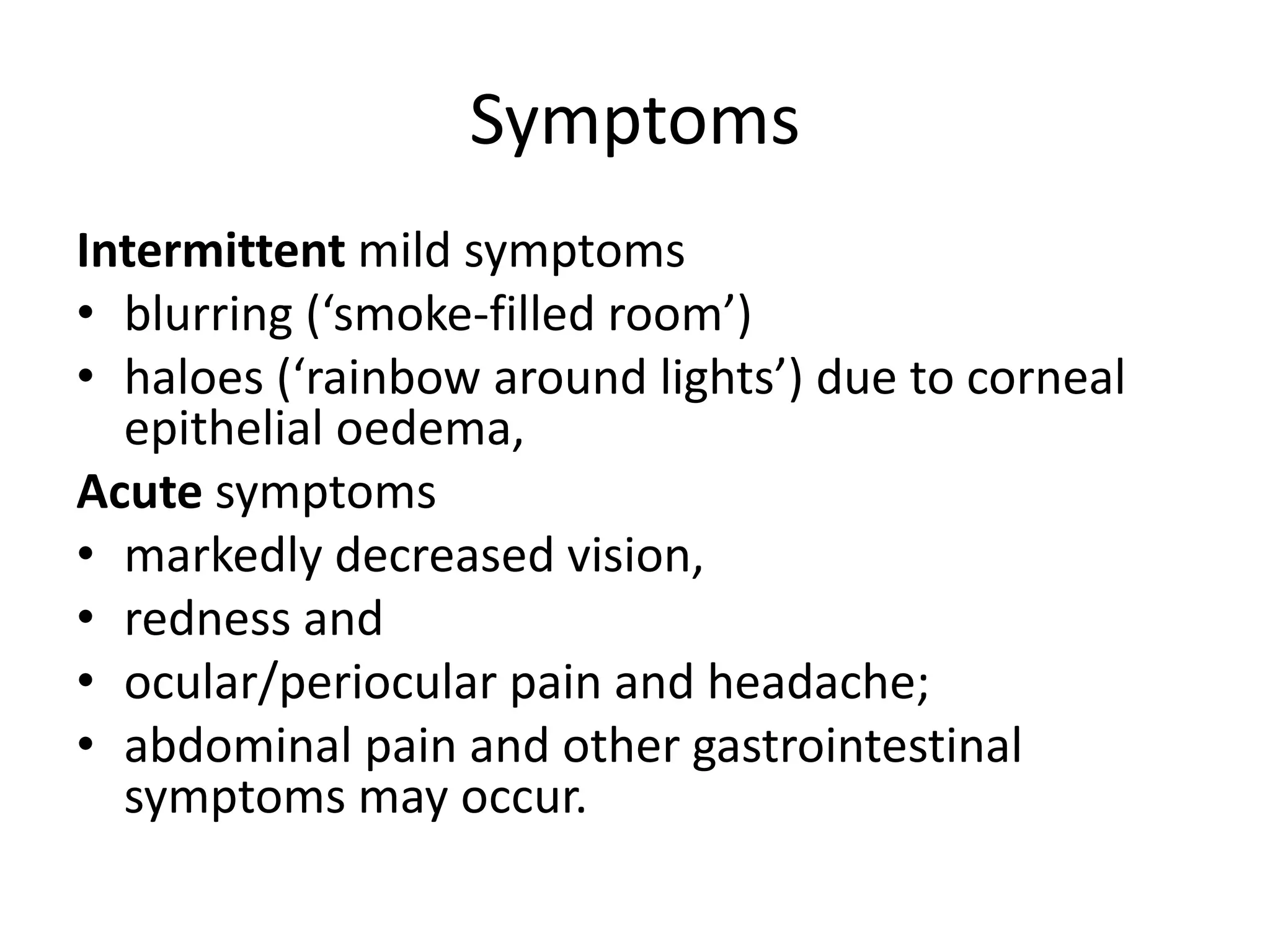
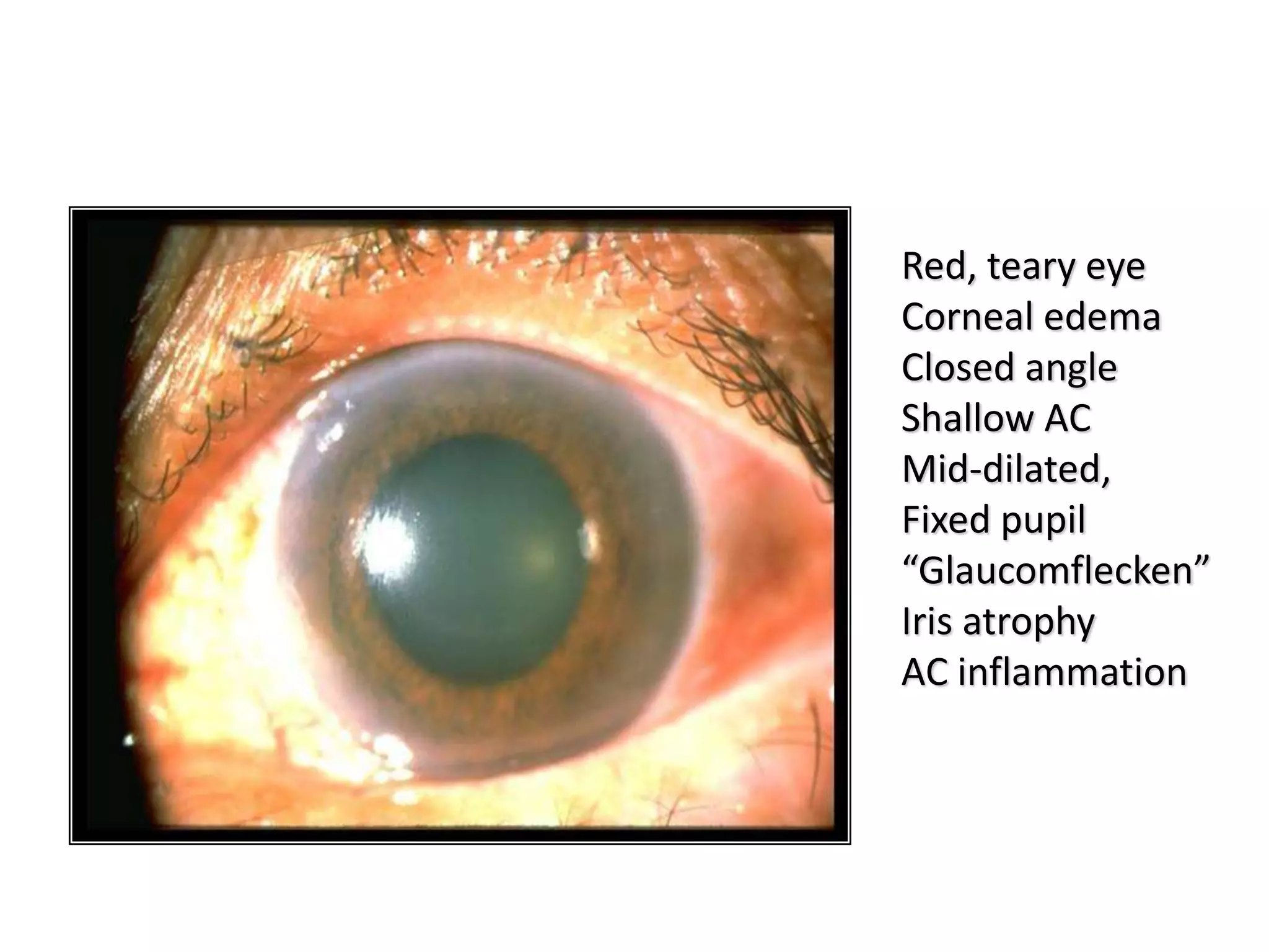
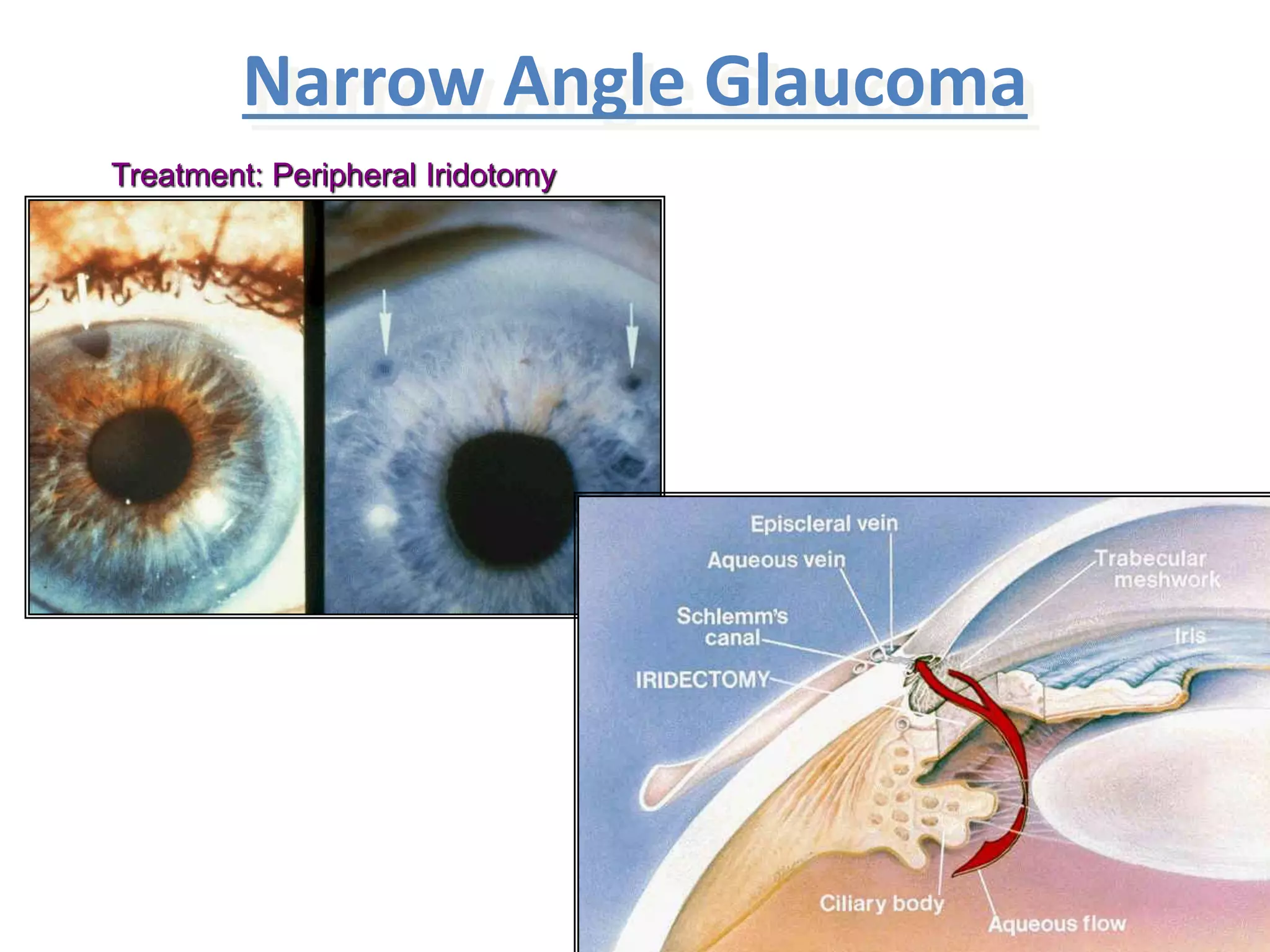
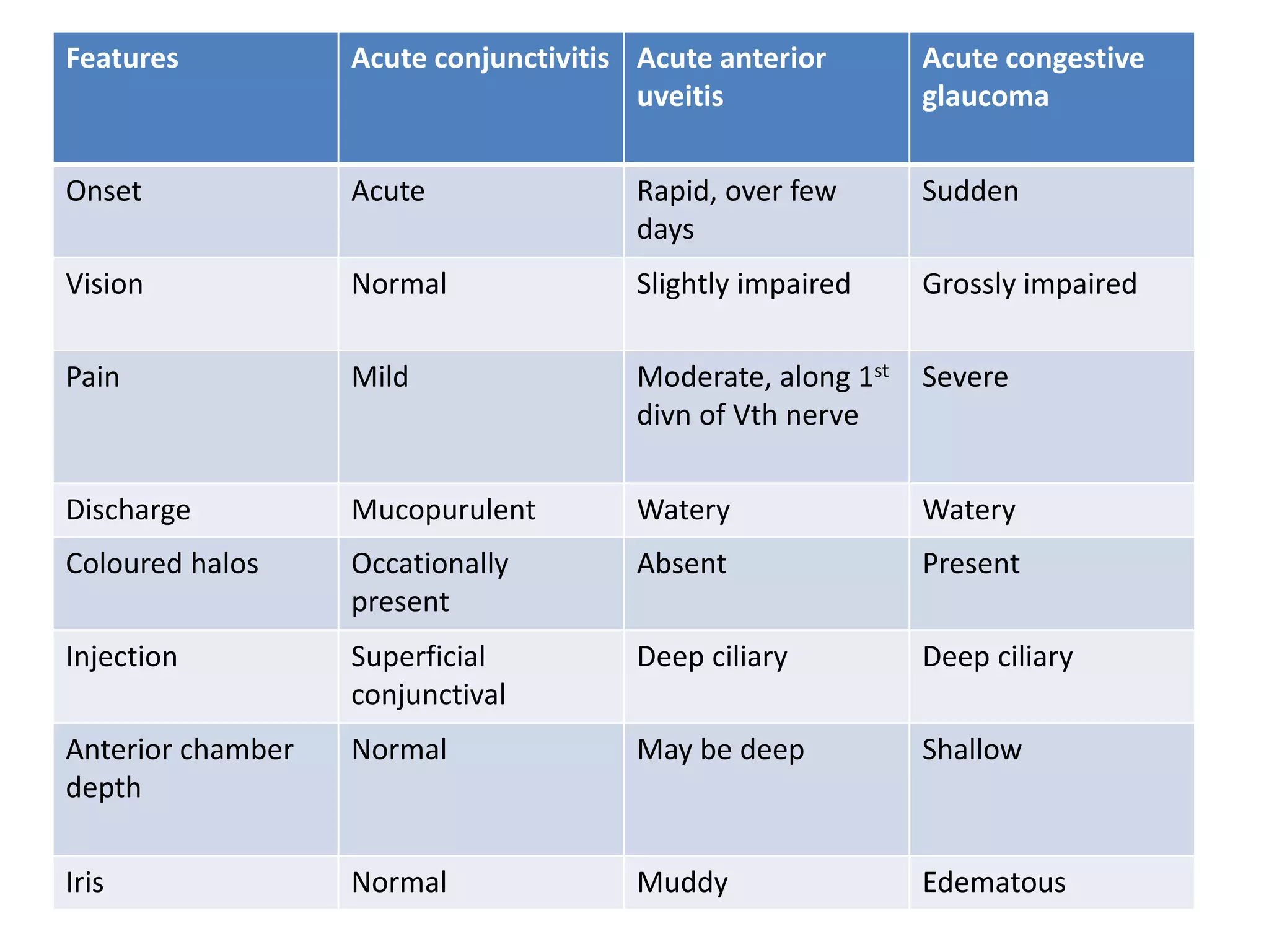
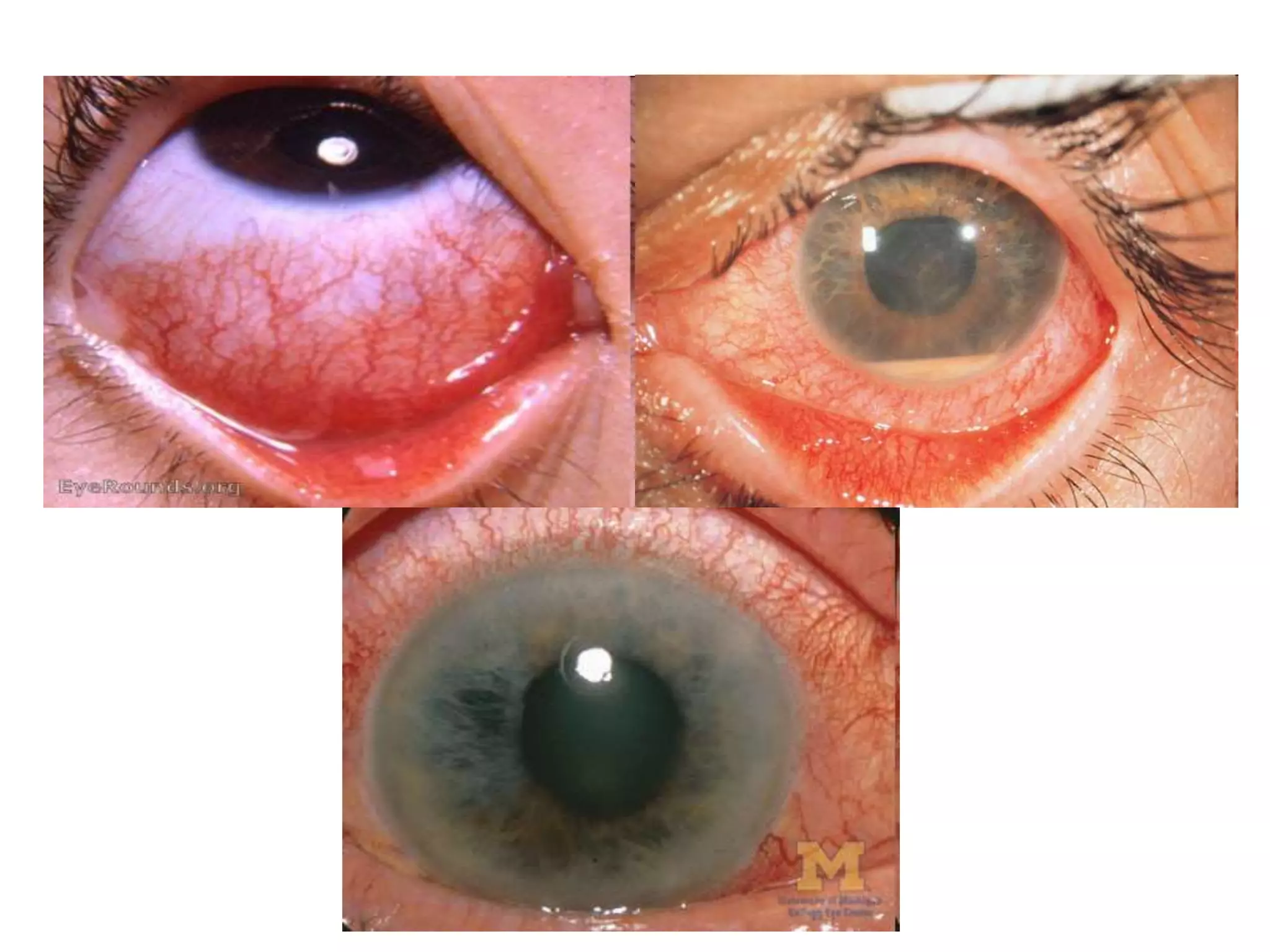
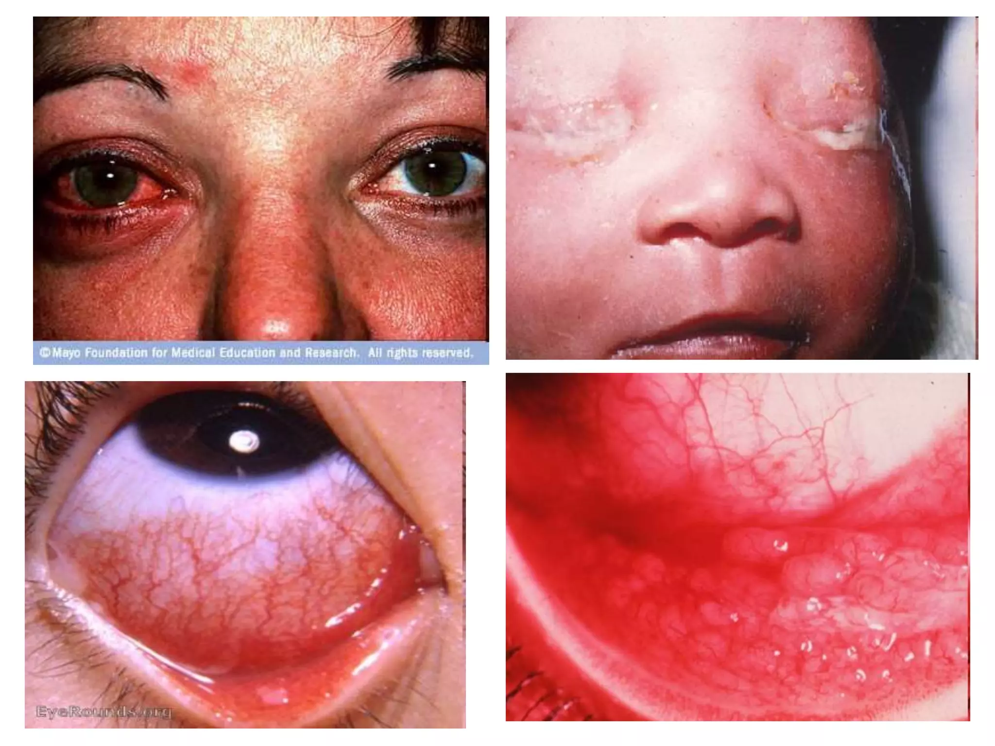
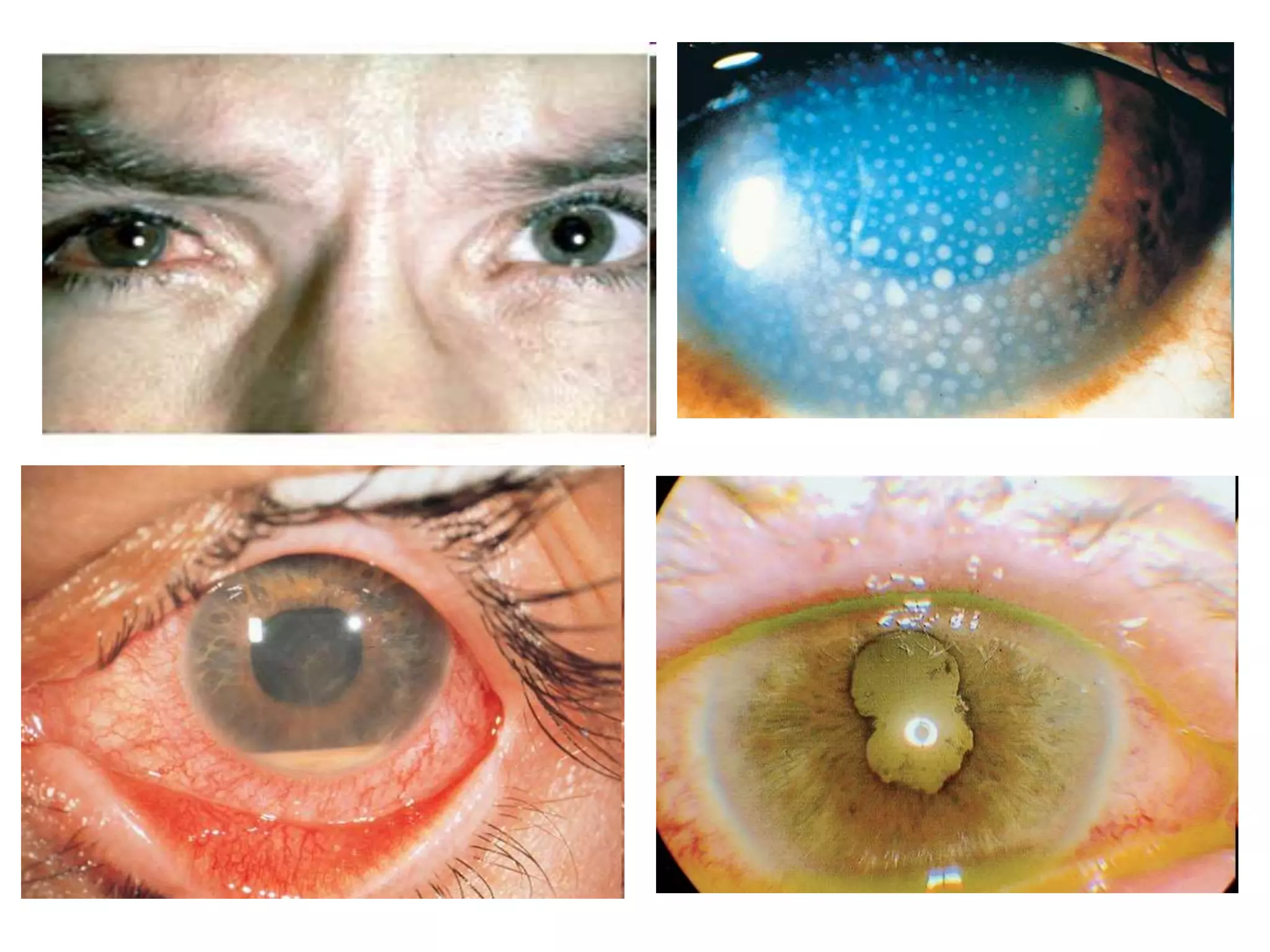
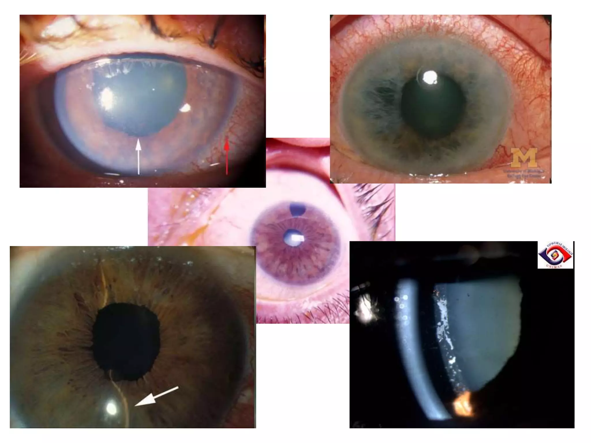
Primary Angle Closure Glaucoma
Dr AR Rajalakshmi
• PACG definition
• Stages
• Clinical presentation
• Management
• Red eye D/D
Angle closure glaucomas
Primary angle-
closure glaucoma
(PACG) with
pupillary block
Movement of aqueous humor from posterior
chamber to anterior chamber restricted at
the point of irido lenticular contact; resulting
in anterior iris bowing and contact with
trabecular meshwork
Acute angle closure
glaucoma
Occurs when I O P rises rapidly as a result of
relatively sudden blockage of the trabecular
meshwork
Subacute angle
closure
( intermittent angle
closure ) glaucoma
Repeated, brief episodes of angle closure
with mild symptoms and elevated I O P, often
a prelude to acute angle closure
Chronic angle
closure glaucoma
I O P elevation caused by variable portions of
anterior chamber angle being permanently
closed by peripheral anterior synechiae
Secondary angle
closure glaucoma
with pupillary
block
Pupillary block occurs as a result of a mechanism
other than the anatomical configuration of the
anterior segment (eg, an intumescent lens or a
secluded pupil ) .
Secondary angle
closure glaucoma
without pupillary
block
Posterior pushing mechanism: lens-iris interface
pushed forward (eg, posterior segment tumor,
scleral buckling procedure, uveal effusion )
Anterior pulling mechanism : anterior segment
process pulling iris forward to form peripheral
anterior synechiae ( e g , iridocorneal endothelial
syndrome, neovascular glaucoma, inflammation)
Plateau iris
syndrome
An anatomical variation in the iris root in which
narrowing of the angle occurs independent of
pupillary block
Risk Factors
• Age. 60 years at presentation.
• Gender. Females > males.
• Race Far Eastern and Indian Asians.
• Family history. Genetic factors are important ;
increased prevalence of angle closure in family
members.
• Refraction. Eyes with ‘pure’ pupillary block are typically
hypermetropic,
• hypermetropia of one dioptre or more are primary
angle closure suspects, so routine gonioscopy should
be considered in all hypermetropes.
• Axial length. Short eyes tend to have a shallow AC.
Precipitating factors include:
• watching television in a darkened room,
• pharmacological mydriasis
• adoption of a semi-prone position (e.g. reading),
• acute emotional stress and
• systemic medication:
– parasympathetic antagonists or sympathetic agonists
including inhalers, motion sickness patches and
cold/flu remedies (mydriatic effect),
– topiramate and other sulfa derivatives (ciliary body
effusion).
Symptoms
Intermittent mild symptoms
• blurring (‘smoke-filled room’)
• haloes (‘rainbow around lights’) due to corneal
epithelial oedema,
Acute symptoms
• markedly decreased vision,
• redness and
• ocular/periocular pain and headache;
• abdominal pain and other gastrointestinal
symptoms may occur.
Chronic presentation
• intraocular pressure is chronically raised
• synechial closure over at least 180°.
• Changes in the optic nerve head and visual field may
or may not be present
• repeated subacute attacks of PACG
• acute PACG persisting for more than a few hours
• asymptomatic or 'creeping' angle closure
Clinical Presentation
Acute primary angle closure
(APAC)
• VA is usually 6/60 to HM.
• IOP is usually very high
(> 50 mmHg).
• Conjunctival hyperaemia
with violaceous
circumcorneal injection.
• Corneal epithelial
oedema
• The AC is shallow, and
aqueous flare present.
• An unreactive mid-dilated
vertically oval pupil is
classic
Red, teary eye
Corneal edema
Closed angle
Shallow AC
Mid-dilated,
Fixed pupil
“Glaucomflecken”
Iris atrophy
AC inflammation
• The fellow eye typically shows an occludable angle; if
not present, secondary causes should be considered
Treatment
PACS
• Laser iridotomy
• If significant ITC persists after iridotomy,
options include
• observation (most),
• laser iridoplasty, and
• If symptomatic cataract is present, lens
extraction usually definitively opens the angle
APAC
Initial treatment
• Supine position to encourage the lens to shift posteriorly
under the influence of gravity.
• Acetazolamide 500 mg is given intravenously if IOP >50
mmHg,
– (Contraindications include sulfonamide allergy and
angle closure secondary to topiramate/other sulfa
derivatives).
• IV mannitol 20% 1 gm/kg body weight
• Timolol 0.5%, and prednisolone 1% or dexamethasone
0.1% to the affected eye, leaving 5 minutes between
each.
• Pilocarpine 2–4% one drop to the affected eye,
repeated after half an hour;
• one drop of 1% into the fellow eye.
• omit pilocarpine until a significant IOP fall, as when
IOP is high ischaemia may compromise its action,
• Analgesia and an antiemetic may be required.
• Assess the other eye
Laser iridotomy
Laser iridotomy: creates a small hole in the iris to
improve flow of aqueous humor into drainage angle.
Narrow Angle Glaucoma
Treatment: Peripheral Iridotomy
Trabeculectomy
Combined mechanism glaucoma
• A patient who has been
successfully treated for a
narrow angle but who
continues to demonstrate
reduced outflow facility
and elevated IOP in the
absence of peripheral
anterior synechiae (PAS).
• the intrinsic resistance of
the trabecular meshwork
to aqueous outflow in
open - angle glaucoma
• A patient who has open-
angle glaucoma but
develops secondary angle
closure from other causes
• the direct anatomical
obstruction of the
filtering meshwork by
synechiae in angle closure
glaucoma
Red eye
Differential diagnosis
• Conjunctivitis
• Acute uveitis
• Acute attack of angle closure glaucoma
• Corneal ulcer
• Scleritis
• Episcleritis
• Trauma
• Subconjunctival hemorrhage
Features Acute conjunctivitis Acute anterior
uveitis
Acute congestive
glaucoma
Onset Acute Rapid, over few
days
Sudden
Vision Normal Slightly impaired Grossly impaired
Pain Mild Moderate, along 1st
divn of Vth nerve
Severe
Discharge Mucopurulent Watery Watery
Coloured halos Occationally
present
Absent Present
Injection Superficial
conjunctival
Deep ciliary Deep ciliary
Anterior chamber
depth
Normal May be deep Shallow
Iris Normal Muddy Edematous
Features Acute conjunctivitis Acute anterior
uveitis
Acute congestive
glaucoma
Pupil Normal size, brisk
reaction
Small, irregular,
sluggish reaction
Vertically oval,non
reacting
IOP Normal Normal, raised, low Markedly raised
Ciliary tenderness Absent Marked Marked
Cornea Clear KPs Edema, haze
Aqueous Clear Flare & cells Flare
Lens Transparent Transparent Opacities
occationally
Vitreous Clear Hazy ant vitr Clear
Constitutional
symptoms
Absent Mild Prostration,
vomitting
• Stages of PACG
• Clinical presentation
• Management
• Red eye D/D

Pacg 04.05.16 - dr.a.r.rajalakshmi

  • 1.
    Primary Angle ClosureGlaucoma Dr AR Rajalakshmi
  • 2.
    • PACG definition •Stages • Clinical presentation • Management • Red eye D/D
  • 4.
    Angle closure glaucomas Primaryangle- closure glaucoma (PACG) with pupillary block Movement of aqueous humor from posterior chamber to anterior chamber restricted at the point of irido lenticular contact; resulting in anterior iris bowing and contact with trabecular meshwork Acute angle closure glaucoma Occurs when I O P rises rapidly as a result of relatively sudden blockage of the trabecular meshwork Subacute angle closure ( intermittent angle closure ) glaucoma Repeated, brief episodes of angle closure with mild symptoms and elevated I O P, often a prelude to acute angle closure Chronic angle closure glaucoma I O P elevation caused by variable portions of anterior chamber angle being permanently closed by peripheral anterior synechiae
  • 5.
    Secondary angle closure glaucoma withpupillary block Pupillary block occurs as a result of a mechanism other than the anatomical configuration of the anterior segment (eg, an intumescent lens or a secluded pupil ) . Secondary angle closure glaucoma without pupillary block Posterior pushing mechanism: lens-iris interface pushed forward (eg, posterior segment tumor, scleral buckling procedure, uveal effusion ) Anterior pulling mechanism : anterior segment process pulling iris forward to form peripheral anterior synechiae ( e g , iridocorneal endothelial syndrome, neovascular glaucoma, inflammation) Plateau iris syndrome An anatomical variation in the iris root in which narrowing of the angle occurs independent of pupillary block
  • 6.
    Risk Factors • Age.60 years at presentation. • Gender. Females > males. • Race Far Eastern and Indian Asians. • Family history. Genetic factors are important ; increased prevalence of angle closure in family members. • Refraction. Eyes with ‘pure’ pupillary block are typically hypermetropic, • hypermetropia of one dioptre or more are primary angle closure suspects, so routine gonioscopy should be considered in all hypermetropes. • Axial length. Short eyes tend to have a shallow AC.
  • 7.
    Precipitating factors include: •watching television in a darkened room, • pharmacological mydriasis • adoption of a semi-prone position (e.g. reading), • acute emotional stress and • systemic medication: – parasympathetic antagonists or sympathetic agonists including inhalers, motion sickness patches and cold/flu remedies (mydriatic effect), – topiramate and other sulfa derivatives (ciliary body effusion).
  • 8.
    Symptoms Intermittent mild symptoms •blurring (‘smoke-filled room’) • haloes (‘rainbow around lights’) due to corneal epithelial oedema, Acute symptoms • markedly decreased vision, • redness and • ocular/periocular pain and headache; • abdominal pain and other gastrointestinal symptoms may occur.
  • 9.
    Chronic presentation • intraocularpressure is chronically raised • synechial closure over at least 180°. • Changes in the optic nerve head and visual field may or may not be present • repeated subacute attacks of PACG • acute PACG persisting for more than a few hours • asymptomatic or 'creeping' angle closure
  • 10.
    Clinical Presentation Acute primaryangle closure (APAC) • VA is usually 6/60 to HM. • IOP is usually very high (> 50 mmHg). • Conjunctival hyperaemia with violaceous circumcorneal injection. • Corneal epithelial oedema • The AC is shallow, and aqueous flare present. • An unreactive mid-dilated vertically oval pupil is classic
  • 11.
    Red, teary eye Cornealedema Closed angle Shallow AC Mid-dilated, Fixed pupil “Glaucomflecken” Iris atrophy AC inflammation
  • 12.
    • The felloweye typically shows an occludable angle; if not present, secondary causes should be considered
  • 13.
    Treatment PACS • Laser iridotomy •If significant ITC persists after iridotomy, options include • observation (most), • laser iridoplasty, and • If symptomatic cataract is present, lens extraction usually definitively opens the angle
  • 14.
    APAC Initial treatment • Supineposition to encourage the lens to shift posteriorly under the influence of gravity. • Acetazolamide 500 mg is given intravenously if IOP >50 mmHg, – (Contraindications include sulfonamide allergy and angle closure secondary to topiramate/other sulfa derivatives). • IV mannitol 20% 1 gm/kg body weight • Timolol 0.5%, and prednisolone 1% or dexamethasone 0.1% to the affected eye, leaving 5 minutes between each.
  • 15.
    • Pilocarpine 2–4%one drop to the affected eye, repeated after half an hour; • one drop of 1% into the fellow eye. • omit pilocarpine until a significant IOP fall, as when IOP is high ischaemia may compromise its action, • Analgesia and an antiemetic may be required. • Assess the other eye
  • 16.
    Laser iridotomy Laser iridotomy:creates a small hole in the iris to improve flow of aqueous humor into drainage angle.
  • 17.
  • 18.
  • 19.
    Combined mechanism glaucoma •A patient who has been successfully treated for a narrow angle but who continues to demonstrate reduced outflow facility and elevated IOP in the absence of peripheral anterior synechiae (PAS). • the intrinsic resistance of the trabecular meshwork to aqueous outflow in open - angle glaucoma • A patient who has open- angle glaucoma but develops secondary angle closure from other causes • the direct anatomical obstruction of the filtering meshwork by synechiae in angle closure glaucoma
  • 20.
  • 21.
    Differential diagnosis • Conjunctivitis •Acute uveitis • Acute attack of angle closure glaucoma • Corneal ulcer • Scleritis • Episcleritis • Trauma • Subconjunctival hemorrhage
  • 22.
    Features Acute conjunctivitisAcute anterior uveitis Acute congestive glaucoma Onset Acute Rapid, over few days Sudden Vision Normal Slightly impaired Grossly impaired Pain Mild Moderate, along 1st divn of Vth nerve Severe Discharge Mucopurulent Watery Watery Coloured halos Occationally present Absent Present Injection Superficial conjunctival Deep ciliary Deep ciliary Anterior chamber depth Normal May be deep Shallow Iris Normal Muddy Edematous
  • 23.
    Features Acute conjunctivitisAcute anterior uveitis Acute congestive glaucoma Pupil Normal size, brisk reaction Small, irregular, sluggish reaction Vertically oval,non reacting IOP Normal Normal, raised, low Markedly raised Ciliary tenderness Absent Marked Marked Cornea Clear KPs Edema, haze Aqueous Clear Flare & cells Flare Lens Transparent Transparent Opacities occationally Vitreous Clear Hazy ant vitr Clear Constitutional symptoms Absent Mild Prostration, vomitting
  • 28.
    • Stages ofPACG • Clinical presentation • Management • Red eye D/D