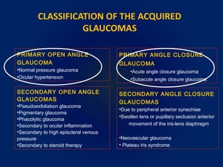
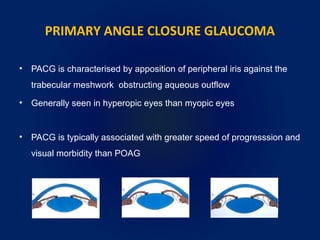
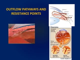
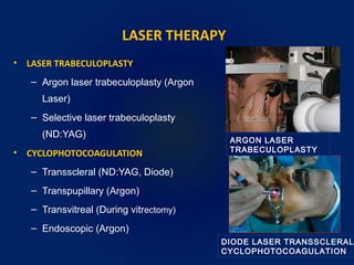
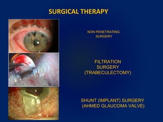
POAG AND PACG
AN INTRODUCTION
JAYENDRA JHA
OPTOMETRIST
C.L GUPTA EYE INSTITUTE
MORADABAD
HISTORICAL ASPECTS
• The glaucoma term is derived from the old greek
word “glaukos” which means “greyish-blue”
• Hippocrates defined glaucoma as “a disease of
the elderly patients in which the pupilla becomes
blue”.
– A person with a swollen cornea and a rapidly
developing cataract and chronic (long-term)
elevated pressure inside the eye
c. 460 BC–c. 380 BC
WHAT IS THE INTRAOCULAR PRESSURE
• Pressure insıde the eye is termed ”intraocular pressure (IOP)”
• Eye pressure is measured in millimeters of mercury (mmHg)
• “Normal eye pressure” is not a stable number(s), it ranges from 10
to 21 mmHg
• Elevated IOP is an eye pressure of “greater than 21 mmHg”
WHAT IS GLAUCOMA
• Currently, glaucoma is defined as “a progressive optic
neuropathy which causes permanent blindness by damaging
the optic nerve and the periferal visual field”.
• Glaucoma affects 3% of the society and the second frequent
reason of permanent blindness (especially in developed
countries).
• The prevalance is higher in elderly population.
RISK FACTORS
• IOP
• AGE
• RACE
• FAMILY HISTORY
• DIABETES MELLITUS
• REFRACTIVE ERROR
• VASCULAR DISEASE
CLASSIFICATION OF GLAUCOMA
• Various classifıcations are available.
• Many classifications are based on etiology, anatomy and clinical
presentation.
• CLASSIFICATION BY THE TIME OF ONSET IS AS;
– Congenital glaucoma
– Acquired glaucoma
• Primary glaucoma
• Secondary glaucoma
CLASSIFICATION OF THE ACQUIRED
GLAUCOMAS
PRIMARY OPEN ANGLE
GLAUCOMA
•Normal pressure glaucoma
•Ocular hypertensıon
SECONDARY OPEN ANGLE
GLAUCOMAS
•Pseudoexfoliatıon glaucoma
•Pigmentary glaucoma
•Phacolytic glaucoma
•Secondary to ocular inflammation
•Secondary to high episcleral venous
pressure
•Secondary to steroid therapy
PRIMARY ANGLE CLOSURE
GLAUCOMA
•Acute angle closure glaucoma
•Subacute angle closure glaucoma
SECONDARY ANGLE CLOSURE
GLAUCOMAS
•Due to peripheral anterior synechiae
•Swollen lens or pupillary seclusion anterior
movement of the iris-lens diaphragm
•Neovascular glaucoma
• Plateau iris syndrome
PRIMARY OPEN ANGLE GLAUCOMA
• POAG is described as optic nerve damage from multiple possible
causes that is chronic and progresses over time
• A loss of optic nerve fibers is characteristic of the disease
• POAG characteristics are open anterior chamber angle, high
intraocular pressure in the eye ,visual field abnormalities and
cupping and atrophy of the optic disc
• The exact cause of POAG is unknown
• The most important (and well known) cause of POAG is increased
IOP
• The cause of the high IOP is generally accepted to be because of an
imbalance in the productıon and drainage of fluid in the eye
(aqueous humor)
• The fluid is continually being produced but cannot be drained
because of the improperly functioning drainage channels (called
trabecular meshwork)
POAG CAUSES ?
RAISING THE IOP!!
PRIMARY ANGLE CLOSURE GLAUCOMA
• PACG is characterised by apposition of peripheral iris against the
trabecular meshwork obstructing aqueous outflow
• Generally seen in hyperopic eyes than myopic eyes
• PACG is typically associated with greater speed of progresssion and
visual morbidity than POAG
OUTFLOW PATHWAYS AND
RESISTANCE POINTS
GLAUCOMATOUS DAMAGE
• The basis of the glaucomatous damage
is the loss of retinal ganglion cells.
• The ganglion cells constituting the
retinal nerve fiber layer and their axons
dies during the glaucomatous damage
process.
SYMPTOMS
• Most cases are asymptomatic until the visual field abnormalities
become prominent and affect central vision.
• Thus, annual routine examinatıon is essential for early
diagnosis.
• In PACG, some patient present acute pain and headache
SIGNS
CHRONIC PRESENTATION
• VA is normal unless advanced stages
• Anterior chamber is shallow
• IOP elevation may be only intermittent
ACUTE ANGLE CLOSURE
• VA is usually 20/200 or HM
• IOP is usually very high (50-100 mmHg)
• Conjunctival hyperaemia, corneal oedema
• Unreactive mid- dilated oval pupil
• Fellow eye generally shows an occludable angle
THE EVALUATION OF GLAUCOMA PATIENTS
• Visual acuity (best corrected)
• Biomicroscopy (clues to specific diagnosis...)
• Measurement of intraocular pressure
– Applanation tonometry
– Noncontact tonometry
• Pachymetry
• Evaluation of the anterior chamber angle
• Perimetry
• Funduscopy
TONOMETRY
• Tonometry is a method used to measure the pressure inside the eye
• Because IOP varies from hour to hour in any individual (diurnal
variation), measurements may be taken at different times of day
(morning and night)
– A difference in pressure between morning and night of 5 mmHg
or more may suggest glaucoma
• A difference in pressure between the two eyes of 3 mmHg or more
may suggest glaucoma
THE TECHNIQUES OF
IOP MEASUREMENTS
APPLANATION TONOMETRY
SCHIOTZ
TONOMETER
PERRKINS HAND
HELD TONOMETER
TONOPEN XLNON CONTACT TONOMETER
PACHYMETRY
Normal central corneal thickness is variable
500-520 microns
 Thinner cornea (CCT < 500 µm) can give
falsely low pressure readings
 Severe glaucoma patients may be failed
diagnose
 A thick cornea (>600 µm) can give falsely
high pressure readings
 Unnecessary treatments !!
GONIOSCOPY
SL:Schwalbe’s lıne
TM:Trabecular Meshwork
SS:Scleral Spur
CBB:Ciliary Body Band
•Gonioscopy is performed to check
the drainage angle of an eye
•A special contact lens(goniolens)
is placed on the eye
•This test is important to
determine if the angles are open,
narrowed, or closed
•Open angle: long term,slowly,
insidious disease
•Close (narrow): risk of PACG.
VISUAL FIELD TESTING
• VF testing to check the patients peripheral vision
• Typically by using an automated visual field machine
• This test is done to rule out any visual defects due to glaucoma
• VF defects may not be apperent until over 40% of the optic nerve
fiber layer has been lost
• VF testıng may need to be repeated
– A low risk of glaucomatous damage, the test may be performed
once a year
– A high risk of glaucomatous damage, test may be performed as
frequently as every 2 months
NORMAL VF
AUTOMATED VISUAL FIELD ANALYZER
STAGES OF GLAUCOMATOUS VISUAL FIELD
DEFECTS
EARLY STAGE MODERATE STAGE END STAGE
OPTIC NERVE HEAD EXAMINATION
• Each optic nerve head is examined for any damage or
abnormalities
• This may require dilation of the pupils to ensure an adequate
examination of the optic nerves
• Fundus photographs,which are pictures of optic disc are taken
for future reference and comparison
• Different imagıing studies may be conducted to document the
status of optic nerve and to detect changes over time
FUNDOSCOPIC CHANGES
NORMAL
OPTIC DISC
GLAUCOMATOUS OPTIC DISCS
CONFOCAL SCANNING LASER
OPHTHALMOSCOPY
HEIDELBERG RETINA TOMOGRAPHY
NORMAL OD GLAUCOMATOUS OD
NORMAL TENSION
GLAUCOMA
• People can have optic nerve
damage without having
elevated IOP
• The main reason of this
condition is vascular
insufficiency (ocular
ischemia)
• People can have elevated
pressures without signs of
optic nerve damage or visual
field loss
• They are considered as a
risk for glaucoma
• These people are known as
glaucoma suspect
OCULAR HYPERTENSION
TWO DIFFERENT SITUATION
GENERAL TREATMENT OPTIONS FOR
GLAUCOMA
• MEDICAL THERAPY
• LASER THERAPY
• SURGICAL THERAPY
THE GOAL OF GLAUCOMA TREATMENT IS
REDUCE THE PRESSURE BEFORE IT CAUSES
GLAUCOMATOUS LOSS OF VISION
•CHOLINERGICS
•Pilocarpıne
•PROSTAGLANDINS
•Latanoprost
•Travoprost
•Bimatoprost
MEDICAL THERAPY
•ADRENERGIC ANTAGONISTS
(BETA BLOCKERS)
•Nonselective
Timolol
Levobunolol
Metipranolol
•Selective
Betaxolol
•ADRENERGIC AGONISTS
(SELECTIVE ALPHA-2 AGONISTS)
•Apraclonidıne
•Brimonidine
•CARBONIC ANHYDRASE INHIBITORS
•Systemic
•Acetozolamide
•Topical
•Dorzolamide
•Brinzolamide
AQUEUS
SUPPRESANTS
OUTFLOW FACILITATIVE
DROGS
FIXED COMBINATIONS
TIMOLOL MALEAT
+
Dorzolamide Latanoprost
+ +
Travoprost
LASER THERAPY
• LASER TRABECULOPLASTY
– Argon laser trabeculoplasty (Argon
Laser)
– Selective laser trabeculoplasty
(ND:YAG)
• CYCLOPHOTOCOAGULATION
– Transscleral (ND:YAG, Diode)
– Transpupillary (Argon)
– Transvitreal (During vitrectomy)
– Endoscopic (Argon)
ARGON LASER
TRABECULOPLASTY
DIODE LASER TRANSSCLERAL
CYCLOPHOTOCOAGULATION
SURGICAL THERAPY
SHUNT (IMPLANT) SURGERY
(AHMED GLAUCOMA VALVE)
FILTRATION
SURGERY
(TRABECULECTOMY)
NON PENETRATING
SURGERY
KEY POINTS
• Glaucoma is progressive Optic neuropathy causes irreversible
vision loss.
• Glaucoma is not treatable entity, only further progression is
restricted by timely intervention.
• Awareness about glaucoma is only the major action to restrict the
progression entity.
• Medical or surgical therapy can only restrict its progression speed.
REFRENCES
• Comprehensive ophthalmology – A K Khurana Sixth Edition
The Health Sciences Publishers
• Clinical Ophthalmology – Jack J Kanski &Brad Bowling Seventh
Edition, Elsevier Saunders
• Shields Textbook of Glaucoma – R. Rand Allingham Sixth Edition
• Educational Sites (Wikipedia, Webmd.com….)
• The Glaucoma Book
THANK YOU

POAG AND PACG

  • 1.
    POAG AND PACG ANINTRODUCTION JAYENDRA JHA OPTOMETRIST C.L GUPTA EYE INSTITUTE MORADABAD
  • 2.
    HISTORICAL ASPECTS • Theglaucoma term is derived from the old greek word “glaukos” which means “greyish-blue” • Hippocrates defined glaucoma as “a disease of the elderly patients in which the pupilla becomes blue”. – A person with a swollen cornea and a rapidly developing cataract and chronic (long-term) elevated pressure inside the eye c. 460 BC–c. 380 BC
  • 3.
    WHAT IS THEINTRAOCULAR PRESSURE • Pressure insıde the eye is termed ”intraocular pressure (IOP)” • Eye pressure is measured in millimeters of mercury (mmHg) • “Normal eye pressure” is not a stable number(s), it ranges from 10 to 21 mmHg • Elevated IOP is an eye pressure of “greater than 21 mmHg”
  • 4.
    WHAT IS GLAUCOMA •Currently, glaucoma is defined as “a progressive optic neuropathy which causes permanent blindness by damaging the optic nerve and the periferal visual field”. • Glaucoma affects 3% of the society and the second frequent reason of permanent blindness (especially in developed countries). • The prevalance is higher in elderly population.
  • 5.
    RISK FACTORS • IOP •AGE • RACE • FAMILY HISTORY • DIABETES MELLITUS • REFRACTIVE ERROR • VASCULAR DISEASE
  • 6.
    CLASSIFICATION OF GLAUCOMA •Various classifıcations are available. • Many classifications are based on etiology, anatomy and clinical presentation. • CLASSIFICATION BY THE TIME OF ONSET IS AS; – Congenital glaucoma – Acquired glaucoma • Primary glaucoma • Secondary glaucoma
  • 7.
    CLASSIFICATION OF THEACQUIRED GLAUCOMAS PRIMARY OPEN ANGLE GLAUCOMA •Normal pressure glaucoma •Ocular hypertensıon SECONDARY OPEN ANGLE GLAUCOMAS •Pseudoexfoliatıon glaucoma •Pigmentary glaucoma •Phacolytic glaucoma •Secondary to ocular inflammation •Secondary to high episcleral venous pressure •Secondary to steroid therapy PRIMARY ANGLE CLOSURE GLAUCOMA •Acute angle closure glaucoma •Subacute angle closure glaucoma SECONDARY ANGLE CLOSURE GLAUCOMAS •Due to peripheral anterior synechiae •Swollen lens or pupillary seclusion anterior movement of the iris-lens diaphragm •Neovascular glaucoma • Plateau iris syndrome
  • 8.
    PRIMARY OPEN ANGLEGLAUCOMA • POAG is described as optic nerve damage from multiple possible causes that is chronic and progresses over time • A loss of optic nerve fibers is characteristic of the disease • POAG characteristics are open anterior chamber angle, high intraocular pressure in the eye ,visual field abnormalities and cupping and atrophy of the optic disc
  • 9.
    • The exactcause of POAG is unknown • The most important (and well known) cause of POAG is increased IOP • The cause of the high IOP is generally accepted to be because of an imbalance in the productıon and drainage of fluid in the eye (aqueous humor) • The fluid is continually being produced but cannot be drained because of the improperly functioning drainage channels (called trabecular meshwork) POAG CAUSES ? RAISING THE IOP!!
  • 10.
    PRIMARY ANGLE CLOSUREGLAUCOMA • PACG is characterised by apposition of peripheral iris against the trabecular meshwork obstructing aqueous outflow • Generally seen in hyperopic eyes than myopic eyes • PACG is typically associated with greater speed of progresssion and visual morbidity than POAG
  • 11.
  • 12.
    GLAUCOMATOUS DAMAGE • Thebasis of the glaucomatous damage is the loss of retinal ganglion cells. • The ganglion cells constituting the retinal nerve fiber layer and their axons dies during the glaucomatous damage process.
  • 13.
    SYMPTOMS • Most casesare asymptomatic until the visual field abnormalities become prominent and affect central vision. • Thus, annual routine examinatıon is essential for early diagnosis. • In PACG, some patient present acute pain and headache
  • 14.
    SIGNS CHRONIC PRESENTATION • VAis normal unless advanced stages • Anterior chamber is shallow • IOP elevation may be only intermittent ACUTE ANGLE CLOSURE • VA is usually 20/200 or HM • IOP is usually very high (50-100 mmHg) • Conjunctival hyperaemia, corneal oedema • Unreactive mid- dilated oval pupil • Fellow eye generally shows an occludable angle
  • 15.
    THE EVALUATION OFGLAUCOMA PATIENTS • Visual acuity (best corrected) • Biomicroscopy (clues to specific diagnosis...) • Measurement of intraocular pressure – Applanation tonometry – Noncontact tonometry • Pachymetry • Evaluation of the anterior chamber angle • Perimetry • Funduscopy
  • 16.
    TONOMETRY • Tonometry isa method used to measure the pressure inside the eye • Because IOP varies from hour to hour in any individual (diurnal variation), measurements may be taken at different times of day (morning and night) – A difference in pressure between morning and night of 5 mmHg or more may suggest glaucoma • A difference in pressure between the two eyes of 3 mmHg or more may suggest glaucoma
  • 17.
    THE TECHNIQUES OF IOPMEASUREMENTS APPLANATION TONOMETRY SCHIOTZ TONOMETER PERRKINS HAND HELD TONOMETER TONOPEN XLNON CONTACT TONOMETER
  • 18.
    PACHYMETRY Normal central cornealthickness is variable 500-520 microns  Thinner cornea (CCT < 500 µm) can give falsely low pressure readings  Severe glaucoma patients may be failed diagnose  A thick cornea (>600 µm) can give falsely high pressure readings  Unnecessary treatments !!
  • 19.
    GONIOSCOPY SL:Schwalbe’s lıne TM:Trabecular Meshwork SS:ScleralSpur CBB:Ciliary Body Band •Gonioscopy is performed to check the drainage angle of an eye •A special contact lens(goniolens) is placed on the eye •This test is important to determine if the angles are open, narrowed, or closed •Open angle: long term,slowly, insidious disease •Close (narrow): risk of PACG.
  • 20.
    VISUAL FIELD TESTING •VF testing to check the patients peripheral vision • Typically by using an automated visual field machine • This test is done to rule out any visual defects due to glaucoma • VF defects may not be apperent until over 40% of the optic nerve fiber layer has been lost • VF testıng may need to be repeated – A low risk of glaucomatous damage, the test may be performed once a year – A high risk of glaucomatous damage, test may be performed as frequently as every 2 months
  • 21.
    NORMAL VF AUTOMATED VISUALFIELD ANALYZER STAGES OF GLAUCOMATOUS VISUAL FIELD DEFECTS EARLY STAGE MODERATE STAGE END STAGE
  • 22.
    OPTIC NERVE HEADEXAMINATION • Each optic nerve head is examined for any damage or abnormalities • This may require dilation of the pupils to ensure an adequate examination of the optic nerves • Fundus photographs,which are pictures of optic disc are taken for future reference and comparison • Different imagıing studies may be conducted to document the status of optic nerve and to detect changes over time
  • 23.
  • 24.
    CONFOCAL SCANNING LASER OPHTHALMOSCOPY HEIDELBERGRETINA TOMOGRAPHY NORMAL OD GLAUCOMATOUS OD
  • 25.
    NORMAL TENSION GLAUCOMA • Peoplecan have optic nerve damage without having elevated IOP • The main reason of this condition is vascular insufficiency (ocular ischemia) • People can have elevated pressures without signs of optic nerve damage or visual field loss • They are considered as a risk for glaucoma • These people are known as glaucoma suspect OCULAR HYPERTENSION TWO DIFFERENT SITUATION
  • 26.
    GENERAL TREATMENT OPTIONSFOR GLAUCOMA • MEDICAL THERAPY • LASER THERAPY • SURGICAL THERAPY THE GOAL OF GLAUCOMA TREATMENT IS REDUCE THE PRESSURE BEFORE IT CAUSES GLAUCOMATOUS LOSS OF VISION
  • 27.
    •CHOLINERGICS •Pilocarpıne •PROSTAGLANDINS •Latanoprost •Travoprost •Bimatoprost MEDICAL THERAPY •ADRENERGIC ANTAGONISTS (BETABLOCKERS) •Nonselective Timolol Levobunolol Metipranolol •Selective Betaxolol •ADRENERGIC AGONISTS (SELECTIVE ALPHA-2 AGONISTS) •Apraclonidıne •Brimonidine •CARBONIC ANHYDRASE INHIBITORS •Systemic •Acetozolamide •Topical •Dorzolamide •Brinzolamide AQUEUS SUPPRESANTS OUTFLOW FACILITATIVE DROGS FIXED COMBINATIONS TIMOLOL MALEAT + Dorzolamide Latanoprost + + Travoprost
  • 28.
    LASER THERAPY • LASERTRABECULOPLASTY – Argon laser trabeculoplasty (Argon Laser) – Selective laser trabeculoplasty (ND:YAG) • CYCLOPHOTOCOAGULATION – Transscleral (ND:YAG, Diode) – Transpupillary (Argon) – Transvitreal (During vitrectomy) – Endoscopic (Argon) ARGON LASER TRABECULOPLASTY DIODE LASER TRANSSCLERAL CYCLOPHOTOCOAGULATION
  • 29.
    SURGICAL THERAPY SHUNT (IMPLANT)SURGERY (AHMED GLAUCOMA VALVE) FILTRATION SURGERY (TRABECULECTOMY) NON PENETRATING SURGERY
  • 30.
    KEY POINTS • Glaucomais progressive Optic neuropathy causes irreversible vision loss. • Glaucoma is not treatable entity, only further progression is restricted by timely intervention. • Awareness about glaucoma is only the major action to restrict the progression entity. • Medical or surgical therapy can only restrict its progression speed.
  • 31.
    REFRENCES • Comprehensive ophthalmology– A K Khurana Sixth Edition The Health Sciences Publishers • Clinical Ophthalmology – Jack J Kanski &Brad Bowling Seventh Edition, Elsevier Saunders • Shields Textbook of Glaucoma – R. Rand Allingham Sixth Edition • Educational Sites (Wikipedia, Webmd.com….) • The Glaucoma Book
  • 32.

Editor's Notes

  • #11 MorbidityMorbidity (from Latin morbidus, meaning &amp;quot;sick, unhealthy&amp;quot;) is a diseased state, disability, or poor health due to any cause.[13] The term may be used to refer to the existence of any form of disease, or to the degree that the health condition affects the patient. Among severely ill patients, the level of morbidity is often measured by ICU scoring systems. Comorbidity is the simultaneous presence of two or more medical conditions, such as schizophrenia and substance abuse.In epidemiology and actuarial science, the term &amp;quot;morbidity rate&amp;quot; can refer to either the incidence rate, or the prevalence of a disease or medical condition. This measure of sickness is contrasted with the mortality rate of a condition, which is the proportion of people dying during a given time interval.