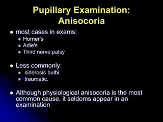
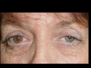
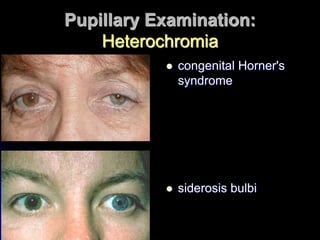
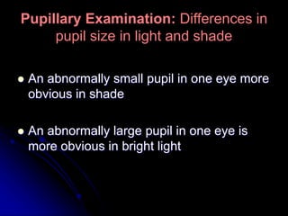
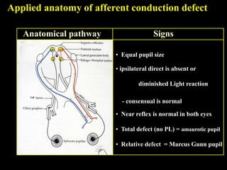
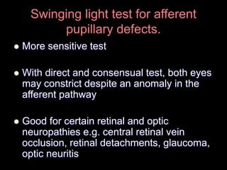
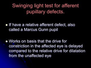
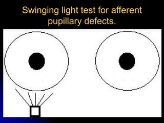
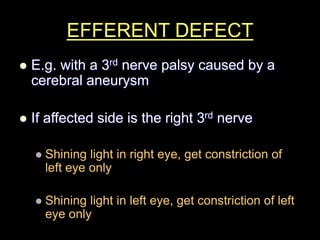
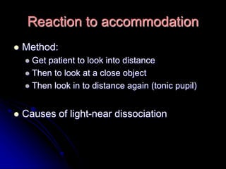
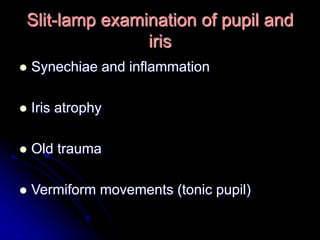
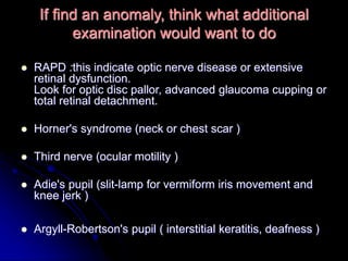
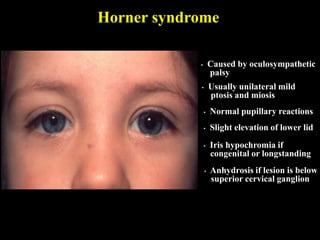
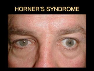
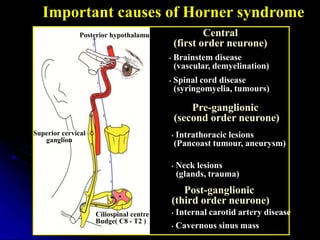
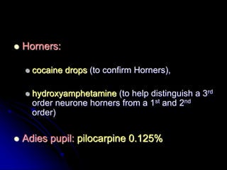
EXAMINATION OF
THE PUPILS
 Anisocoria (unequal pupil sizes)
 Heterochromia (difference in iris colour)
Pupillary Examination: Observe
Pupillary Examination:
Anisocoria
 most cases in exams:
 Horner's
 Adie's
 Third nerve palsy
 Less commonly:
 siderosis bulbi
 traumatic.
 Although physiological anisocoria is the most
common cause, it seldoms appear in an
examination
Pupillary Examination:
Heterochromia
 congenital Horner's
syndrome
 siderosis bulbi
Pupillary Examination: Differences in
pupil size in light and shade
 An abnormally small pupil in one eye more
obvious in shade
 An abnormally large pupil in one eye is
more obvious in bright light
PUPIL REFLEXES
Reaction to direct and consensual light
 Should know already
 Shine light in right eye once looking for
constriction in right eye
 Shine light in right eye again to look for
consensual reaction in left eye
 Do same for other eye
Why is the above technique frowned upon
by the examiner?
Answer to the question:
 Miosis occurs with accommodation.
 To see clearly the pupil reaction to light,
the patient should be instructed to look at
a distant object to reduce accommodation.
 By standing in front of the patient, the
candidate stimulates
accommodation and hence miosis.
What is the neurological
pathway for the pupil reflex?
Applied anatomy of afferent conduction defect
Anatomical pathway Signs
• Equal pupil size
• ipsilateral direct is absent or
diminished Light reaction
- consensual is normal
• Near reflex is normal in both eyes
• Total defect (no PL) = amaurotic pupil
• Relative defect = Marcus Gunn pupil
3rd
Swinging light test for afferent
pupillary defects.
 More sensitive test
 With direct and consensual test, both eyes
may constrict despite an anomaly in the
afferent pathway
 Good for certain retinal and optic
neuropathies e.g. central retinal vein
occlusion, retinal detachments, glaucoma,
optic neuritis
Swinging light test for afferent
pupillary defects.
 If have a relative afferent defect, also
called a Marcus Gunn pupil
 Works on basis that the drive for
constriction in the affected eye is delayed
compared to the relative drive for dilatation
from the unaffected eye
Swinging light test for afferent
pupillary defects.
EFFERENT DEFECT
 E.g. with a 3rd nerve palsy caused by a
cerebral aneurysm
 If affected side is the right 3rd nerve
 Shining light in right eye, get constriction of
left eye only
 Shining light in left eye, get constriction of left
eye only
Reaction to accommodation
 Method:
 Get patient to look into distance
 Then to look at a close object
 Then look in to distance again (tonic pupil)
 Causes of light-near dissociation
Slit-lamp examination of pupil and
iris
 Synechiae and inflammation
 Iris atrophy
 Old trauma
 Vermiform movements (tonic pupil)
If find an anomaly, think what additional
examination would want to do
 RAPD :this indicate optic nerve disease or extensive
retinal dysfunction.
Look for optic disc pallor, advanced glaucoma cupping or
total retinal detachment.
 Horner's syndrome (neck or chest scar )
 Third nerve (ocular motility )
 Adie's pupil (slit-lamp for vermiform iris movement and
knee jerk )
 Argyll-Robertson's pupil ( interstitial keratitis, deafness )
Horner syndrome
• Caused by oculosympathetic
palsy
• Usually unilateral mild
ptosis and miosis
• Slight elevation of lower lid
• Normal pupillary reactions
• Iris hypochromia if
congenital or longstanding
• Anhydrosis if lesion is below
superior cervical ganglion
HORNER’S SYNDROME
Important causes of Horner syndrome
Central
(first order neurone)
• Brainstem disease
(vascular, demyelination)
• Spinal cord disease
(syringomyelia, tumours)
Pre-ganglionic
(second order neurone)
• Intrathoracic lesions
(Pancoast tumour, aneurysm)
• Neck lesions
(glands, trauma)
Post-ganglionic
(third order neurone)
• Internal carotid artery disease
• Cavernous sinus mass
Posterior hypothalamus
Ciliospinal centre of
Budge( C8 - T2 )
Superior cervical
ganglion
PHARMACOLOGICAL TESTS
FOR PUPIL DEFECTS
 Horners:
 cocaine drops (to confirm Horners),
 hydroxyamphetamine (to help distinguish a 3rd
order neurone horners from a 1st and 2nd
order)
 Adies pupil: pilocarpine 0.125%

Pupil examination

  • 1.
  • 2.
     Anisocoria (unequalpupil sizes)  Heterochromia (difference in iris colour) Pupillary Examination: Observe
  • 3.
    Pupillary Examination: Anisocoria  mostcases in exams:  Horner's  Adie's  Third nerve palsy  Less commonly:  siderosis bulbi  traumatic.  Although physiological anisocoria is the most common cause, it seldoms appear in an examination
  • 5.
    Pupillary Examination: Heterochromia  congenitalHorner's syndrome  siderosis bulbi
  • 6.
    Pupillary Examination: Differencesin pupil size in light and shade  An abnormally small pupil in one eye more obvious in shade  An abnormally large pupil in one eye is more obvious in bright light
  • 7.
  • 8.
    Reaction to directand consensual light  Should know already  Shine light in right eye once looking for constriction in right eye  Shine light in right eye again to look for consensual reaction in left eye  Do same for other eye
  • 9.
    Why is theabove technique frowned upon by the examiner?
  • 10.
    Answer to thequestion:  Miosis occurs with accommodation.  To see clearly the pupil reaction to light, the patient should be instructed to look at a distant object to reduce accommodation.  By standing in front of the patient, the candidate stimulates accommodation and hence miosis.
  • 11.
    What is theneurological pathway for the pupil reflex?
  • 12.
    Applied anatomy ofafferent conduction defect Anatomical pathway Signs • Equal pupil size • ipsilateral direct is absent or diminished Light reaction - consensual is normal • Near reflex is normal in both eyes • Total defect (no PL) = amaurotic pupil • Relative defect = Marcus Gunn pupil 3rd
  • 13.
    Swinging light testfor afferent pupillary defects.  More sensitive test  With direct and consensual test, both eyes may constrict despite an anomaly in the afferent pathway  Good for certain retinal and optic neuropathies e.g. central retinal vein occlusion, retinal detachments, glaucoma, optic neuritis
  • 14.
    Swinging light testfor afferent pupillary defects.  If have a relative afferent defect, also called a Marcus Gunn pupil  Works on basis that the drive for constriction in the affected eye is delayed compared to the relative drive for dilatation from the unaffected eye
  • 15.
    Swinging light testfor afferent pupillary defects.
  • 16.
    EFFERENT DEFECT  E.g.with a 3rd nerve palsy caused by a cerebral aneurysm  If affected side is the right 3rd nerve  Shining light in right eye, get constriction of left eye only  Shining light in left eye, get constriction of left eye only
  • 17.
    Reaction to accommodation Method:  Get patient to look into distance  Then to look at a close object  Then look in to distance again (tonic pupil)  Causes of light-near dissociation
  • 18.
    Slit-lamp examination ofpupil and iris  Synechiae and inflammation  Iris atrophy  Old trauma  Vermiform movements (tonic pupil)
  • 19.
    If find ananomaly, think what additional examination would want to do  RAPD :this indicate optic nerve disease or extensive retinal dysfunction. Look for optic disc pallor, advanced glaucoma cupping or total retinal detachment.  Horner's syndrome (neck or chest scar )  Third nerve (ocular motility )  Adie's pupil (slit-lamp for vermiform iris movement and knee jerk )  Argyll-Robertson's pupil ( interstitial keratitis, deafness )
  • 20.
    Horner syndrome • Causedby oculosympathetic palsy • Usually unilateral mild ptosis and miosis • Slight elevation of lower lid • Normal pupillary reactions • Iris hypochromia if congenital or longstanding • Anhydrosis if lesion is below superior cervical ganglion
  • 21.
  • 22.
    Important causes ofHorner syndrome Central (first order neurone) • Brainstem disease (vascular, demyelination) • Spinal cord disease (syringomyelia, tumours) Pre-ganglionic (second order neurone) • Intrathoracic lesions (Pancoast tumour, aneurysm) • Neck lesions (glands, trauma) Post-ganglionic (third order neurone) • Internal carotid artery disease • Cavernous sinus mass Posterior hypothalamus Ciliospinal centre of Budge( C8 - T2 ) Superior cervical ganglion
  • 23.
  • 24.
     Horners:  cocainedrops (to confirm Horners),  hydroxyamphetamine (to help distinguish a 3rd order neurone horners from a 1st and 2nd order)  Adies pupil: pilocarpine 0.125%