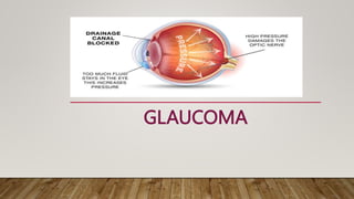
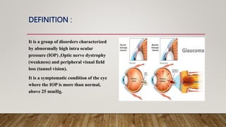
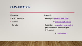
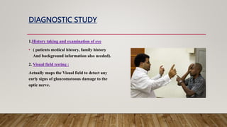
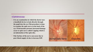
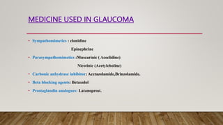
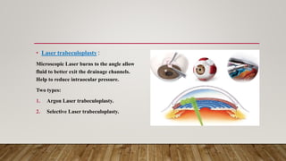
GLAUCOMA
INTRODUCTION
Glaucoma is an eye disease where the eye's Optic nerve is damaged. It is one of the
leading cause of blindness in the world. As of the year 2006 , 66.8 million people are
now estimated to have glaucoma with 6.7 million bilaterally blind from this disease .
DEFINITION :
It is a group of disorders characterized
by abnormally high intra ocular
pressure (IOP) ,Optic nerve dystrophy
(weakness) and peripheral visual field
loss (tunnel vision).
It is a symptomatic condition of the eye
where the IOP is more than normal,
above 25 mmHg.
ANATOMY AND PHYSIOLOGY
• The eye has 3 chambers, the anterior chamber in front of the iris, the posterior chamber
between the iris and the lens, and the vitreous chamber behind the lens.
• Intraocular pressure is maintained by a balance between inflow and outflow of the
aqueous humour, the fluid which nourishes the transparent structures of the eye.
Aqueous humour is produced and secreted by the ciliary body, a gland behind the iris of
the eye. Aqueous humour enters the anterior chamber of the eye through the pupil, and
leaves by passing through the trabecular meshwork in the iridocorneal angle of the
anterior chamber and back into venous circulation through the canal of Schlemm.
• The optic nerve is a bundle of nerve
fibers.
• It carries visual information from the
retina to the brain.
CLASSIFICATION
Congenital :
• True Congenital
• Infantile
• Juvenile
Acquired :
• Primary- ☆ primary open angle
☆ primary angle closure
• Secondary- ☆secondary open angle (
pre – trabecular, trabecular ,post
trabecular)
☆ Angle closure
• True congenital :
Occur when IOP increases during intrauterine period. Child is born with ocular
enlargement. Occur 40% of cases.
• Infantile :
Occur during 3rd birthday.
• Juvenile:
Occur during 3-16 years of life.
PRIMARY OPEN ANGLE GLAUCOMA
• Also called as open angle glaucoma or Chronic simple glaucoma , Results from the
overproduction of aqueous humour through trabecular Mesh work results in
increased IOP and damage to optic nerve, results in loss of vision .
• Clinical features:
Mild ache in the eye.
Headache
Increased IOP
Corneal edema.
PRIMARY ANGLE CLOSURE GLAUCOMA
• It is also called as primary closed angle glaucoma. This is due to the close angle between iris and
cornea, this results obstruction to the out flow of aqueous humour.
• Clinical features:
Pain and redness in eye
Increased IOP.
Blurred vision
Headache
Nausea, vomiting.
SECONDARY GLAUCOMA
• Secondary glaucoma occurs as a result of either disease within the eye such as uveitis,
inflammation, trauma, previous eye surgery, DM etc.
CAUSE AND RISK FACTORS
1. Age : older people mainly age 50 or above.
2. Race : African Americans are more likely to develop glaucoma at a younger age.
3. Family history : Family history of glaucoma increases chances of developing glaucoma.
4. Myopia: may increases pathological ocular complications causing glaucoma.
RISK FACTORS:
1. History of steroid use
2. Eye trauma (blunt trauma)
3. Diabetes
4. Ocular surgery like cataract surgery
PATHOPHYSIOLOGY
CLINICAL MANIFESTATIONS
Angle closure glaucoma:
• Seeing halos around light.
• Vision loss
• Redness in eye
• Eye feels swollen
• Eye pain
• Upset stomach or vomiting.
Open angle glaucoma:
• Most people have no symptoms until they
begin to loss vision
• Gradual loss of peripheral (side) vision
Congenital glaucoma:
• Cloudiness of the front of the eye
• Enlargement of one or both eyes
• Red eye
• Sensitivity to light
• Tearing
DIAGNOSTIC STUDY
1.History taking and examination of eye
• ( patients medical history, family history
And background information also needed).
2. Visual field testing :
Actually maps the Visual field to detect any
early signs of glaucomatoous damage to the
optic nerve.
3.Tonometry :
• Determine the pressure in the eye by
measuring the tone or firmness of its
surface.
• The firmer the tone of the surface of the
eye, the higher pressure reading.
• Normal pressure 12-22 mmHg.
4.Opthalmoscopy:
• It is an examination in which the doctor uses
a handheld device to look directly through
the pupil into the eye.This procedure is done
to examine the optic nerve at the back of the
eye. If intraocular pressure increased damage
occur to optic nerve called Cupping which is
an indentation of the optic disc.
• Pale Surface of the nerve can occur due to
poor blood supply & due to increase IOP.
5.Gonioscopy :
• It is used to seeing the interior of the eye
from different directions.
• The purpose of this test to examine the
drainage angle and drainage area of the
eye.
6.Pachymetry :
It is a relatively new test being used for the diagnosis and treatment of glaucoma.
Pachymetry determine the thickness of the cornea.
7. Slit lamp test :
A special microscope called a Slit lamp is used to examine the structure of the eye.
MANAGEMENT OF GLAUCOMA
Medical management:
1. Eye drops
2. Oral medication
Surgical management :
1. Laser iridotomy
2. Laser trabeculoplasty
3. Laser cyclocryopexy
4. Trabeculectomy
5. Aqueous shunt device
6. Viscocanalostomy.
Eye drops:
Eye drops are used to reduce Eye pressure by either increasing the eye‘s ability to drain or by
decreasing the amount of fluid it produce.
The medicine which are used –
• Beta blocker: cause little discomfort and work by decreasing the production of aqueous fluid.
• Prostaglandin : increase drainage of fluid.
• Alpha- stimulator are used with other medications to lower eye pressure.
• Miotics : increases fluid drainage, they also cause the pupil to constrict which can cause blurred
vision.
MEDICINE USED IN GLAUCOMA
• Sympathomimetics : clonidine
Epinephrine
• Parasympathomimetics :Muscarinic ( Aceclidine)
Nicotinic (Acetylcholine)
• Carbonic anhydrase inhibitor: Acetazolamide,Brinzolamide.
• Beta blocking agents: Betaxolol
• Prostaglandin analogues: Latanoprost.
SURGICAL MANAGEMENT
• Laser iridotomy:
Making a hole in the colored part of the eye
to allow fluid to drain normally in eyes with
narrow or closed angle.
• Laser trabeculoplasty :
Microscopic Laser burns to the angle allow
fluid to better exit the drainage channels.
Help to reduce intraocular pressure.
Two types:
1. Argon Laser trabeculoplasty.
2. Selective Laser trabeculoplasty.
Laser cyclocryopexy :
• It is another form of laser treatment generally
reserved for patients with severe forms of
glaucoma with poor visual potential.
• This therapy destroys the cell that make the
fluid, there by reducing the eye pressure.
Trabeculectomy:
• It is a delicate microsurgical procedure
used to treat glaucoma. In this operation
small piece of the clogged toabecular
meshwork is removed to create an opening
and a new drainage Pathway is made for
the fluid to exit the eye.
Aqueous shunt device:
• This are artificial drainage device used to
lower the eye pressure.
• This procedure may be performed as an
alternative to trabeculectomy in patients
with certain types of glaucoma.
Viscocanalostomy :
It is an alternative surgical procedure used to
lower eye pressure. It involves removing a
piece of the sclera(eyewall) to leave only a
thin membrane of tissue through which
aqueous fluid can more easily drain.
POST-OPERATIVE COMPLICATIONS
Shallow anterior chamber-
• Pupillary block
• Over filtration
• Malignant glaucoma
• Bacterial infection
• Failure of filtration
NURSING MANAGEMENT
1. Acute pain related to pathophysiology process and surgical correction.
2. Self care deficit related to visual acuity deficit.
3. Risk for injury related to visual acuity deficit.
Presentation (8).pptx

Presentation (8).pptx

  • 1.
  • 2.
    INTRODUCTION Glaucoma is aneye disease where the eye's Optic nerve is damaged. It is one of the leading cause of blindness in the world. As of the year 2006 , 66.8 million people are now estimated to have glaucoma with 6.7 million bilaterally blind from this disease .
  • 3.
    DEFINITION : It isa group of disorders characterized by abnormally high intra ocular pressure (IOP) ,Optic nerve dystrophy (weakness) and peripheral visual field loss (tunnel vision). It is a symptomatic condition of the eye where the IOP is more than normal, above 25 mmHg.
  • 4.
    ANATOMY AND PHYSIOLOGY •The eye has 3 chambers, the anterior chamber in front of the iris, the posterior chamber between the iris and the lens, and the vitreous chamber behind the lens. • Intraocular pressure is maintained by a balance between inflow and outflow of the aqueous humour, the fluid which nourishes the transparent structures of the eye. Aqueous humour is produced and secreted by the ciliary body, a gland behind the iris of the eye. Aqueous humour enters the anterior chamber of the eye through the pupil, and leaves by passing through the trabecular meshwork in the iridocorneal angle of the anterior chamber and back into venous circulation through the canal of Schlemm.
  • 6.
    • The opticnerve is a bundle of nerve fibers. • It carries visual information from the retina to the brain.
  • 7.
    CLASSIFICATION Congenital : • TrueCongenital • Infantile • Juvenile Acquired : • Primary- ☆ primary open angle ☆ primary angle closure • Secondary- ☆secondary open angle ( pre – trabecular, trabecular ,post trabecular) ☆ Angle closure
  • 8.
    • True congenital: Occur when IOP increases during intrauterine period. Child is born with ocular enlargement. Occur 40% of cases. • Infantile : Occur during 3rd birthday. • Juvenile: Occur during 3-16 years of life.
  • 9.
    PRIMARY OPEN ANGLEGLAUCOMA • Also called as open angle glaucoma or Chronic simple glaucoma , Results from the overproduction of aqueous humour through trabecular Mesh work results in increased IOP and damage to optic nerve, results in loss of vision . • Clinical features: Mild ache in the eye. Headache Increased IOP Corneal edema.
  • 11.
    PRIMARY ANGLE CLOSUREGLAUCOMA • It is also called as primary closed angle glaucoma. This is due to the close angle between iris and cornea, this results obstruction to the out flow of aqueous humour. • Clinical features: Pain and redness in eye Increased IOP. Blurred vision Headache Nausea, vomiting.
  • 13.
    SECONDARY GLAUCOMA • Secondaryglaucoma occurs as a result of either disease within the eye such as uveitis, inflammation, trauma, previous eye surgery, DM etc.
  • 14.
    CAUSE AND RISKFACTORS 1. Age : older people mainly age 50 or above. 2. Race : African Americans are more likely to develop glaucoma at a younger age. 3. Family history : Family history of glaucoma increases chances of developing glaucoma. 4. Myopia: may increases pathological ocular complications causing glaucoma. RISK FACTORS: 1. History of steroid use 2. Eye trauma (blunt trauma) 3. Diabetes 4. Ocular surgery like cataract surgery
  • 15.
  • 17.
    CLINICAL MANIFESTATIONS Angle closureglaucoma: • Seeing halos around light. • Vision loss • Redness in eye • Eye feels swollen • Eye pain • Upset stomach or vomiting.
  • 18.
    Open angle glaucoma: •Most people have no symptoms until they begin to loss vision • Gradual loss of peripheral (side) vision
  • 20.
    Congenital glaucoma: • Cloudinessof the front of the eye • Enlargement of one or both eyes • Red eye • Sensitivity to light • Tearing
  • 21.
    DIAGNOSTIC STUDY 1.History takingand examination of eye • ( patients medical history, family history And background information also needed). 2. Visual field testing : Actually maps the Visual field to detect any early signs of glaucomatoous damage to the optic nerve.
  • 22.
    3.Tonometry : • Determinethe pressure in the eye by measuring the tone or firmness of its surface. • The firmer the tone of the surface of the eye, the higher pressure reading. • Normal pressure 12-22 mmHg.
  • 23.
    4.Opthalmoscopy: • It isan examination in which the doctor uses a handheld device to look directly through the pupil into the eye.This procedure is done to examine the optic nerve at the back of the eye. If intraocular pressure increased damage occur to optic nerve called Cupping which is an indentation of the optic disc. • Pale Surface of the nerve can occur due to poor blood supply & due to increase IOP.
  • 24.
    5.Gonioscopy : • Itis used to seeing the interior of the eye from different directions. • The purpose of this test to examine the drainage angle and drainage area of the eye.
  • 25.
    6.Pachymetry : It isa relatively new test being used for the diagnosis and treatment of glaucoma. Pachymetry determine the thickness of the cornea. 7. Slit lamp test : A special microscope called a Slit lamp is used to examine the structure of the eye.
  • 26.
    MANAGEMENT OF GLAUCOMA Medicalmanagement: 1. Eye drops 2. Oral medication Surgical management : 1. Laser iridotomy 2. Laser trabeculoplasty 3. Laser cyclocryopexy 4. Trabeculectomy 5. Aqueous shunt device 6. Viscocanalostomy.
  • 27.
    Eye drops: Eye dropsare used to reduce Eye pressure by either increasing the eye‘s ability to drain or by decreasing the amount of fluid it produce. The medicine which are used – • Beta blocker: cause little discomfort and work by decreasing the production of aqueous fluid. • Prostaglandin : increase drainage of fluid. • Alpha- stimulator are used with other medications to lower eye pressure. • Miotics : increases fluid drainage, they also cause the pupil to constrict which can cause blurred vision.
  • 28.
    MEDICINE USED INGLAUCOMA • Sympathomimetics : clonidine Epinephrine • Parasympathomimetics :Muscarinic ( Aceclidine) Nicotinic (Acetylcholine) • Carbonic anhydrase inhibitor: Acetazolamide,Brinzolamide. • Beta blocking agents: Betaxolol • Prostaglandin analogues: Latanoprost.
  • 29.
    SURGICAL MANAGEMENT • Laseriridotomy: Making a hole in the colored part of the eye to allow fluid to drain normally in eyes with narrow or closed angle.
  • 30.
    • Laser trabeculoplasty: Microscopic Laser burns to the angle allow fluid to better exit the drainage channels. Help to reduce intraocular pressure. Two types: 1. Argon Laser trabeculoplasty. 2. Selective Laser trabeculoplasty.
  • 31.
    Laser cyclocryopexy : •It is another form of laser treatment generally reserved for patients with severe forms of glaucoma with poor visual potential. • This therapy destroys the cell that make the fluid, there by reducing the eye pressure.
  • 32.
    Trabeculectomy: • It isa delicate microsurgical procedure used to treat glaucoma. In this operation small piece of the clogged toabecular meshwork is removed to create an opening and a new drainage Pathway is made for the fluid to exit the eye.
  • 33.
    Aqueous shunt device: •This are artificial drainage device used to lower the eye pressure. • This procedure may be performed as an alternative to trabeculectomy in patients with certain types of glaucoma.
  • 34.
    Viscocanalostomy : It isan alternative surgical procedure used to lower eye pressure. It involves removing a piece of the sclera(eyewall) to leave only a thin membrane of tissue through which aqueous fluid can more easily drain.
  • 35.
    POST-OPERATIVE COMPLICATIONS Shallow anteriorchamber- • Pupillary block • Over filtration • Malignant glaucoma • Bacterial infection • Failure of filtration
  • 36.
    NURSING MANAGEMENT 1. Acutepain related to pathophysiology process and surgical correction. 2. Self care deficit related to visual acuity deficit. 3. Risk for injury related to visual acuity deficit.