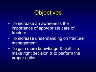
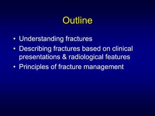
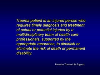
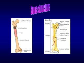
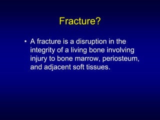
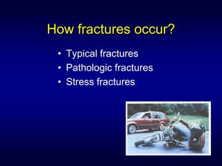
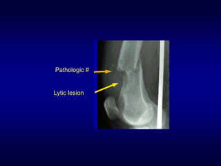
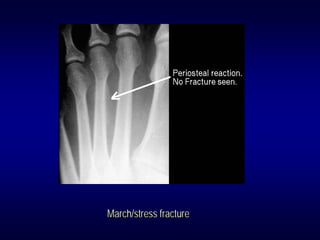
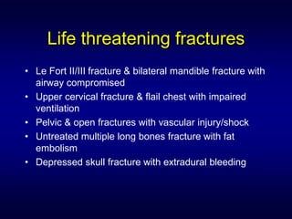
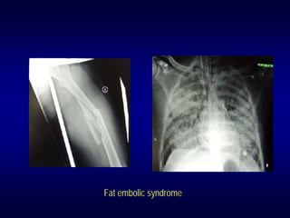
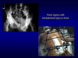
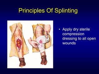
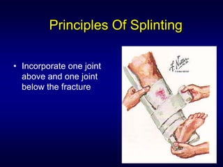
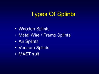
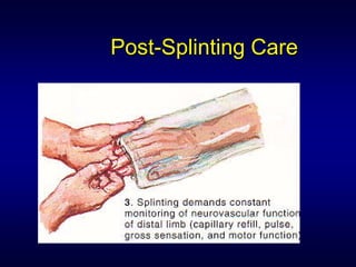
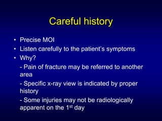
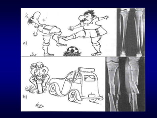
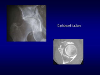
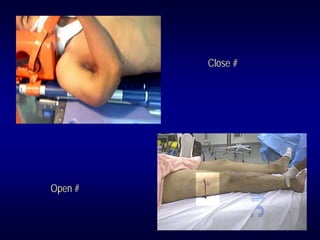
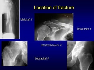
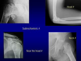
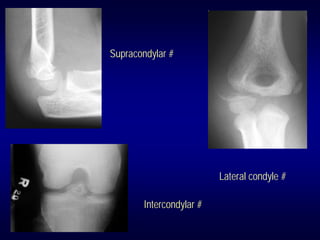
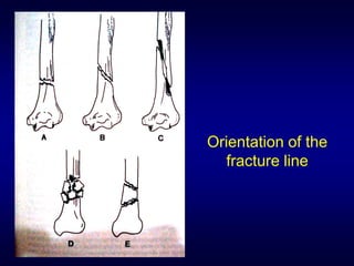
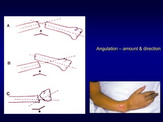
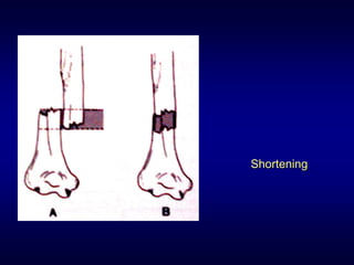
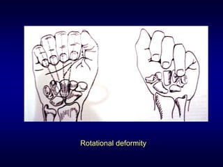
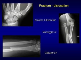
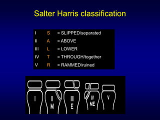
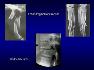
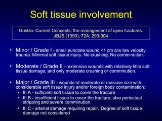
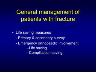
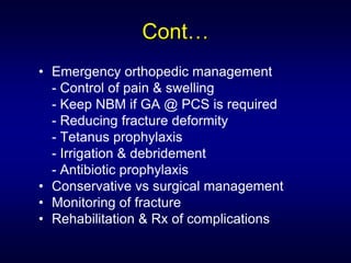
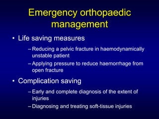
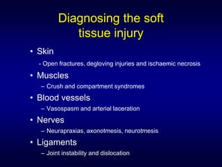
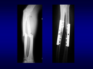
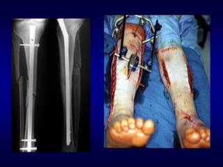
POP application course
      18th December 2006


An Overview on
Fractures Care
    Dr. Rashidi Ahmad
 MD USM, MMED USM, FADUSM
     Lecturer/Emergentist
     USM Health Campus
Objectives
• To increase an awareness the
  importance of appropriate care of
  fracture
• To increase understanding on fracture
  management
• To gain more knowledge & skill – to
  make right decision & to perform the
  proper action
Outline
• Understanding fractures
• Describing fractures based on clinical
  presentations & radiological features
• Principles of fracture management
Trauma patient is an injured person who
requires timely diagnosis and treatment
of actual or potential injuries by a
multidisciplinary team of health care
professionals, supported by the
appropriate resources, to diminish or
eliminate the risk of death or permanent
disability.

                      Europian Trauma Life Support
Fracture?
• A fracture is a disruption in the
  integrity of a living bone involving
  injury to bone marrow, periosteum,
  and adjacent soft tissues.
How fractures occur?
 • Typical fractures
 • Pathologic fractures
 • Stress fractures
Pathologic #


Lytic lesion
March/stress fracture
Life threatening fractures
• Le Fort II/III fracture & bilateral mandible fracture with
  airway compromised
• Upper cervical fracture & flail chest with impaired
  ventilation
• Pelvic & open fractures with vascular injury/shock
• Untreated multiple long bones fracture with fat
  embolism
• Depressed skull fracture with extradural bleeding
Fat embolic syndrome
Pelvic injuries with
intrabdominal injury & shock
Limb threatening
• Fractures with Compartmental
  Syndrome @ Volkmann’s ischemia
• Fractures with neurovascular injuries/
  avascular necrosis
• Open fractures with infection
Volkmann’s ischemic
contracture


   Compartmental syndrome
Avascular necrosis
Osteomyelitis
Factors affecting fracture healing

 • The energy transfer of the injury
 • The tissue response
   –   Two bone ends in opposition or compressed
   –   Micro-movement or no movement
   –   BS (scaphoid, talus, femoral and humeral head)
   –   NS
   –   No infection
 • The patient
 • The method of treatment
Goals of fracture treatment
• Restore the patient to optimal functional state

• Prevent fracture and soft-tissue complications

• Get the fracture to heal, and in a position
  which will produce optimal functional recovery

• Rehabilitate the patient as early as possible
Prehospital care
• First aid principles
• Preliminary splinting/sling of the injured
  extremity
  - reduces pain
  - reduces damage to nerve & vessels
  - reduces risk of conversion to open fracture
  - facilitates transportation & x-ray taking.
Principles Of Splinting

               • Apply dry sterile
                 compression
                 dressing to all open
                 wounds
Principles Of Splinting

• Incorporate one joint
  above and one joint
  below the fracture
Types Of Splints

•   Wooden Splints
•   Metal Wire / Frame Splints
•   Air Splints
•   Vacuum Splints
•   MAST suit
Post-Splinting Care
Prehospital reduction
• Prehospital reduction of deformity – by
  advice of physician
• Obvious fracture along the shaft of a
  long bone with a neurovascular deficit –
  longitudinal traction
• Deformity near a joint – possibility of
  dislocation
Careful history
• Precise MOI
• Listen carefully to the patient’s symptoms
• Why?
  - Pain of fracture may be referred to another
  area
  - Specific x-ray view is indicated by proper
  history
  - Some injuries may not be radiologically
  apparent on the 1st day
High-energy injury
Low energy injury
Dashboard fracture
Chance # @ lap seat belt #
Physical examination
• Inspection for swelling, discoloration,
  deformity
• Assessment of active & passive ROM of
  the joints proximal & distal to the injury
• Palpation for tenderness
• Verification of neurovascular status
Radiologic evaluation
• X-ray is an important adjunct
• Ordered based on Hx & PE
• 2 views – AP & lateral
• 2 joints – above & below the shaft
  fracture
• In children with injury near the joint –
  bilateral x-rays for comparison
• Repeat x-rays after 1 – 2 weeks to show
  callus in doubtful fractures
Describing fractures
•   Open versus closed
•   Location of the fracture
•   Orientation of the fracture line
•   Displacement & separation
•   Angulation
•   Shortening
•   Rotational deformity
•   Fracture – dislocation/subluxation
•   Salter fractures
•   Fragmentation
•   Soft tissue involvement
Close #


Open #
Location of fracture

Midshaft #


                                        Distal third #


                  Intertrochanteric #


             Subcapital #
Head #


Subtrochanteric #


                      Neck #


   Near the head #
Supracondylar #


                          Lateral condyle #

        Intercondylar #
Orientation of the
  fracture line
Torus #


          Greenstick #
Bowing #
Displaced & separation
Minimally displaced distal radius fracture


Comminuted proximal- third femoral
fracture with significant displacement
Angulation – amount & direction
Shortening
Rotational deformity
Fracture - dislocation


Bennet’s # dislocation


        Monteggia’s #


          Galeazzi’s #
Salter Harris classification

I     S   = SLIPPED/separated
II    A   = ABOVE
III   L   = LOWER
IV    T   = THROUGH/together
V     R   = RAMMED/ruined
Salter Harris Type I
Salter Harris Type II
Salter Harris Type III
Salter Harris Type IV
Salter Harris Type V
Intraarticular fracure


             Rolando #


Barton’s #
Fragmentation
The Muller AO Comprehensive Classification of Fractures

- A multi-fragmentary fracture: several breaks (>2
  fragments) in the bone

- Wedge fractures: spiral (low energy) @ bending
  (high energy

- The complex multi-fragmentary fracture: segmental
  fracture in which there is no contact between the
  proximal & distal fragments, no bone shortening.

- Simple fractures are spiral, oblique, or transverse.
A multi-fragmentary fracture


Wedge fractures
Soft tissue involvement
    Gustilo. Current Concepts: the management of open fractures.
                      JBJS (1990); 72A; 299-304


• Minor / Grade I - small punctate wound <1 cm a/w low velocity
  trauma. Minimal soft tissue injury. No crushing. No comminution.

• Moderate / Grade II – extensive wounds with relatively little soft
  tissue damage, and only moderate crushing or comminution.

• Major / Grade III - wounds of moderate or massive size with
  considerable soft tissue injury and/or foreign body contamination:
   • III A - sufficient soft tissue to cover the fracture
   • III B - insufficient tissue to cover the fracture; also periosteal
     stripping and severe comminution
   • III C - arterial damage requiring repair. Degree of soft tissue
     damage not considered
Crush injury


        Degloving injury
Bomb blast injury


Gunshot #
General management of
   patients with fracture

• Life saving measures
  - Primary & secondary survey
  - Emergency orthopaedic involvement
         –Life saving
         –Complication saving
Cont…
• Emergency orthopedic management
  - Control of pain & swelling
  - Keep NBM if GA @ PCS is required
  - Reducing fracture deformity
  - Tetanus prophylaxis
  - Irrigation & debridement
  - Antibiotic prophylaxis
• Conservative vs surgical management
• Monitoring of fracture
• Rehabilitation & Rx of complications
Emergency orthopaedic
        management
• Life saving measures
    – Reducing a pelvic fracture in haemodynamically
      unstable patient
    – Applying pressure to reduce haemorrhage from
      open fracture

• Complication saving
    – Early and complete diagnosis of the extent of
      injuries
    – Diagnosing and treating soft-tissue injuries
Diagnosing the soft
            tissue injury
• Skin
  - Open fractures, degloving injuries and ischaemic necrosis

• Muscles
  – Crush and compartment syndromes
• Blood vessels
  – Vasospasm and arterial laceration
• Nerves
  – Neurapraxias, axonotmesis, neurotmesis
• Ligaments
  – Joint instability and dislocation
Treating the soft tissue injury
• All severe soft tissue injuries………require urgent
  treatment
     – Open fractures , Vascular injuries, Nerve injuries,
       Compartment syndromes, Fracture/dislocations
  • After the treatment of the soft tissue injury the
    fracture requires rigid fixation
  • A severe soft-tissue injury will delay fracture
    healing
Treating the fracture
• Purpose: to reduce, hold & maintain the # in a suitable
  alignment
• Does the fracture require reduction? Displaced?
• Methods: CMR method by ACCROCHAGE and
  continuous traction (skin & skeletal traction)
• What is acceptable # alignment?
• Consider: age, site, weight bearing, shortening,
  angulation & rotation
Risk benefit

                           Operative   Non-operative

Rehabilitation               Rapid         Slow
Risk of joint stiffness      Low           Present
Risk of malunion             Low           Present
Risk of non-union            Present       Present
Speed of healing             Slow          Rapid
Risk of infection            Present       Low
Cost                         ?             ?
Treating the fracture
• How are we going to hold the reduction?
      – Semi-rigid (Plaster)
      – Rigid (Internal fixation)


   • What treatment plan will we follow?
      – When can the patient load the injured limb?
      – When can the patient be allowed to move the
        joints?
      – How long will we have to immobilise the fracture
        for?
Indications for operative treatment
• General trend toward operative treatment last
  30 yrs
     – Improved implants and antibiotic prophylaxis, use
       of closed and minimally invasive methods


• Current absolute indications:
     – Polytrauma, displaced intra-articular fractures
     – Open #’s, #’s with vascular injury or compartment
       syndrome
     – Pathological #’s, Non-unions
Indications for operative treatment

• Current relative indications:-
     – Loss of position with closed method
     – Poor functional result with non-anatomical
       reduction
     – Displaced fractures with poor blood supply
     – Economic and medical indications
When is the fracture healed?
• Clinically
                         Upper limb   Lower limb
     Adult               6-8 weeks    12-16 weeks
     Child               3-4 weeks    6-8 weeks


• Radiologically
     – Bridging callus formation
     – Remodelling
Rehabilitation
• Restoring the patient as close to pre-injury
  functional level as possible
• Approach needs to be:-
     –Pragmatic with realistic targets
     –Multidisciplinary: Physiotherapist,
      Occupational therapist, District nurse,
      GP, Social worker
Summary
• Fractures are a/w mortality & morbidity
• Fractures care starts from the onset till fully
  recover
• Primary survey + resuscitation are the
  PRIORITY
• Do not underestimate the benefit of
  reassurance, pain management & splinting
• Multidiscipline approach
Final message
Dr. Rashidi Ahmad
        MD USM, MMED USM, FADUSM
Pensyarah/Pakar Perubatan kecemasan & Trauma
        Jabatan perubatan Kecemasan
       Pusat Pengajian Sains Perubatan
       USM Kampus Kesihatan, Malaysia

         shidee_ahmad@yahoo.com
               +609 7663244

Fracture care

  • 1.
    POP application course 18th December 2006 An Overview on Fractures Care Dr. Rashidi Ahmad MD USM, MMED USM, FADUSM Lecturer/Emergentist USM Health Campus
  • 2.
    Objectives • To increasean awareness the importance of appropriate care of fracture • To increase understanding on fracture management • To gain more knowledge & skill – to make right decision & to perform the proper action
  • 3.
    Outline • Understanding fractures •Describing fractures based on clinical presentations & radiological features • Principles of fracture management
  • 4.
    Trauma patient isan injured person who requires timely diagnosis and treatment of actual or potential injuries by a multidisciplinary team of health care professionals, supported by the appropriate resources, to diminish or eliminate the risk of death or permanent disability. Europian Trauma Life Support
  • 6.
    Fracture? • A fractureis a disruption in the integrity of a living bone involving injury to bone marrow, periosteum, and adjacent soft tissues.
  • 7.
    How fractures occur? • Typical fractures • Pathologic fractures • Stress fractures
  • 8.
  • 9.
  • 10.
    Life threatening fractures •Le Fort II/III fracture & bilateral mandible fracture with airway compromised • Upper cervical fracture & flail chest with impaired ventilation • Pelvic & open fractures with vascular injury/shock • Untreated multiple long bones fracture with fat embolism • Depressed skull fracture with extradural bleeding
  • 11.
  • 12.
  • 13.
    Limb threatening • Fractureswith Compartmental Syndrome @ Volkmann’s ischemia • Fractures with neurovascular injuries/ avascular necrosis • Open fractures with infection
  • 14.
  • 15.
  • 16.
  • 17.
    Factors affecting fracturehealing • The energy transfer of the injury • The tissue response – Two bone ends in opposition or compressed – Micro-movement or no movement – BS (scaphoid, talus, femoral and humeral head) – NS – No infection • The patient • The method of treatment
  • 18.
    Goals of fracturetreatment • Restore the patient to optimal functional state • Prevent fracture and soft-tissue complications • Get the fracture to heal, and in a position which will produce optimal functional recovery • Rehabilitate the patient as early as possible
  • 19.
    Prehospital care • Firstaid principles • Preliminary splinting/sling of the injured extremity - reduces pain - reduces damage to nerve & vessels - reduces risk of conversion to open fracture - facilitates transportation & x-ray taking.
  • 20.
    Principles Of Splinting • Apply dry sterile compression dressing to all open wounds
  • 21.
    Principles Of Splinting •Incorporate one joint above and one joint below the fracture
  • 22.
    Types Of Splints • Wooden Splints • Metal Wire / Frame Splints • Air Splints • Vacuum Splints • MAST suit
  • 23.
  • 24.
    Prehospital reduction • Prehospitalreduction of deformity – by advice of physician • Obvious fracture along the shaft of a long bone with a neurovascular deficit – longitudinal traction • Deformity near a joint – possibility of dislocation
  • 25.
    Careful history • PreciseMOI • Listen carefully to the patient’s symptoms • Why? - Pain of fracture may be referred to another area - Specific x-ray view is indicated by proper history - Some injuries may not be radiologically apparent on the 1st day
  • 26.
  • 27.
  • 31.
  • 32.
    Chance # @lap seat belt #
  • 33.
    Physical examination • Inspectionfor swelling, discoloration, deformity • Assessment of active & passive ROM of the joints proximal & distal to the injury • Palpation for tenderness • Verification of neurovascular status
  • 34.
    Radiologic evaluation • X-rayis an important adjunct • Ordered based on Hx & PE • 2 views – AP & lateral • 2 joints – above & below the shaft fracture • In children with injury near the joint – bilateral x-rays for comparison • Repeat x-rays after 1 – 2 weeks to show callus in doubtful fractures
  • 35.
    Describing fractures • Open versus closed • Location of the fracture • Orientation of the fracture line • Displacement & separation • Angulation • Shortening • Rotational deformity • Fracture – dislocation/subluxation • Salter fractures • Fragmentation • Soft tissue involvement
  • 36.
  • 37.
    Location of fracture Midshaft# Distal third # Intertrochanteric # Subcapital #
  • 38.
    Head # Subtrochanteric # Neck # Near the head #
  • 39.
    Supracondylar # Lateral condyle # Intercondylar #
  • 40.
    Orientation of the fracture line
  • 41.
    Torus # Greenstick #
  • 42.
  • 43.
  • 44.
    Minimally displaced distalradius fracture Comminuted proximal- third femoral fracture with significant displacement
  • 45.
  • 46.
  • 47.
  • 48.
    Fracture - dislocation Bennet’s# dislocation Monteggia’s # Galeazzi’s #
  • 49.
    Salter Harris classification I S = SLIPPED/separated II A = ABOVE III L = LOWER IV T = THROUGH/together V R = RAMMED/ruined
  • 50.
  • 51.
  • 52.
  • 53.
  • 54.
  • 55.
    Intraarticular fracure Rolando # Barton’s #
  • 56.
    Fragmentation The Muller AOComprehensive Classification of Fractures - A multi-fragmentary fracture: several breaks (>2 fragments) in the bone - Wedge fractures: spiral (low energy) @ bending (high energy - The complex multi-fragmentary fracture: segmental fracture in which there is no contact between the proximal & distal fragments, no bone shortening. - Simple fractures are spiral, oblique, or transverse.
  • 57.
  • 58.
    Soft tissue involvement Gustilo. Current Concepts: the management of open fractures. JBJS (1990); 72A; 299-304 • Minor / Grade I - small punctate wound <1 cm a/w low velocity trauma. Minimal soft tissue injury. No crushing. No comminution. • Moderate / Grade II – extensive wounds with relatively little soft tissue damage, and only moderate crushing or comminution. • Major / Grade III - wounds of moderate or massive size with considerable soft tissue injury and/or foreign body contamination: • III A - sufficient soft tissue to cover the fracture • III B - insufficient tissue to cover the fracture; also periosteal stripping and severe comminution • III C - arterial damage requiring repair. Degree of soft tissue damage not considered
  • 59.
    Crush injury Degloving injury
  • 60.
  • 61.
    General management of patients with fracture • Life saving measures - Primary & secondary survey - Emergency orthopaedic involvement –Life saving –Complication saving
  • 62.
    Cont… • Emergency orthopedicmanagement - Control of pain & swelling - Keep NBM if GA @ PCS is required - Reducing fracture deformity - Tetanus prophylaxis - Irrigation & debridement - Antibiotic prophylaxis • Conservative vs surgical management • Monitoring of fracture • Rehabilitation & Rx of complications
  • 63.
    Emergency orthopaedic management • Life saving measures – Reducing a pelvic fracture in haemodynamically unstable patient – Applying pressure to reduce haemorrhage from open fracture • Complication saving – Early and complete diagnosis of the extent of injuries – Diagnosing and treating soft-tissue injuries
  • 64.
    Diagnosing the soft tissue injury • Skin - Open fractures, degloving injuries and ischaemic necrosis • Muscles – Crush and compartment syndromes • Blood vessels – Vasospasm and arterial laceration • Nerves – Neurapraxias, axonotmesis, neurotmesis • Ligaments – Joint instability and dislocation
  • 65.
    Treating the softtissue injury • All severe soft tissue injuries………require urgent treatment – Open fractures , Vascular injuries, Nerve injuries, Compartment syndromes, Fracture/dislocations • After the treatment of the soft tissue injury the fracture requires rigid fixation • A severe soft-tissue injury will delay fracture healing
  • 66.
    Treating the fracture •Purpose: to reduce, hold & maintain the # in a suitable alignment • Does the fracture require reduction? Displaced? • Methods: CMR method by ACCROCHAGE and continuous traction (skin & skeletal traction) • What is acceptable # alignment? • Consider: age, site, weight bearing, shortening, angulation & rotation
  • 67.
    Risk benefit Operative Non-operative Rehabilitation Rapid Slow Risk of joint stiffness Low Present Risk of malunion Low Present Risk of non-union Present Present Speed of healing Slow Rapid Risk of infection Present Low Cost ? ?
  • 68.
    Treating the fracture •How are we going to hold the reduction? – Semi-rigid (Plaster) – Rigid (Internal fixation) • What treatment plan will we follow? – When can the patient load the injured limb? – When can the patient be allowed to move the joints? – How long will we have to immobilise the fracture for?
  • 71.
    Indications for operativetreatment • General trend toward operative treatment last 30 yrs – Improved implants and antibiotic prophylaxis, use of closed and minimally invasive methods • Current absolute indications: – Polytrauma, displaced intra-articular fractures – Open #’s, #’s with vascular injury or compartment syndrome – Pathological #’s, Non-unions
  • 72.
    Indications for operativetreatment • Current relative indications:- – Loss of position with closed method – Poor functional result with non-anatomical reduction – Displaced fractures with poor blood supply – Economic and medical indications
  • 73.
    When is thefracture healed? • Clinically Upper limb Lower limb Adult 6-8 weeks 12-16 weeks Child 3-4 weeks 6-8 weeks • Radiologically – Bridging callus formation – Remodelling
  • 74.
    Rehabilitation • Restoring thepatient as close to pre-injury functional level as possible • Approach needs to be:- –Pragmatic with realistic targets –Multidisciplinary: Physiotherapist, Occupational therapist, District nurse, GP, Social worker
  • 75.
    Summary • Fractures area/w mortality & morbidity • Fractures care starts from the onset till fully recover • Primary survey + resuscitation are the PRIORITY • Do not underestimate the benefit of reassurance, pain management & splinting • Multidiscipline approach
  • 76.
  • 77.
    Dr. Rashidi Ahmad MD USM, MMED USM, FADUSM Pensyarah/Pakar Perubatan kecemasan & Trauma Jabatan perubatan Kecemasan Pusat Pengajian Sains Perubatan USM Kampus Kesihatan, Malaysia shidee_ahmad@yahoo.com +609 7663244