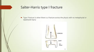
FRACTURE
JMAYANGA
2018
DEFINITION
 An interruption in the continuity of the bone which may be a
complete break or an incomplete break.
 Fragility fractures are common; 1 in 2 women over 50 years of age will suffer one,
as will 1 in 5 men.
 Globally, during year 2000, there were an estimated 9 million new fragility fractures,
of which 1.6 million were at the hip, 1.7 million at the wrist, 0.7 million at the
humerus and 1.4 million symptomatic vertebral fractures.
ESTIMATED NUMBER OF FRACTURES (IN THOUSANDS) AT THE SITES SHOWN
IN MEN AND WOMEN AGED 50 YEARS OR MORE IN 2000 BY WORLD HEALTH
ORGANISATION REGIONS
 Region Hip Spine Forearm HumerusOther All sites Percentage
 Africa 8 12 16 6 33 75 0.8
 Americas 311 214 248 111 521 1406 15.7
 Southeast Asia 221 253 306 121 660 1562 17.4
 Europe 620 490 574 250 119 3119 34.8
 Eastern Mediterranean 35 43 52 21 109 261 2.9
 Western Pacific 432 405 464 197 1039 2536 28.6
 Total 1627 1416 1660 706 3550 8959 100
CLASSIFICATION OF FRACTURES
 Why classify fractures?
 Classification or description of fractures is only used when the classification or
description is useful in providing treatment or outcomes.
 Types of classifications
 Anatomic description
 AO classification
 Salter-Harris classification
 Gustillo-Anderson open fracture classification
 Fracture specific classifications
Anatomic description of fractures
 Type
 Comminution
 Location
 Displacement
ANATOMIC DESCRIPTION - TYPE
 Type is the overall fracture pattern
 Examples are:
 Simple
 Spiral
 segmental
ANATOMIC DESCRIPTION -
COMMINUTION
 Comminution is the measure of the number of pieces of broken bone.
ANATOMIC DESCRIPTION - LOCATION
 Location is the anatomic location of the fracture usually described by giving the
bone involved and location on the bone
 Examples are: distal radial shaft, proximal 1/3 humeral shaft, intraarticular distal
tibial
ANATOMIC DESCRIPTION -
DISPLACEMENT
 Displacement is the amount the pieces of a fracture have moved from their normal
location.
 Subdivided into 3 sub-categories:
 translation,
 angulation, and
 shortening
Anatomic description?
Anatomic description
 Simple, transverse,
 non-communited
 midshaft radial and ulnar fracture with 30 degrees apex radial angulation.
AO Classification
AO Classification
1st number = long bone
2nd number = bone segment
Letter = fracture type (A,B,C)
Then 3rd & 4th numbers classify fracture group
& subgroup
Salter-Harris Classification
 Only used for pediatric fractures that involve the growth plate (physis)
 Five types (I-V)
Salter-Harris type I fracture
 Type I fracture is when there is a fracture across the physis with no metaphysial or
epiphysial injury
Salter-Harris type II fracture
 Type II fracture is when there is a fracture across the physis which extends into the
metaphysis
Salter-Harris type III fracture
 Type III fracture is when there is a fracture across the physis which extends into the
epiphysis
Salter-Harris type IV fracture
 Type IV fracture is when there is a fracture through metaphysis, physis, and
epiphysis
Salter-Harris type V fracture
 Type V fracture is when there is a crush injury to the physis
Gustillo classification
 The Gustillo classification is used to classify open fracture - ones in which the skin
has been disrupted
 Three grades that try to quantify the amount of soft tissue damage associated with
the fracture, level of contamination.
Open Fracture grade I
 wound less than 1 cm w/ minimal soft tissue injury
 wound bed is clean
 bone injury is simple w/ minimal comminution
Open fracture grade II
 wound is greater than 1 cm w/moderate soft tissue injury
 wound bed is moderately contaminated
 fracture contains moderate comminution
Open Fracture grade IIIA
 wound greater than 10 cm w/ crushed tissue and contamination
 soft tissue coverage of bone is usually possible, intact periosteum
Open fracture grade IIIB
 wound greater than 10 cm w/ crushed tissue and contamination, periostal
stripping, bone damage
 soft tissue is inadequate and requires regional or free flap
Open fracture grade IIIC
 is an open fracture in which there is a major vascular injury requiring repair for limb
salvage
Open fracture & Infection
Gustilo Type Infection %
 Grad I 0-2 %
 Grad II 2-5 %
 Grad III A 5-10 %
 Grad III B 10-50 %
 Grad III C 25-50 %
CLINICAL PRESENTATION
OF
FRACTURES
SYMPTOMS
 A history of injury, followed by inability to use the injured limb.
 The fracture may not always be at the site of the injury. Eg : A blow to the knee
and its varied effects.
 If a fracture occurs with trivial trauma, or spontaneously, suspect a pathological
lesion.
 Pain
 Bruising
 Swelling
 These are common symptoms but they do not distinguish a fracture from a soft-
tissueinjury.
SYMPTOMS – CONT….
 Deformity – More suggestive of a fracture.
 Enquire about symptoms of associated injuries:
 Pain and swelling elsewhere (it is a common mistake to get distracted by the main injury, particularly
if it is severe),
 Numbness or Loss of movement.
 Skin pallor or cyanosis.
 Blood in the urine.
 Abdominal pain.
 Difficulty with breathing.
 Transient loss of consciousness.
 Ask about previous injuries, or any other musculoskeletal abnormality that might cause confusion
when the x-ray is seen.
 Finally, a general medical history is important, in preparation for anesthesia or operation.
GENERAL SIGNS
 First follow the ABCs: look for, and if necessary attend to,
 Airway obstruction,
 Breathing problems,
 Circulatory problems
 Cervical spine injury.
 Secondary survey – Examine the main injury- ascertain the type of fracture, classify, plan a
management protocol and look out for complications.
 It will also be necessary to exclude other previously unsuspected injuries.
LOCAL SIGNS
LOOK • For Swelling, bruising and deformity
• Examine whether the skin is intact
• Note also the posture of the distal extremity and the color of the skin.
FEEL • The injured part is gently palpated for localized tenderness.
• The common and characteristic associated injuries should also be felt
for, even if the patient does not complain of them.
• Vascular and peripheral nerve abnormalities should be tested for both
before and after treatment.
MOVE • Crepitus and abnormal movement may be present.
• More important to ascertain if the patient can move the joints distal to the
injury.
X-RAY INVESTIGATION
• X-ray examination is mandatory.
• Rule of twos:
Two views – A fracture or a dislocation may not be seen
on a single x-ray film, and at least two views
(anteroposterior and lateral) must be taken.
Two limbs – In children, the appearance of immature
epiphyses may confuse the diagnosis of a fracture; x-rays
of the uninjured limb are needed for comparison.
Two joints – In the forearm or leg, one bone may be fractured and angulated.
Angulation, however, is impossible unless the other bone is also broken, or a joint
dislocated. The joints above and below the fracture must both be included on the x-ray
films.
 Two injuries – Severe force often causes injuries at more than one level. Eg: In fractures
of the calcaneum or femur it is important to also x-ray the pelvis and spine.
 Two occasions – Some fractures are notoriously difficult to detect soon after injury, but
another x-ray examination a week or two later may show the lesion. Eg: Undisplaced
fractures of the distal end of the clavicle, scaphoid, femoral neck and lateral malleolus, and
also stress fractures and physeal injuries.
SPECIAL IMAGING
 Computed tomography (CT) may be helpful in lesions of the spine or for
complex joint fractures; help in accurate visualization of fractures in ‘difficult’ sites such as
the calcaneum or acetabulum.
 Magnetic resonance imaging (MRI) may be the only way of showing whether
a fractured vertebra is threatening to compress the spinal cord.
 Radioisotope scanning is helpful in diagnosing a suspected stress fracture or other
undisplaced fractures.
HEALING OF FEATURES
 Stage of haematoma
 Stage of cellular proliferation
 Stage of callus formation
 Stage of consolidation
 State of remodelling
PRINCIPLES OF MANAGEMENT
 General condition
 Temporary stabilization
 Definitive treatment
 Reduce
o Closed reduction
o Open reduction
 Hold
 Exercise
TEMPORARY STABILISATION
CLOSED REDUCTION
HOLD/ MAINTAIN REDUCTION
 Traction
 Slab/ Cast
 Brace
MAINTAIN
 Fixation
 Internal Fixation
 Screws
 K wires
 Plates and screws
 Nails
 External Fixation
TENSION BAND WIRING
INTERLOCKING NAIL
PLATES AND SCREWS
Extramedullary internal
fixation Intramedullary internal
fixation
OPEN FRACTURES
PRINCIPLES
 IV antibiotics,
 Tetanus prophylaxis
 emergent irrigation & debridement
 skeletal stabilization
 soft tissue coverage
PHYSIOTHERAPY DURING
IMMOBILIZATION
 Reduce edema – to prevent the adhesion formation
 Assist the maintenance of the circulation – active exercise either by static or
isotonic muscle activity
 Maintain muscle function by active or static contraction
 Maintain joint range where possible
 Maintain as much function as allowed by the particular injury and the fixation
 Teach the patient how to use special appliances such as crutches, sticks, frames,
and how to care for these or any other apparatus
PHYSIOTHERAPY AFTER THE
REMOVAL OF FIXATION
 To reduce any swelling
 To regain full range of joint movement
 To regain full muscle power
 To re-educate full function
THANK YOU

FRACTURE.pptx

  • 1.
  • 2.
    DEFINITION  An interruptionin the continuity of the bone which may be a complete break or an incomplete break.  Fragility fractures are common; 1 in 2 women over 50 years of age will suffer one, as will 1 in 5 men.  Globally, during year 2000, there were an estimated 9 million new fragility fractures, of which 1.6 million were at the hip, 1.7 million at the wrist, 0.7 million at the humerus and 1.4 million symptomatic vertebral fractures.
  • 3.
    ESTIMATED NUMBER OFFRACTURES (IN THOUSANDS) AT THE SITES SHOWN IN MEN AND WOMEN AGED 50 YEARS OR MORE IN 2000 BY WORLD HEALTH ORGANISATION REGIONS  Region Hip Spine Forearm HumerusOther All sites Percentage  Africa 8 12 16 6 33 75 0.8  Americas 311 214 248 111 521 1406 15.7  Southeast Asia 221 253 306 121 660 1562 17.4  Europe 620 490 574 250 119 3119 34.8  Eastern Mediterranean 35 43 52 21 109 261 2.9  Western Pacific 432 405 464 197 1039 2536 28.6  Total 1627 1416 1660 706 3550 8959 100
  • 4.
    CLASSIFICATION OF FRACTURES Why classify fractures?  Classification or description of fractures is only used when the classification or description is useful in providing treatment or outcomes.  Types of classifications  Anatomic description  AO classification  Salter-Harris classification  Gustillo-Anderson open fracture classification  Fracture specific classifications
  • 5.
    Anatomic description offractures  Type  Comminution  Location  Displacement
  • 6.
    ANATOMIC DESCRIPTION -TYPE  Type is the overall fracture pattern  Examples are:  Simple  Spiral  segmental
  • 7.
    ANATOMIC DESCRIPTION - COMMINUTION Comminution is the measure of the number of pieces of broken bone.
  • 8.
    ANATOMIC DESCRIPTION -LOCATION  Location is the anatomic location of the fracture usually described by giving the bone involved and location on the bone  Examples are: distal radial shaft, proximal 1/3 humeral shaft, intraarticular distal tibial
  • 9.
    ANATOMIC DESCRIPTION - DISPLACEMENT Displacement is the amount the pieces of a fracture have moved from their normal location.  Subdivided into 3 sub-categories:  translation,  angulation, and  shortening
  • 10.
  • 11.
    Anatomic description  Simple,transverse,  non-communited  midshaft radial and ulnar fracture with 30 degrees apex radial angulation.
  • 12.
    AO Classification AO Classification 1stnumber = long bone 2nd number = bone segment Letter = fracture type (A,B,C) Then 3rd & 4th numbers classify fracture group & subgroup
  • 13.
    Salter-Harris Classification  Onlyused for pediatric fractures that involve the growth plate (physis)  Five types (I-V)
  • 14.
    Salter-Harris type Ifracture  Type I fracture is when there is a fracture across the physis with no metaphysial or epiphysial injury
  • 15.
    Salter-Harris type IIfracture  Type II fracture is when there is a fracture across the physis which extends into the metaphysis
  • 16.
    Salter-Harris type IIIfracture  Type III fracture is when there is a fracture across the physis which extends into the epiphysis
  • 17.
    Salter-Harris type IVfracture  Type IV fracture is when there is a fracture through metaphysis, physis, and epiphysis
  • 18.
    Salter-Harris type Vfracture  Type V fracture is when there is a crush injury to the physis
  • 19.
    Gustillo classification  TheGustillo classification is used to classify open fracture - ones in which the skin has been disrupted  Three grades that try to quantify the amount of soft tissue damage associated with the fracture, level of contamination.
  • 20.
    Open Fracture gradeI  wound less than 1 cm w/ minimal soft tissue injury  wound bed is clean  bone injury is simple w/ minimal comminution
  • 21.
    Open fracture gradeII  wound is greater than 1 cm w/moderate soft tissue injury  wound bed is moderately contaminated  fracture contains moderate comminution
  • 22.
    Open Fracture gradeIIIA  wound greater than 10 cm w/ crushed tissue and contamination  soft tissue coverage of bone is usually possible, intact periosteum
  • 23.
    Open fracture gradeIIIB  wound greater than 10 cm w/ crushed tissue and contamination, periostal stripping, bone damage  soft tissue is inadequate and requires regional or free flap
  • 24.
    Open fracture gradeIIIC  is an open fracture in which there is a major vascular injury requiring repair for limb salvage
  • 25.
    Open fracture &Infection Gustilo Type Infection %  Grad I 0-2 %  Grad II 2-5 %  Grad III A 5-10 %  Grad III B 10-50 %  Grad III C 25-50 %
  • 26.
  • 27.
    SYMPTOMS  A historyof injury, followed by inability to use the injured limb.  The fracture may not always be at the site of the injury. Eg : A blow to the knee and its varied effects.  If a fracture occurs with trivial trauma, or spontaneously, suspect a pathological lesion.  Pain  Bruising  Swelling  These are common symptoms but they do not distinguish a fracture from a soft- tissueinjury.
  • 28.
    SYMPTOMS – CONT…. Deformity – More suggestive of a fracture.  Enquire about symptoms of associated injuries:  Pain and swelling elsewhere (it is a common mistake to get distracted by the main injury, particularly if it is severe),  Numbness or Loss of movement.  Skin pallor or cyanosis.  Blood in the urine.  Abdominal pain.  Difficulty with breathing.  Transient loss of consciousness.  Ask about previous injuries, or any other musculoskeletal abnormality that might cause confusion when the x-ray is seen.  Finally, a general medical history is important, in preparation for anesthesia or operation.
  • 29.
    GENERAL SIGNS  Firstfollow the ABCs: look for, and if necessary attend to,  Airway obstruction,  Breathing problems,  Circulatory problems  Cervical spine injury.  Secondary survey – Examine the main injury- ascertain the type of fracture, classify, plan a management protocol and look out for complications.  It will also be necessary to exclude other previously unsuspected injuries.
  • 30.
    LOCAL SIGNS LOOK •For Swelling, bruising and deformity • Examine whether the skin is intact • Note also the posture of the distal extremity and the color of the skin. FEEL • The injured part is gently palpated for localized tenderness. • The common and characteristic associated injuries should also be felt for, even if the patient does not complain of them. • Vascular and peripheral nerve abnormalities should be tested for both before and after treatment.
  • 31.
    MOVE • Crepitusand abnormal movement may be present. • More important to ascertain if the patient can move the joints distal to the injury.
  • 32.
    X-RAY INVESTIGATION • X-rayexamination is mandatory. • Rule of twos: Two views – A fracture or a dislocation may not be seen on a single x-ray film, and at least two views (anteroposterior and lateral) must be taken. Two limbs – In children, the appearance of immature epiphyses may confuse the diagnosis of a fracture; x-rays of the uninjured limb are needed for comparison.
  • 33.
    Two joints –In the forearm or leg, one bone may be fractured and angulated. Angulation, however, is impossible unless the other bone is also broken, or a joint dislocated. The joints above and below the fracture must both be included on the x-ray films.  Two injuries – Severe force often causes injuries at more than one level. Eg: In fractures of the calcaneum or femur it is important to also x-ray the pelvis and spine.  Two occasions – Some fractures are notoriously difficult to detect soon after injury, but another x-ray examination a week or two later may show the lesion. Eg: Undisplaced fractures of the distal end of the clavicle, scaphoid, femoral neck and lateral malleolus, and also stress fractures and physeal injuries.
  • 34.
    SPECIAL IMAGING  Computedtomography (CT) may be helpful in lesions of the spine or for complex joint fractures; help in accurate visualization of fractures in ‘difficult’ sites such as the calcaneum or acetabulum.  Magnetic resonance imaging (MRI) may be the only way of showing whether a fractured vertebra is threatening to compress the spinal cord.  Radioisotope scanning is helpful in diagnosing a suspected stress fracture or other undisplaced fractures.
  • 35.
    HEALING OF FEATURES Stage of haematoma  Stage of cellular proliferation  Stage of callus formation  Stage of consolidation  State of remodelling
  • 36.
    PRINCIPLES OF MANAGEMENT General condition  Temporary stabilization  Definitive treatment  Reduce o Closed reduction o Open reduction  Hold  Exercise
  • 37.
  • 38.
  • 39.
    HOLD/ MAINTAIN REDUCTION Traction  Slab/ Cast  Brace
  • 40.
    MAINTAIN  Fixation  InternalFixation  Screws  K wires  Plates and screws  Nails  External Fixation
  • 41.
  • 42.
    INTERLOCKING NAIL PLATES ANDSCREWS Extramedullary internal fixation Intramedullary internal fixation
  • 43.
    OPEN FRACTURES PRINCIPLES  IVantibiotics,  Tetanus prophylaxis  emergent irrigation & debridement  skeletal stabilization  soft tissue coverage
  • 44.
    PHYSIOTHERAPY DURING IMMOBILIZATION  Reduceedema – to prevent the adhesion formation  Assist the maintenance of the circulation – active exercise either by static or isotonic muscle activity  Maintain muscle function by active or static contraction  Maintain joint range where possible  Maintain as much function as allowed by the particular injury and the fixation  Teach the patient how to use special appliances such as crutches, sticks, frames, and how to care for these or any other apparatus
  • 45.
    PHYSIOTHERAPY AFTER THE REMOVALOF FIXATION  To reduce any swelling  To regain full range of joint movement  To regain full muscle power  To re-educate full function
  • 46.