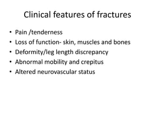
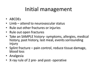
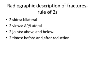
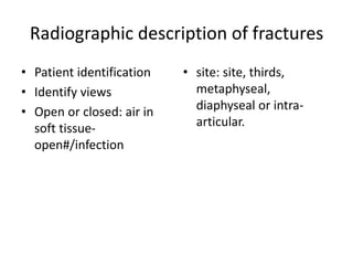
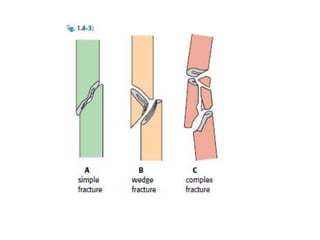
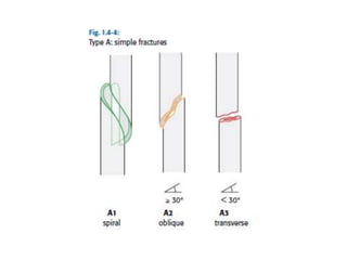
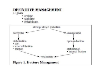
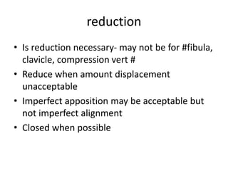
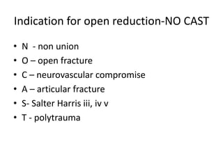
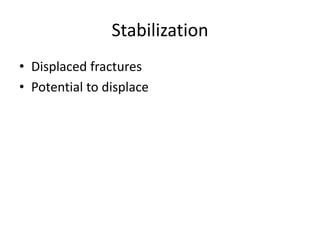
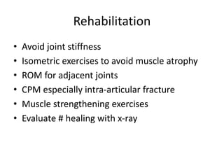
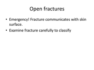
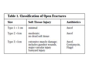
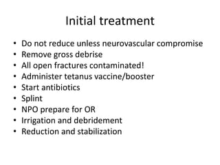
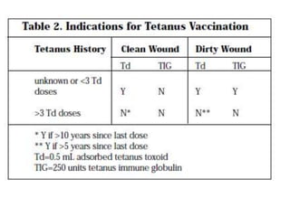
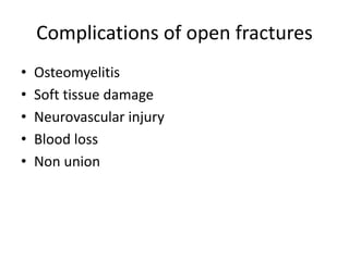
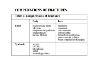
Basic principles of fracture
management
Nyiiro Francis MBchB, MMed/Ortho,
FCS(ECSA), Fellow Pediatric Ortho (uoT)
Consultant Orthopaedic Surgeon
CASE
• Middle aged man picked by police from the
middle of the road, reportedly knocked by
speeding vehicle.
• Unable to walk, leg deformed, in a lot of pain
and extremities cold
• Pulse 100 b/min, Respiratory rate 30 b/min
What is a fracture
• Cracking or breaking of hard object or material
• Continuity of bone is broken
What we need to achieve
• Able to identify an injured patient
• Identify life threatening conditions
• Identify specific injuries
• Emergency treatment of injured patient
• Initial management of injured patient
Mechanism- remember process
leading to fracture
• Traumatic –(high)
• Pathological (low energy)-tumor, metabolic
bone disease, osteopenia, infection
• Stress fractures – repetitive mechanic loading
Clinical features of fractures
• Pain /tenderness
• Loss of function- skin, muscles and bones
• Deformity/leg length discrepancy
• Abnormal mobility and crepitus
• Altered neurovascular status
Initial management
• ABCDEs
• Limb – attend to neurovascular status
• Rule out other fractures or injuries
• Rule out open fractures
• Take an SAMPLE history- symptoms, allergies, medical
history, past history, last meal, events sorrounding
injury
• Splint fracture – pain control, reduce tissue damage,
blood loss
• Analgesia
• X-ray rule of 2 pre- and post- operative
Radiographic description of fractures-
rule of 2s
• 2 sides: bilateral
• 2 views: AP/Lateral
• 2 joints: above and below
• 2 times: before and after reduction
Radiographic description of fractures
• Patient identification
• Identify views
• Open or closed: air in
soft tissue-
open#/infection
• site: site, thirds,
metaphyseal,
diaphyseal or intra-
articular.
type
• Spiral: rotational force, low energy
• Oblique: angular or rotational force
• Transverse: direct force, high energy
• Comminuted: (>2pcs) – direct force, high
energy
Soft tissue
• Cast/splint
• Calcification
• Gas /air
• Foreign bodies
Displacement –distal wrt proximal
• Aposition/translation
• Angulation
• Rotation
• shortening
reduction
• Is reduction necessary- may not be for #fibula,
clavicle, compression vert #
• Reduce when amount displacement
unacceptable
• Imperfect apposition may be acceptable but
not imperfect alignment
• Closed when possible
Indication for open reduction-NO CAST
• N - non union
• O – open fracture
• C – neurovascular compromise
• A – articular fracture
• S- Salter Harris iii, iv v
• T - polytrauma
Stabilization
• Displaced fractures
• Potential to displace
External fixation
• Splints/tape
• Casts
• Traction
• External fixator
Internal fixation
• Percutaneous pinning
• Extra medullary fixation
• Intra medullary fixation
Rehabilitation
• Avoid joint stiffness
• Isometric exercises to avoid muscle atrophy
• ROM for adjacent joints
• CPM especially intra-articular fracture
• Muscle strengthening exercises
• Evaluate # healing with x-ray
Open fractures
• Emergency! Fracture communicates with skin
surface.
• Examine fracture carefully to classify
Initial treatment
• Do not reduce unless neurovascular compromise
• Remove gross debrise
• All open fractures contaminated!
• Administer tetanus vaccine/booster
• Start antibiotics
• Splint
• NPO prepare for OR
• Irrigation and debridement
• Reduction and stabilization
Initial treatment
• Go to OR within 6 hrs preferrably
• Re-exam for possible repeat I&D within 48 hrs.
Complications of open fractures
• Osteomyelitis
• Soft tissue damage
• Neurovascular injury
• Blood loss
• Non union
basic fracture management JUNE.pptx

basic fracture management JUNE.pptx

  • 1.
    Basic principles offracture management Nyiiro Francis MBchB, MMed/Ortho, FCS(ECSA), Fellow Pediatric Ortho (uoT) Consultant Orthopaedic Surgeon
  • 2.
    CASE • Middle agedman picked by police from the middle of the road, reportedly knocked by speeding vehicle. • Unable to walk, leg deformed, in a lot of pain and extremities cold • Pulse 100 b/min, Respiratory rate 30 b/min
  • 3.
    What is afracture • Cracking or breaking of hard object or material • Continuity of bone is broken
  • 4.
    What we needto achieve • Able to identify an injured patient • Identify life threatening conditions • Identify specific injuries • Emergency treatment of injured patient • Initial management of injured patient
  • 5.
    Mechanism- remember process leadingto fracture • Traumatic –(high) • Pathological (low energy)-tumor, metabolic bone disease, osteopenia, infection • Stress fractures – repetitive mechanic loading
  • 6.
    Clinical features offractures • Pain /tenderness • Loss of function- skin, muscles and bones • Deformity/leg length discrepancy • Abnormal mobility and crepitus • Altered neurovascular status
  • 7.
    Initial management • ABCDEs •Limb – attend to neurovascular status • Rule out other fractures or injuries • Rule out open fractures • Take an SAMPLE history- symptoms, allergies, medical history, past history, last meal, events sorrounding injury • Splint fracture – pain control, reduce tissue damage, blood loss • Analgesia • X-ray rule of 2 pre- and post- operative
  • 8.
    Radiographic description offractures- rule of 2s • 2 sides: bilateral • 2 views: AP/Lateral • 2 joints: above and below • 2 times: before and after reduction
  • 9.
    Radiographic description offractures • Patient identification • Identify views • Open or closed: air in soft tissue- open#/infection • site: site, thirds, metaphyseal, diaphyseal or intra- articular.
  • 10.
    type • Spiral: rotationalforce, low energy • Oblique: angular or rotational force • Transverse: direct force, high energy • Comminuted: (>2pcs) – direct force, high energy
  • 13.
    Soft tissue • Cast/splint •Calcification • Gas /air • Foreign bodies
  • 14.
    Displacement –distal wrtproximal • Aposition/translation • Angulation • Rotation • shortening
  • 16.
    reduction • Is reductionnecessary- may not be for #fibula, clavicle, compression vert # • Reduce when amount displacement unacceptable • Imperfect apposition may be acceptable but not imperfect alignment • Closed when possible
  • 17.
    Indication for openreduction-NO CAST • N - non union • O – open fracture • C – neurovascular compromise • A – articular fracture • S- Salter Harris iii, iv v • T - polytrauma
  • 18.
  • 19.
    External fixation • Splints/tape •Casts • Traction • External fixator
  • 20.
    Internal fixation • Percutaneouspinning • Extra medullary fixation • Intra medullary fixation
  • 21.
    Rehabilitation • Avoid jointstiffness • Isometric exercises to avoid muscle atrophy • ROM for adjacent joints • CPM especially intra-articular fracture • Muscle strengthening exercises • Evaluate # healing with x-ray
  • 22.
    Open fractures • Emergency!Fracture communicates with skin surface. • Examine fracture carefully to classify
  • 24.
    Initial treatment • Donot reduce unless neurovascular compromise • Remove gross debrise • All open fractures contaminated! • Administer tetanus vaccine/booster • Start antibiotics • Splint • NPO prepare for OR • Irrigation and debridement • Reduction and stabilization
  • 25.
    Initial treatment • Goto OR within 6 hrs preferrably • Re-exam for possible repeat I&D within 48 hrs.
  • 27.
    Complications of openfractures • Osteomyelitis • Soft tissue damage • Neurovascular injury • Blood loss • Non union