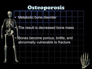
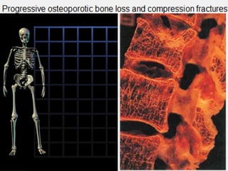
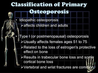
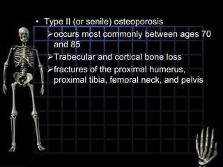
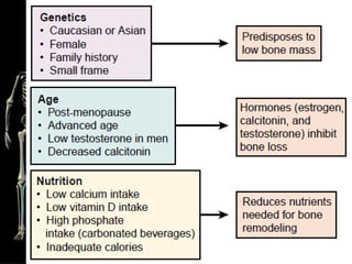
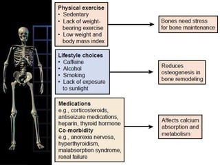
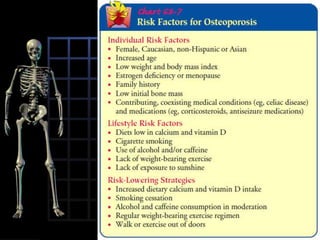
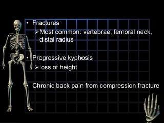
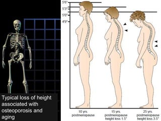
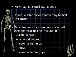
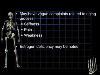
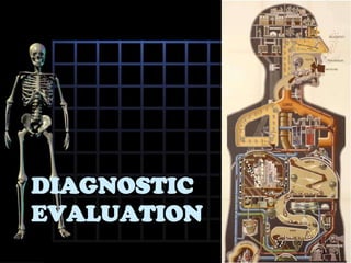
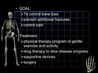
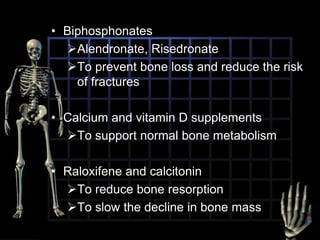
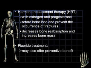
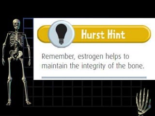
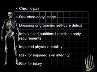
OSTEOPOROSIS
OVERVIEW
OsteoporosisMetabolic bone disorderThe result is decreased bone mass Bones become porous, brittle, and abnormally vulnerable to fracture
Primary
Secondary to an underlying disease
Classification of Primary OsteoporosisIdiopathic osteoporosis affects children and adultsType I (or postmenopausal) osteoporosis Usually affects females ages 51 to 75
Related to the loss of estrogen's protective effect on bone
Results in trabecular bone loss and some cortical bone loss
Vertebral and wrist fractures are common. Type II (or senile) osteoporosis occurs most commonly between ages 70 and 85
Trabecular and cortical bone loss
fractures of the proximal humerus, proximal tibia, femoral neck, and pelvis
Pathophysiology and etiology
The rate of bone resorption increases over the rate of bone formationloss of bone massCalcium and phosphate salts are lostporous, brittle bonesOccurs most commonly in postmenopausal women
COmplications
FracturesMost common: vertebrae, femoral neck, distal radiusProgressive kyphosisloss of height Chronic back pain from compression fracture
Typical loss of height associated with  osteoporosis and aging
Clinical manifestations
Asymptomatic until later stagesFracture after minor trauma may be first indicationMost frequent fractures associated with osteoporosis include fractures of:distal radiusvertebral bodiesproximal humerusPelvisproximal femur (hip)
May have vague complaints related to aging processStiffness
Pain
WeaknessEstrogen deficiency may be noted
Diagnostic evaluation
Differential diagnosisexclude other causes of rarefying bone disease, especially those that affect the spine
metastatic carcinoma, advanced multiple myelomaX-rays show changes only after 30% to 60% loss of boneDEXADual-energy X-ray absorptiometry
Shows decreased bone mineral density
T score -2.5 or worse
Serum and urine calcium levelsnormalSerum bone GLA-proteinMarker for bone turnover
ElevatedBone biopsy Shows thin, porous, otherwise normal boneSpine Computed Tomography Scan Shows demineralization
Quantitative computed tomography can evaluate bone density
Less available and more expensive than DEXAMANAGEMENT
GOAL:To control bone loss
prevent additional fractures
control pain
Treatment:
physical therapy program of gentle exercise and activity
drug therapy to slow disease progress
supportive devices
surgeryMedications
BiphosphonatesAlendronate, Risedronate
To prevent bone loss and reduce the risk of fracturesCalcium and vitamin D supplements To support normal bone metabolism
Raloxifene and calcitonin
To reduce bone resorption
To slow the decline in bone mass
Supportive devices and surgery
Back braceSupports weakened vertebraeSurgery open reduction and internal fixation
to correct pathologic fractures of the femur
Reduction and Immobilization
with a cast
Required in Colles' fracture
4 to 10 weeksLifestyle modification
DietAdequate intake of dietary calcium
Decreased alcohol consumption and caffeine useRegular ExerciseSmoking cessation
Hormonal and fluoride treatment

Osteoporosis