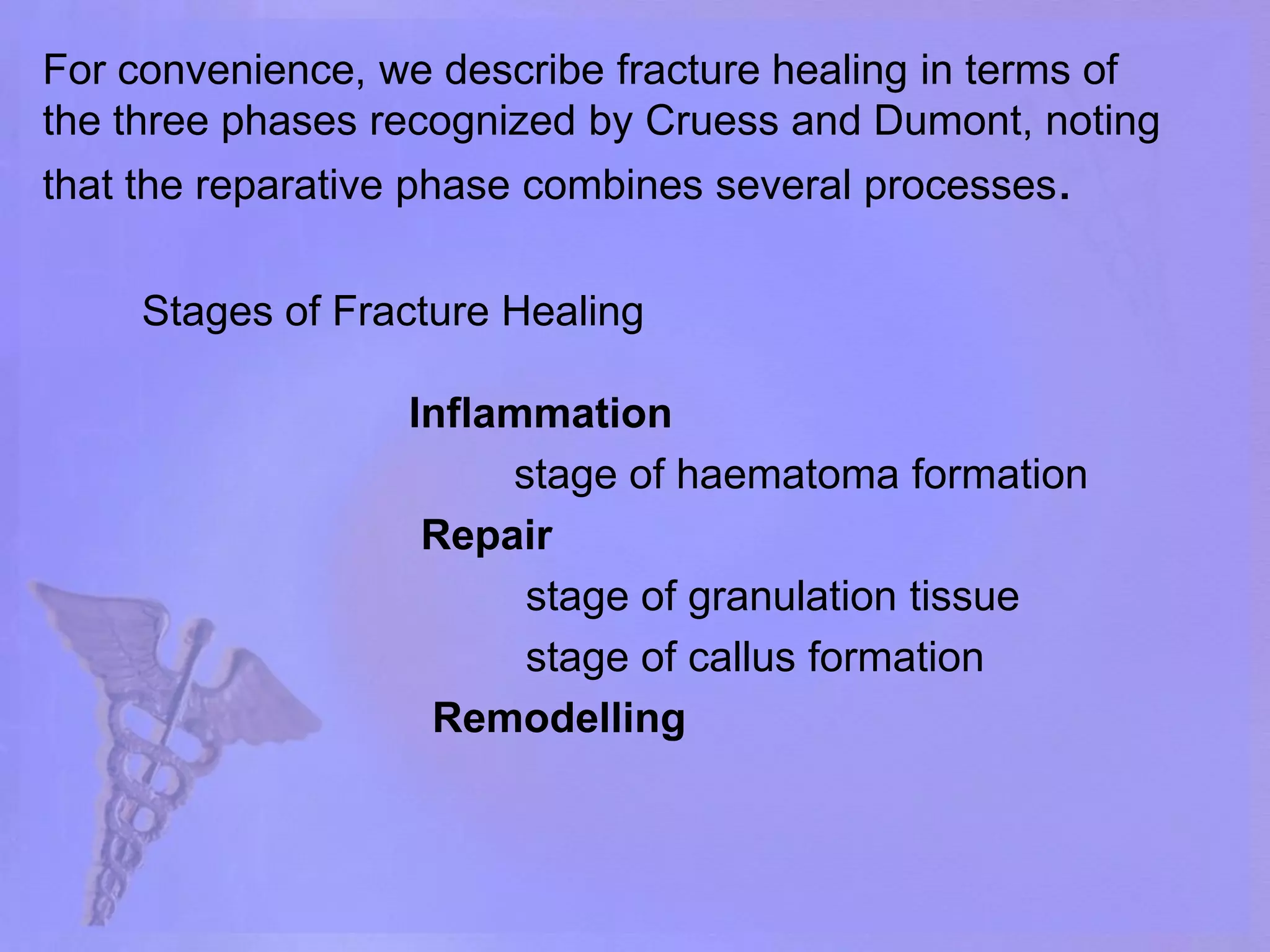
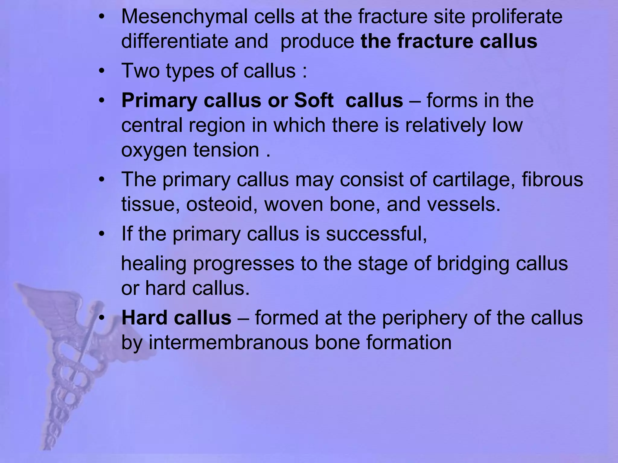
SlideShare a Scribd company logo
Fracture Healing DR HARDIK PAWAR  DNB ORTHO RESIDENT.                     CARE HOSPITAL ,                    HYDERABAD.
Types of  Bone Lamellar Bone      -   Orderly cellular distribution Collagen fibers arranged in parallel layers Normal adult bone Woven Bone or immature bone (non-lamellar)	 Randomly oriented collagen fibers In adults, seen at sites of  fracture healing,  tendon or ligament attachment and in pathological conditions
Lamellar Bone Cortical bone - Comprised of osteons (Haversian systems) runs longitudinally Osteons communicate with medullary cavity by Volkmann’s canals that run horizontally
Haversian System osteon osteocyte Volkmann’s canal Haversian canal Picture courtesy Gwen Childs, PhD.
Woven Bone Coarse with random orientation    Weaker than   lamellar bone Normally remodeled to lamellar bone Figure from Rockwood and Green’s: Fractures in Adults, 4th ed
Bone Composition Cells Osteocytes Osteoblasts Osteoclasts Extracellular Matrix Organic (35%) Collagen (type I) 90% Osteocalcin, osteonectin, proteoglycans, glycosaminoglycans, lipids (ground substance) Inorganic (65%) Primarily hydroxyapatite Ca5(PO4)3(OH)2
Osteoblasts Derived from mesenchymal stem cells Line the surface of the bone and produce osteoid Immediate precursor is fibroblast-like preosteoblasts Picture courtesy Gwen Childs, PhD.
Osteocytes Osteoblasts surrounded by bone matrix  trapped in lacunae Function poorly understood  regulating bone metabolism in response to stress and strain Picture courtesy Gwen Childs, PhD.
Osteocyte Network Osteocyte lacunae are connected by canaliculi Osteocytes are interconnected by long cell processes that project through the canaliculi Preosteoblasts also have connections via canaliculi with the osteocytes Network probably facilitates response of bone  to mechanical and chemical factors
Osteoclasts Derived from hematopoietic stem cells (monocyte precursor cells) Multinucleated cells whose function is bone resorption Reside in bone resorption pits (Howship’s lacunae) Parathyroid hormone stimulates receptors on osteoblasts that activate osteoclastic bone resorption Picture courtesy Gwen Childs, PhD.
Components of Bone Formation Cortex Periosteum Bone marrow Soft tissue
FRACTURE Fracture is a break in the structural continuity of bone . It may be a crack , a crumpling or a splintering of the cortex.
TYPES OF FRACTURES ON BASIS OF ETIOLOGY               -  Traumatic fracture             -  pathologic fractures due to some                           diseases             -  stress fracture  ON BASIS OF DISPLACEMEMT             - undisplaced             - displaced                        translation ( shift ) angulation ( tilt )                        rotation      ( twist )
ON BASIS OF RELATIONSHIP WITH EXTERNAL ENVIRONMENT                      - simple / closed fracture                     - open fracture ON BASIS OF PATTERN             - transverse             - oblique              - spiral             - comminuted             - segmental
Prerequisites for Bone Healing Adequate blood supply Adequate mechanical stability
Mechanisms of Bone Formation Cutting Cones Intramembranous Bone Formation Endochondral Bone Formation
Cutting Cones Primarily a mechanism to remodel bone Osteoclasts at the front of the cutting cone remove bone Trailing osteoblasts lay down new bone Courtesy Drs. Charles Schwab and Bruce Martin
Intramembranous (Periosteal) Bone Formation Mechanism by which a long bone grows in width Osteoblasts differentiate directly from preosteoblasts and lay down seams of osteoid Does NOT involve cartilage . It mainly forms cancellous bone.
Intramembranous Bone Formation Picture courtesy Gwen Childs, PhD.
Endochondral Bone Formation Mechanism by which a long bone grows in length Osteoblasts line a cartilage precursor The chondrocytes hypertrophy, degenerate and calcify (area of low oxygen tension) Vascular invasion of the cartilage occurs followed by ossification (increasing oxygen tension)
Endochondral Bone Formation Picture courtesy Gwen Childs, PhD.
Blood Supply Long bones have four blood supplies Nutrient artery (intramedullary) Periosteal vessels Metaphyseal vessels Epiphysial vessels Periosteal vessels Nutrient  artery Metaphyseal vessels Figure adapted from Rockwood and Green, 5th Ed
Nutrient Artery Normally the major blood supply for the diaphyseal cortex (80 to 85%) Enters the long bone via a nutrient foramen  Forms medullary arteries up and down the bone
Periosteal Vessels Arise from the capillary-rich periosteum Supply outer 15 to 20% of cortex normally Capable of supplying a much greater proportion of the cortex in the event of injury to the medullary blood supply
Metaphyseal Vessels Arise from periarticular vessels Penetrate the thin cortex in the metaphyseal region and anastomose with the medullary blood supply
Vascular Response in Fracture Repair Fracture stimulates the release of growth factors that promote angiogenesis and vasodilation Blood flow is increased substantially to the fracture site Peaks at two weeks after fracture
Fracture healing  Introduction  Fracture healing is a complex process that  requires the recruitment of appropriate cells (fibroblasts, macrophages, chondroblasts, osteoblasts, osteoclasts) and the subsequent expression of the appropriate genes (genes that control matrix production and organization, growth factors, transcription factors) at the right time and in the right anatomical location
HISTORY In 1975, Cruess and Dumont proposed that fracture healing may be considered to consist of three overlapping phases: an inflammatory phase, a reparative phase, and a remodeling phase  In 1989, FROST proposed the stages of fracture healing      five stages.                       stage of haematoma                       stage of granulation tissue                        stage of callus                       stage of modelling                       stage of remodelling
For convenience, we describe fracture healing in terms of the three phases recognized by Cruess and Dumont, noting that the reparative phase combines several processes.                        Inflammation                                 stage of haematoma formation Repair                                  stage of granulation tissue                                   stage of callus formation Remodelling Stages of Fracture Healing
Duration The inflammatory phase peaks within 48 hours and is quite diminished by 1 week after fracture. The reparative phase becomes activated within the first few days after fracture and persists for 2-3 months. The remodelling phase lasts for many years
Inflammation inflammatory phase is identical to the typical inflammatory response of most tissues to traumatic injury.  Vasodilation and hyperemia, presumably mediated by histamines, prostaglandins, and various cytokines, accompany invasion of the injury site by neutrophils, basophils, and phagocytes that participate in clearing away necrotic debris.
Disruption of blood vessels in the bone, marrow, periosteum, and surrounding tissue disruption at the time of injury results in the extravasation of blood at the fracture site and the formation of a hematoma Local vessels thrombose causing bony necrosis at the edges of the fracture  Increased capillary permeability results in a local inflammatory milieu Osteoinductive growth factors stimulate the proliferation and differentiation of mesenchymal stem cells
Reparative phase The reparative phase, which usually begins 4 or 5 days after injury, is characterized by the invasion of pluripotentialmesenchymal cells, which differentiate into fibroblasts, chondroblasts, and osteoblasts and form a soft primary fracture callus. Proliferation of blood vessels (angiogenesis) within the periosteal tissues and marrow space helps route the appropriate cells to the fracture site and contributes to the formation of a bed of granulation tissue.
Mesenchymal cells at the fracture site proliferate  differentiate and  produce the fracture callus Two types of callus :  Primary callus or Soft  callus – forms in the central region in which there is relatively low oxygen tension . The primary callus may consist of cartilage, fibrous tissue, osteoid, woven bone, and vessels. If the primary callus is successful,     healing progresses to the stage of bridging callus             or hard callus. Hard callus – formed at the periphery of the callus by intermembranous bone formation
Periosteal callus forms along the periphery of the fracture site Intramembranous ossification initiated by preosteoblasts Intramedullary callus forms in the center of the fracture site Endochondral ossification at the site of the fracture hematoma Chemical and mechanical factors stimulate callus formation and mineralization
Repair Figure from Brighton, et al, JBJS-A, 1991.
Remodeling The biochemical composition of the fracture callus matrix changes as repair progresses.  The cells replace the fibrin clot with a loose fibrous matrix containing glycosaminoglycans, proteoglycans, and types I and III collagen In many regions they convert this tissue to more dense fibrocartilage or hyaline-like cartilage.  With formation of hyaline-like cartilage, type II collagen, cartilage-specific proteoglycan and link protein content increase.
Woven bone is gradually converted to lamellar bone Medullary cavity is reconstituted Stability of the fracture fragments progressively increases .  eventually clinical union occurs that is, the fracture site becomes stable and pain-free. Radiographic union occurs when plain radiographs show bone trabeculae or cortical bone crossing the fracture site, and often occurs later than clinical union . Despite successful fracture healing, the bone density of the involved limb may be decreased for years
Types for Bone Healing Direct (primary) bone healing Indirect (secondary) bone healing
Direct Bone Healing Mechanism of bone healing seen when there is no motion at the fracture site (i.e. absolute stability) Does not involve formation of fracture callus Osteoblasts originate from endothelial and perivascular cells
Components of Direct Bone Healing Contact Healing Direct contact between the fracture ends allows healing to be with lamellar bone immediately  Gap Healing Gaps less than 200-500 microns are primarily filled with woven bone that is subsequently remodeled into lamellar bone Larger gaps are healed by indirect bone healing (partially filled with fibrous tissue that undergoes secondary ossification)
Direct Bone Healing Figure from http://www.vetmed.ufl.edu/sacs/notes
Indirect Bone Healing Mechanism for healing in fractures that have some motion, but not enough to disrupt the healing process. Bridging periosteal (soft) callus and medullary (hard) callus re-establish structural continuity Callus subsequently undergoes endochondral ossification Process fairly rapid - weeks
VARIABLES THAT INFLUENCE THE FRACTURE HEALING
I.    Systemic factors   or patient variables         A.    Age   B.    Activity level including                            1.  immobilization   C.    Nutritional status   D.    Hormonal factors                      1.    Growth hormone             2.    Corticosteroids             3.    Others (thyroid, estrogen, androgen,  calcitonin, parathyroid hormone,                                       prostaglandins)        E.   Cigarette smoking    
   E.    Diseases: diabetes,anemia,neuropathies,        tabesdorsalis   F.    Vitamin deficiencies: A, C, D, K   G.    Drugs: nonsteroidalantiinflammatory drugs                                     (NSAIDs), anticoagulants, factor XIII,                            calcium channel blockers (verapamil),  cytotoxins, diphosphonates, phenytoin                          (Dilantin), sodium fluoride, tetracycline   H.    Other substances (nicotine, alcohol)   I.      Hyperoxia   J.    Systemic growth factors   K.    Environmental temperature   L.    Central nervous system trauma
 II.  Local factors    or tissue variables           A.    Factors independent of injury, treatment, or                     complications                       1.    Type of bone                                        cortical or cancellous              2.    Abnormal bone                                                      a.   Radiation necrosis                                              b.   Infection                                              c.   Tumors and other                                                           pathological conditions                  3.    Denervation   
 B.    Factors depending on injury  or injury variables               1.    Degree of local damage                               a.    Compound fracture                       b.    Comminution of fracture                            c.    segmental fractures                       d.    Velocity of injury                       e.    Low circulatory levels of vitamin K1            2.    Extent of disruption of vascular supply to                     bone, its fragments (macrovascular osteonecrosis), or soft tissues;             3.    Type and location of fracture (one or two                    bones,  e.g., tibia and fibula or tibia alone            4.    Loss of bone            5.    Soft-tissue interposition            6.    Local growth factors            7.    Intraarticular fractures
C.    Factors depending on treatment           OR                 treatment   variables           1.    Extent of surgical trauma (blood supply, heat)     2.    Implant-induced altered blood flow     3.    Degree and kind of rigidity of internal or                   external fixation and the influence of timing     4.    Degree, duration, and direction of load-                 induced deformation of bone and soft tissues     5.    Apposition of fracture fragments (gap,                  displacement, overdistraction)     6.    Factors stimulating posttraumatic  osteogenesis (bone grafts, bone                morphogenetic protein, electrical stimulation,               surgical technique, intermittent venous stasis .
D.    Factors associated with complications                       1.    Infection              2.    Venous stasis              3.    Metal allergy
Local Regulation of Bone Healing Growth factors                  Transforming growth factor                  Bone morphogenetic proteins                  Fibroblast growth factors                  Platelet-derived growth factors                 Insulin-like growth factors Cytokines                   Interleukin-1,-4,-6,-11, macrophage  and granulocyte/macrophage (GM) colony-stimulating factors (CSFs) and Tumor Necrosis Factor Prostaglandins/Leukotrienes Hormones Growth factor antagonists
Transforming Growth Factor Super-family of growth factors (~34 members) Acts on serine/threoninekinase cell wall receptors Promotes proliferation and differentiation of mesenchymal precursors for osteoblasts, osteoclasts and chondrocytes Stimulates both enchondral and intramembranous bone formation Induces synthesis of cartilage-specific proteoglycans and type II collagen Stimulates collagen synthesis by osteoblasts
Bone Morphogenetic Proteins Osteoinductive proteins initially isolated from demineralized bone matrix Induce cell differentiation BMP-3 (osteogenin) is an extremely potent inducer of mesenchymal tissue differentiation into bone Promote endochondral ossification BMP-2 and BMP-7 induce endochondral bone formation in segmental defects Regulate extracellular matrix production BMP-1 is an enzyme that cleaves the carboxy termini of procollagens I, II and III
Bone Morphogenetic Proteins  These are included in the TGF-β family Except BMP-1 Sixteen different BMP’s have been identified BMP2-7,9 are osteoinductive BMP2,6, & 9 may be the most potent in osteoblastic differentiation Involved in progenitor cell transformation to pre-osteoblasts Work through the intracellular Smad pathway Follow a dose/response ratio
Timing and Function of Growth Factors Table from Dimitriou, et al., Injury, 2005
Clinical Use of BMP’s Used at doses between 10x & 1000x native levels Negligible risk of excessive bone formation rhBMP-2 used in “fresh” open fractures to enhance healing and reduce need for secondary procedures after unreamedIM nailin It is found that application of rhBMP-2 decreases infection rate in Type IIIA & B open fractures. BMP-7 approved for use in recalcitrant nonunions in patients for whom autografting is not a good option (i.e. medically unstable, previous harvesting of all iliac crest sites, etc.)
BMP Future Directions BMP-2  Increased fusion rate in spinal fusion  BMP-7 equally effective as ICBG in nonunions (small series: need larger studies) Must be applied locally because of rapid systemic clearance
BMP Antagonists May have important role in bone formation Noggin Extra-cellular inhibitor Competes with BMP-2 for receptors BMP-13 found to limit differentiation of mesenchymalstromal cells Inhibits osteogenic differentiation
Fibroblast Growth Factors Both acidic (FGF-1) and basic (FGF-2) forms  Increase proliferation of chondrocytes and osteoblasts Enhance callus formation FGF-2 stimulates angiogenesis
Platelet-Derived Growth Factor A dimer of the products of two genes, PDGF-A and PDGF-B PDGF-BB and PDGF-AB are the predominant forms found in the circulation Stimulates bone cell growth Mitogen for cells of mesenchymal origin Increases type I collagen synthesis by increasing the number of osteoblasts PDGF-BB stimulates bone resorption by increasing the number of osteoclasts
Insulin-like Growth Factor Two types:  IGF-I and IGF-II Synthesized by multiple tissues IGF-I production in the liver is stimulated by Growth Hormone Stimulates bone collagen and matrix synthesis Stimulates replication of osteoblasts Inhibits bone collagen degradation
Cytokines  Interleukin-1,-4,-6,-11, macrophage and granulocyte/macrophage (GM) colony-stimulating factors (CSFs) and Tumor Necrosis Factor Stimulate bone resorption IL-1 is the most potent IL-1 and IL-6 synthesis is decreased by estrogen May be mechanism for post-menopausal bone resorption Peak during 1st 24 hours then again during remodeling Regulate endochondral bone formation
Specific Factor Stimulation of Osteoblasts and Osteoclasts Cytokine		 Bone Formation		Bone Resorption IL-1				+				+++ TNF-α				+				+++ TNF-β				+				+++ TGF-α				--				+++ TGF-β				++				++ PDGF				++				++ IGF-1				+++				0 IGF-2				+++				0 FGF				+++				0
Prostaglandins / Leukotrienes Effect on bone resorption is species dependent and their overall effects in humans unknown Prostaglandins of the E series Stimulate osteoblastic bone formation Inhibit activity of isolated osteoclasts Leukotrienes Stimulate osteoblastic bone formation Enhance the capacity of isolated osteoclasts to form resorption pits
Hormones Estrogen Stimulates fracture healing through receptor mediated mechanism Modulates release of a specific inhibitor of IL-1 Thyroid hormones Thyroxine and triiodothyronine stimulate osteoclastic bone resorption Glucocorticoids Inhibit calcium absorption from the gut causing increased PTH and therefore increased osteoclastic bone resorption
Hormones (cont.) Parathyroid Hormone Intermittent exposure stimulates Osteoblasts Increased bone formation Growth Hormone Mediated through IGF-1 (Somatomedin-C) Increases callus formation and fracture strength
Vascular Factors Metalloproteinases Degrade cartilage and bones to allow invasion of vessels Angiogenic factors Vascular-endothelial growth factors Mediate neo-angiogenesis & endothelial-cell specific mitogens Angiopoietin (1&2) Regulate formation of larger vessels and branches
Electromagnetic Field Electromagnetic (EM) devices are based on Wolff’s Law that bone responds to mechanical stress:  In vitro bone deformation produces piezoelectric currents and streaming potentials. Exogenous EM fields may stimulate bone growth and repair by the same mechanism Clinical efficacy very controversial No studies have shown PEMF to be effective in “gap healing” or pseudarthrosis
Types of EM Devices Microamperes Direct electrical current Capacitively coupled electric fields Pulsed electromagnetic fields (PEMF)
PEMF Approved by the FDA for the treatment of non-unions Efficacy of bone stimulation appears to be frequency dependant Extremely low frequency (ELF) sinusoidal electric fields in the physiologic range are most effective (15 to 30 Hz range) Specifically, PEMF signals in the 20 to 30 Hz range (postural muscle activity) appear more effective than those below 10 Hz (walking)
Ultrasound Low-intensity ultrasound is approved by the FDA for stimulating healing of fresh fractures Modulates signal transduction, increases gene expression, increases blood flow, enhances bone remodeling and increases callus torsional strength in animal models
Ultrasound Human clinical trials show a decreased time of healing in fresh fractures treated nonoperatively Four level 1 studies show a decrease in healing time up to 38%  Has also been shown to decrease the healing time in smokers potentially reversing the ill effects of smoking
Summary Fracture healing is influenced by many variables including mechanical stability, electrical environment, biochemical factors and blood flow Our ability to enhance fracture healing will increase as we better understand the interaction between these variables  If you would like to volunteer as an author for the Resident Slide Project or recommend updates to any of the following slides, please send an e-mail to hardikpawar103@gmail.com

More Related Content

What's hot

Bone grafts and bone grafts substitutes
Bone grafts and bone grafts substitutesBone grafts and bone grafts substitutes
Bone grafts and bone grafts substitutes
siddharth438
 
Bone cement,Cementing Generations, Complications & Recent advances
Bone cement,Cementing Generations, Complications & Recent advancesBone cement,Cementing Generations, Complications & Recent advances
Bone cement,Cementing Generations, Complications & Recent advances
Sameer Ashar
 
Fracture healing by dr.v.r.vignesh
Fracture healing by dr.v.r.vigneshFracture healing by dr.v.r.vignesh
Fracture healing by dr.v.r.vignesh
Vignesh Ramaiyah
 
Salvage of bone defects
Salvage of bone defectsSalvage of bone defects
Salvage of bone defects
fathi neana
 
Ilizarov External fixator
Ilizarov External fixatorIlizarov External fixator
Ilizarov External fixatorAbdullah Mamun
 
Masquelet technique for management of large bone defects.
Masquelet technique for management of large bone defects.Masquelet technique for management of large bone defects.
Masquelet technique for management of large bone defects.
Kushi Rithvic
 
Non union by rv ppt
Non union  by rv ppt Non union  by rv ppt
Non union by rv ppt
ravi varma
 
Multi ligamentous knee injury
Multi ligamentous knee injuryMulti ligamentous knee injury
Multi ligamentous knee injury
Jose Austine
 
Peri prosthetic fractures
Peri prosthetic fracturesPeri prosthetic fractures
Peri prosthetic fractures
Anshul Sethi
 
Masquelet Technique
Masquelet TechniqueMasquelet Technique
Basic Principles of Fracture Management
Basic Principles of Fracture ManagementBasic Principles of Fracture Management
Basic Principles of Fracture Management
Pathrose Parathuvayalil Group
 
Tibial plateau fractures
Tibial plateau fracturesTibial plateau fractures
Tibial plateau fractures
Ponnilavan Ponz
 
Nonunion femoral neck fractures
Nonunion femoral neck fracturesNonunion femoral neck fractures
Nonunion femoral neck fractures
Rajesh Raj
 
Chronic osteomyelitis
Chronic  osteomyelitisChronic  osteomyelitis
Chronic osteomyelitis
PG (MS) orthopaedics @ KBNIMS
 
Osteotomies around hip by dr rohit kumar
Osteotomies around hip by dr rohit kumarOsteotomies around hip by dr rohit kumar
Osteotomies around hip by dr rohit kumarDr Rohit Kumar
 
Tribology in orthopaedics seminar
Tribology in orthopaedics seminarTribology in orthopaedics seminar
Tribology in orthopaedics seminar
ujjalrajbangshi
 
Damage control orthopaedic surgery
Damage control orthopaedic surgeryDamage control orthopaedic surgery
Damage control orthopaedic surgery
Mohamed Abulsoud
 
Management of TendoAchillis rupture
Management of TendoAchillis ruptureManagement of TendoAchillis rupture
Management of TendoAchillis ruptureAnkur Mittal
 
non union and malunion final.pptx
non union and malunion final.pptxnon union and malunion final.pptx
non union and malunion final.pptx
RAdhavan
 

What's hot (20)

Bone grafts and bone grafts substitutes
Bone grafts and bone grafts substitutesBone grafts and bone grafts substitutes
Bone grafts and bone grafts substitutes
 
Bone cement,Cementing Generations, Complications & Recent advances
Bone cement,Cementing Generations, Complications & Recent advancesBone cement,Cementing Generations, Complications & Recent advances
Bone cement,Cementing Generations, Complications & Recent advances
 
Fracture healing by dr.v.r.vignesh
Fracture healing by dr.v.r.vigneshFracture healing by dr.v.r.vignesh
Fracture healing by dr.v.r.vignesh
 
Salvage of bone defects
Salvage of bone defectsSalvage of bone defects
Salvage of bone defects
 
Ilizarov External fixator
Ilizarov External fixatorIlizarov External fixator
Ilizarov External fixator
 
Masquelet technique for management of large bone defects.
Masquelet technique for management of large bone defects.Masquelet technique for management of large bone defects.
Masquelet technique for management of large bone defects.
 
Non union by rv ppt
Non union  by rv ppt Non union  by rv ppt
Non union by rv ppt
 
Multi ligamentous knee injury
Multi ligamentous knee injuryMulti ligamentous knee injury
Multi ligamentous knee injury
 
Peri prosthetic fractures
Peri prosthetic fracturesPeri prosthetic fractures
Peri prosthetic fractures
 
Masquelet Technique
Masquelet TechniqueMasquelet Technique
Masquelet Technique
 
Basic Principles of Fracture Management
Basic Principles of Fracture ManagementBasic Principles of Fracture Management
Basic Principles of Fracture Management
 
Tibial plateau fractures
Tibial plateau fracturesTibial plateau fractures
Tibial plateau fractures
 
Nonunion femoral neck fractures
Nonunion femoral neck fracturesNonunion femoral neck fractures
Nonunion femoral neck fractures
 
Fracture healing
Fracture  healingFracture  healing
Fracture healing
 
Chronic osteomyelitis
Chronic  osteomyelitisChronic  osteomyelitis
Chronic osteomyelitis
 
Osteotomies around hip by dr rohit kumar
Osteotomies around hip by dr rohit kumarOsteotomies around hip by dr rohit kumar
Osteotomies around hip by dr rohit kumar
 
Tribology in orthopaedics seminar
Tribology in orthopaedics seminarTribology in orthopaedics seminar
Tribology in orthopaedics seminar
 
Damage control orthopaedic surgery
Damage control orthopaedic surgeryDamage control orthopaedic surgery
Damage control orthopaedic surgery
 
Management of TendoAchillis rupture
Management of TendoAchillis ruptureManagement of TendoAchillis rupture
Management of TendoAchillis rupture
 
non union and malunion final.pptx
non union and malunion final.pptxnon union and malunion final.pptx
non union and malunion final.pptx
 

Viewers also liked

fracture healing by DR ROHIT KUMAR
 fracture healing by DR ROHIT KUMAR fracture healing by DR ROHIT KUMAR
fracture healing by DR ROHIT KUMARDr Rohit Kumar
 
Maintainance & replacement fluid therapy pediatrics AG
Maintainance & replacement fluid therapy pediatrics AGMaintainance & replacement fluid therapy pediatrics AG
Maintainance & replacement fluid therapy pediatrics AG
Akshay Golwalkar
 
Hip dislocation class
Hip dislocation classHip dislocation class
Hip dislocation class
DrHarpreet Bhatia
 
Congenital dislocation of hip_UTSAV
Congenital dislocation of hip_UTSAVCongenital dislocation of hip_UTSAV
Congenital dislocation of hip_UTSAVUtsav Agrawal
 
Upper limb fractures (part2)
Upper limb fractures (part2)Upper limb fractures (part2)
Upper limb fractures (part2)
Apoorv Jain
 
Proximal tibia fracture
Proximal tibia fractureProximal tibia fracture
Proximal tibia fracturevisheshrohatgi
 
Maintenance fluid calculation
Maintenance fluid calculationMaintenance fluid calculation
Maintenance fluid calculation
johngeorge123
 
collection of blood sample and preanalytical errors
collection of blood sample and preanalytical errorscollection of blood sample and preanalytical errors
collection of blood sample and preanalytical errors
Utkarsh Sharma
 
Lower limb fractures part 1 (for UGs)
Lower limb fractures part 1 (for UGs)Lower limb fractures part 1 (for UGs)
Lower limb fractures part 1 (for UGs)
Apoorv Jain
 
Surgical site infection 2015
Surgical site infection 2015Surgical site infection 2015
Surgical site infection 2015
Philippine Hospital Infection Control Society
 
Pelvic and acetabular fractures
Pelvic and acetabular fracturesPelvic and acetabular fractures
Pelvic and acetabular fractures
Sidharth Baheti
 
Surgical Site Infection by Doctor Saleem Plastic Surgeon
Surgical Site Infection by Doctor Saleem Plastic Surgeon Surgical Site Infection by Doctor Saleem Plastic Surgeon
Surgical Site Infection by Doctor Saleem Plastic Surgeon
Muhammad Saleem
 
Fractures, bone healing & principles of tx. of fractures
Fractures, bone healing & principles of tx. of fracturesFractures, bone healing & principles of tx. of fractures
Fractures, bone healing & principles of tx. of fracturesSimba Syed
 
Amputations by Dr. Sunny Agarwal
Amputations by Dr. Sunny AgarwalAmputations by Dr. Sunny Agarwal
Amputations by Dr. Sunny Agarwal
Sunny Agarwal
 
Principles of fracture management Saseendar
Principles of fracture management SaseendarPrinciples of fracture management Saseendar
Principles of fracture management Saseendar
Dr Saseendar MD
 
Principles in fractures management
Principles in fractures managementPrinciples in fractures management
Principles in fractures management
Isa Basuki
 
Upperlimb fractures bpt
Upperlimb fractures bptUpperlimb fractures bpt
Upperlimb fractures bptvaruntandra
 
Protein energy malnutrition
Protein energy malnutritionProtein energy malnutrition
Protein energy malnutrition
DrBabu Meena
 
Fractures and Dislocations of Upper Limb
Fractures and Dislocations of Upper LimbFractures and Dislocations of Upper Limb
Fractures and Dislocations of Upper Limb
Mohammad AlSofyani
 

Viewers also liked (20)

fracture healing by DR ROHIT KUMAR
 fracture healing by DR ROHIT KUMAR fracture healing by DR ROHIT KUMAR
fracture healing by DR ROHIT KUMAR
 
Maintainance & replacement fluid therapy pediatrics AG
Maintainance & replacement fluid therapy pediatrics AGMaintainance & replacement fluid therapy pediatrics AG
Maintainance & replacement fluid therapy pediatrics AG
 
Hip dislocation class
Hip dislocation classHip dislocation class
Hip dislocation class
 
Congenital dislocation of hip_UTSAV
Congenital dislocation of hip_UTSAVCongenital dislocation of hip_UTSAV
Congenital dislocation of hip_UTSAV
 
Upper limb fractures (part2)
Upper limb fractures (part2)Upper limb fractures (part2)
Upper limb fractures (part2)
 
Proximal tibia fracture
Proximal tibia fractureProximal tibia fracture
Proximal tibia fracture
 
Maintenance fluid calculation
Maintenance fluid calculationMaintenance fluid calculation
Maintenance fluid calculation
 
collection of blood sample and preanalytical errors
collection of blood sample and preanalytical errorscollection of blood sample and preanalytical errors
collection of blood sample and preanalytical errors
 
Lower limb fractures part 1 (for UGs)
Lower limb fractures part 1 (for UGs)Lower limb fractures part 1 (for UGs)
Lower limb fractures part 1 (for UGs)
 
Surgical site infection 2015
Surgical site infection 2015Surgical site infection 2015
Surgical site infection 2015
 
Pelvic and acetabular fractures
Pelvic and acetabular fracturesPelvic and acetabular fractures
Pelvic and acetabular fractures
 
Surgical Site Infection by Doctor Saleem Plastic Surgeon
Surgical Site Infection by Doctor Saleem Plastic Surgeon Surgical Site Infection by Doctor Saleem Plastic Surgeon
Surgical Site Infection by Doctor Saleem Plastic Surgeon
 
Fractures, bone healing & principles of tx. of fractures
Fractures, bone healing & principles of tx. of fracturesFractures, bone healing & principles of tx. of fractures
Fractures, bone healing & principles of tx. of fractures
 
Amputations by Dr. Sunny Agarwal
Amputations by Dr. Sunny AgarwalAmputations by Dr. Sunny Agarwal
Amputations by Dr. Sunny Agarwal
 
Principles of fracture management Saseendar
Principles of fracture management SaseendarPrinciples of fracture management Saseendar
Principles of fracture management Saseendar
 
Principles in fractures management
Principles in fractures managementPrinciples in fractures management
Principles in fractures management
 
Pelvic fractures
Pelvic fracturesPelvic fractures
Pelvic fractures
 
Upperlimb fractures bpt
Upperlimb fractures bptUpperlimb fractures bpt
Upperlimb fractures bpt
 
Protein energy malnutrition
Protein energy malnutritionProtein energy malnutrition
Protein energy malnutrition
 
Fractures and Dislocations of Upper Limb
Fractures and Dislocations of Upper LimbFractures and Dislocations of Upper Limb
Fractures and Dislocations of Upper Limb
 

Similar to Fracture healing

Fracture healing
Fracture healingFracture healing
Fracture healing
MONTHER ALKHAWLANY
 
Fracture healing
Fracture healing Fracture healing
Fracture healing
Diwakar vasudev
 
Stages of Bone healing and madalities to enhance bone healing
Stages of Bone healing and madalities to enhance bone healing Stages of Bone healing and madalities to enhance bone healing
Stages of Bone healing and madalities to enhance bone healing
Surya Vijay Singh
 
Fractures...types and healing of fractures
Fractures...types and healing of fracturesFractures...types and healing of fractures
Fractures...types and healing of fractures
Dr ashwani panchal
 
Fracture healing
Fracture healingFracture healing
Fracture healing
Vibhuti Nautiyal
 
Bone healing
Bone healingBone healing
Bone healing
Bone healing Bone healing
Bone healing
Dr.Noreen
 
Bone and fracture healing
Bone and fracture healingBone and fracture healing
Bone and fracture healing
Praveen Kumar Reddy Gorantla
 
Fracture Healing (Dr Arinze) [Autosaved].pdf
Fracture Healing (Dr Arinze) [Autosaved].pdfFracture Healing (Dr Arinze) [Autosaved].pdf
Fracture Healing (Dr Arinze) [Autosaved].pdf
Arizona29
 
Fracture healing By Dr Baijnath Agrahari
Fracture healing By Dr Baijnath AgrahariFracture healing By Dr Baijnath Agrahari
Fracture healing By Dr Baijnath Agrahari
Baijnath Agrahari
 
Anatomy of bone, General orthopedics and fracture healing
Anatomy of bone, General orthopedics and fracture healingAnatomy of bone, General orthopedics and fracture healing
Anatomy of bone, General orthopedics and fracture healing
adityarana242502
 
Phsiology of fractures
Phsiology of fracturesPhsiology of fractures
Phsiology of fracturesAnkur Mittal
 
Fracture healing and wound healing
Fracture healing and wound healingFracture healing and wound healing
Fracture healing and wound healingShermil Sayd
 
bone healing.pptx
bone healing.pptxbone healing.pptx
bone healing.pptx
Manjula N
 
Fracture Healing, Complications & factors promotes the fracture healing.pptx
Fracture Healing, Complications & factors promotes the fracture healing.pptxFracture Healing, Complications & factors promotes the fracture healing.pptx
Fracture Healing, Complications & factors promotes the fracture healing.pptx
DrAdnanMughal1
 
Fracture healing, complication & factors promotes the fracture healing
Fracture healing, complication & factors promotes the fracture healingFracture healing, complication & factors promotes the fracture healing
Fracture healing, complication & factors promotes the fracture healing
Adnan Sabir
 
Orthopaedics Surgery Tutor.Com
Orthopaedics Surgery Tutor.ComOrthopaedics Surgery Tutor.Com
Orthopaedics Surgery Tutor.Com
Deep Deep
 
Healing of fracture
Healing  of  fractureHealing  of  fracture
Healing of fracture
Dr. Armaan Singh
 

Similar to Fracture healing (20)

Fracture healing
Fracture healingFracture healing
Fracture healing
 
Fracture healing
Fracture healing Fracture healing
Fracture healing
 
Stages of Bone healing and madalities to enhance bone healing
Stages of Bone healing and madalities to enhance bone healing Stages of Bone healing and madalities to enhance bone healing
Stages of Bone healing and madalities to enhance bone healing
 
Fractures...types and healing of fractures
Fractures...types and healing of fracturesFractures...types and healing of fractures
Fractures...types and healing of fractures
 
Fracture healing
Fracture healingFracture healing
Fracture healing
 
Bone healing
Bone healingBone healing
Bone healing
 
Bone healing
Bone healing Bone healing
Bone healing
 
Fracture healing srinath
Fracture healing   srinathFracture healing   srinath
Fracture healing srinath
 
Bone and fracture healing
Bone and fracture healingBone and fracture healing
Bone and fracture healing
 
Fracture Healing (Dr Arinze) [Autosaved].pdf
Fracture Healing (Dr Arinze) [Autosaved].pdfFracture Healing (Dr Arinze) [Autosaved].pdf
Fracture Healing (Dr Arinze) [Autosaved].pdf
 
Fracture healing By Dr Baijnath Agrahari
Fracture healing By Dr Baijnath AgrahariFracture healing By Dr Baijnath Agrahari
Fracture healing By Dr Baijnath Agrahari
 
Anatomy of bone, General orthopedics and fracture healing
Anatomy of bone, General orthopedics and fracture healingAnatomy of bone, General orthopedics and fracture healing
Anatomy of bone, General orthopedics and fracture healing
 
Epiphyseal injury
Epiphyseal injuryEpiphyseal injury
Epiphyseal injury
 
Phsiology of fractures
Phsiology of fracturesPhsiology of fractures
Phsiology of fractures
 
Fracture healing and wound healing
Fracture healing and wound healingFracture healing and wound healing
Fracture healing and wound healing
 
bone healing.pptx
bone healing.pptxbone healing.pptx
bone healing.pptx
 
Fracture Healing, Complications & factors promotes the fracture healing.pptx
Fracture Healing, Complications & factors promotes the fracture healing.pptxFracture Healing, Complications & factors promotes the fracture healing.pptx
Fracture Healing, Complications & factors promotes the fracture healing.pptx
 
Fracture healing, complication & factors promotes the fracture healing
Fracture healing, complication & factors promotes the fracture healingFracture healing, complication & factors promotes the fracture healing
Fracture healing, complication & factors promotes the fracture healing
 
Orthopaedics Surgery Tutor.Com
Orthopaedics Surgery Tutor.ComOrthopaedics Surgery Tutor.Com
Orthopaedics Surgery Tutor.Com
 
Healing of fracture
Healing  of  fractureHealing  of  fracture
Healing of fracture
 

Recently uploaded

special B.ed 2nd year old paper_20240531.pdf
special B.ed 2nd year old paper_20240531.pdfspecial B.ed 2nd year old paper_20240531.pdf
special B.ed 2nd year old paper_20240531.pdf
Special education needs
 
Additional Benefits for Employee Website.pdf
Additional Benefits for Employee Website.pdfAdditional Benefits for Employee Website.pdf
Additional Benefits for Employee Website.pdf
joachimlavalley1
 
Language Across the Curriculm LAC B.Ed.
Language Across the  Curriculm LAC B.Ed.Language Across the  Curriculm LAC B.Ed.
Language Across the Curriculm LAC B.Ed.
Atul Kumar Singh
 
Model Attribute Check Company Auto Property
Model Attribute  Check Company Auto PropertyModel Attribute  Check Company Auto Property
Model Attribute Check Company Auto Property
Celine George
 
Basic phrases for greeting and assisting costumers
Basic phrases for greeting and assisting costumersBasic phrases for greeting and assisting costumers
Basic phrases for greeting and assisting costumers
PedroFerreira53928
 
The geography of Taylor Swift - some ideas
The geography of Taylor Swift - some ideasThe geography of Taylor Swift - some ideas
The geography of Taylor Swift - some ideas
GeoBlogs
 
Phrasal Verbs.XXXXXXXXXXXXXXXXXXXXXXXXXX
Phrasal Verbs.XXXXXXXXXXXXXXXXXXXXXXXXXXPhrasal Verbs.XXXXXXXXXXXXXXXXXXXXXXXXXX
Phrasal Verbs.XXXXXXXXXXXXXXXXXXXXXXXXXX
MIRIAMSALINAS13
 
Overview on Edible Vaccine: Pros & Cons with Mechanism
Overview on Edible Vaccine: Pros & Cons with MechanismOverview on Edible Vaccine: Pros & Cons with Mechanism
Overview on Edible Vaccine: Pros & Cons with Mechanism
DeeptiGupta154
 
How libraries can support authors with open access requirements for UKRI fund...
How libraries can support authors with open access requirements for UKRI fund...How libraries can support authors with open access requirements for UKRI fund...
How libraries can support authors with open access requirements for UKRI fund...
Jisc
 
aaaaaaaaaaaaaaaaaaaaaaaaaaaaaaaaaaaaaaaaa
aaaaaaaaaaaaaaaaaaaaaaaaaaaaaaaaaaaaaaaaaaaaaaaaaaaaaaaaaaaaaaaaaaaaaaaaaaaaaaaaaa
aaaaaaaaaaaaaaaaaaaaaaaaaaaaaaaaaaaaaaaaa
siemaillard
 
Fish and Chips - have they had their chips
Fish and Chips - have they had their chipsFish and Chips - have they had their chips
Fish and Chips - have they had their chips
GeoBlogs
 
The French Revolution Class 9 Study Material pdf free download
The French Revolution Class 9 Study Material pdf free downloadThe French Revolution Class 9 Study Material pdf free download
The French Revolution Class 9 Study Material pdf free download
Vivekanand Anglo Vedic Academy
 
Synthetic Fiber Construction in lab .pptx
Synthetic Fiber Construction in lab .pptxSynthetic Fiber Construction in lab .pptx
Synthetic Fiber Construction in lab .pptx
Pavel ( NSTU)
 
MARUTI SUZUKI- A Successful Joint Venture in India.pptx
MARUTI SUZUKI- A Successful Joint Venture in India.pptxMARUTI SUZUKI- A Successful Joint Venture in India.pptx
MARUTI SUZUKI- A Successful Joint Venture in India.pptx
bennyroshan06
 
Digital Tools and AI for Teaching Learning and Research
Digital Tools and AI for Teaching Learning and ResearchDigital Tools and AI for Teaching Learning and Research
Digital Tools and AI for Teaching Learning and Research
Vikramjit Singh
 
Chapter 3 - Islamic Banking Products and Services.pptx
Chapter 3 - Islamic Banking Products and Services.pptxChapter 3 - Islamic Banking Products and Services.pptx
Chapter 3 - Islamic Banking Products and Services.pptx
Mohd Adib Abd Muin, Senior Lecturer at Universiti Utara Malaysia
 
aaaaaaaaaaaaaaaaaaaaaaaaaaaaaaaaaaaaaaaaaaaaaaaaaaaaaaa
aaaaaaaaaaaaaaaaaaaaaaaaaaaaaaaaaaaaaaaaaaaaaaaaaaaaaaaaaaaaaaaaaaaaaaaaaaaaaaaaaaaaaaaaaaaaaaaaaaaaaaaaaaaaaa
aaaaaaaaaaaaaaaaaaaaaaaaaaaaaaaaaaaaaaaaaaaaaaaaaaaaaaa
siemaillard
 
PART A. Introduction to Costumer Service
PART A. Introduction to Costumer ServicePART A. Introduction to Costumer Service
PART A. Introduction to Costumer Service
PedroFerreira53928
 
How to Create Map Views in the Odoo 17 ERP
How to Create Map Views in the Odoo 17 ERPHow to Create Map Views in the Odoo 17 ERP
How to Create Map Views in the Odoo 17 ERP
Celine George
 
2024.06.01 Introducing a competency framework for languag learning materials ...
2024.06.01 Introducing a competency framework for languag learning materials ...2024.06.01 Introducing a competency framework for languag learning materials ...
2024.06.01 Introducing a competency framework for languag learning materials ...
Sandy Millin
 

Recently uploaded (20)

special B.ed 2nd year old paper_20240531.pdf
special B.ed 2nd year old paper_20240531.pdfspecial B.ed 2nd year old paper_20240531.pdf
special B.ed 2nd year old paper_20240531.pdf
 
Additional Benefits for Employee Website.pdf
Additional Benefits for Employee Website.pdfAdditional Benefits for Employee Website.pdf
Additional Benefits for Employee Website.pdf
 
Language Across the Curriculm LAC B.Ed.
Language Across the  Curriculm LAC B.Ed.Language Across the  Curriculm LAC B.Ed.
Language Across the Curriculm LAC B.Ed.
 
Model Attribute Check Company Auto Property
Model Attribute  Check Company Auto PropertyModel Attribute  Check Company Auto Property
Model Attribute Check Company Auto Property
 
Basic phrases for greeting and assisting costumers
Basic phrases for greeting and assisting costumersBasic phrases for greeting and assisting costumers
Basic phrases for greeting and assisting costumers
 
The geography of Taylor Swift - some ideas
The geography of Taylor Swift - some ideasThe geography of Taylor Swift - some ideas
The geography of Taylor Swift - some ideas
 
Phrasal Verbs.XXXXXXXXXXXXXXXXXXXXXXXXXX
Phrasal Verbs.XXXXXXXXXXXXXXXXXXXXXXXXXXPhrasal Verbs.XXXXXXXXXXXXXXXXXXXXXXXXXX
Phrasal Verbs.XXXXXXXXXXXXXXXXXXXXXXXXXX
 
Overview on Edible Vaccine: Pros & Cons with Mechanism
Overview on Edible Vaccine: Pros & Cons with MechanismOverview on Edible Vaccine: Pros & Cons with Mechanism
Overview on Edible Vaccine: Pros & Cons with Mechanism
 
How libraries can support authors with open access requirements for UKRI fund...
How libraries can support authors with open access requirements for UKRI fund...How libraries can support authors with open access requirements for UKRI fund...
How libraries can support authors with open access requirements for UKRI fund...
 
aaaaaaaaaaaaaaaaaaaaaaaaaaaaaaaaaaaaaaaaa
aaaaaaaaaaaaaaaaaaaaaaaaaaaaaaaaaaaaaaaaaaaaaaaaaaaaaaaaaaaaaaaaaaaaaaaaaaaaaaaaaa
aaaaaaaaaaaaaaaaaaaaaaaaaaaaaaaaaaaaaaaaa
 
Fish and Chips - have they had their chips
Fish and Chips - have they had their chipsFish and Chips - have they had their chips
Fish and Chips - have they had their chips
 
The French Revolution Class 9 Study Material pdf free download
The French Revolution Class 9 Study Material pdf free downloadThe French Revolution Class 9 Study Material pdf free download
The French Revolution Class 9 Study Material pdf free download
 
Synthetic Fiber Construction in lab .pptx
Synthetic Fiber Construction in lab .pptxSynthetic Fiber Construction in lab .pptx
Synthetic Fiber Construction in lab .pptx
 
MARUTI SUZUKI- A Successful Joint Venture in India.pptx
MARUTI SUZUKI- A Successful Joint Venture in India.pptxMARUTI SUZUKI- A Successful Joint Venture in India.pptx
MARUTI SUZUKI- A Successful Joint Venture in India.pptx
 
Digital Tools and AI for Teaching Learning and Research
Digital Tools and AI for Teaching Learning and ResearchDigital Tools and AI for Teaching Learning and Research
Digital Tools and AI for Teaching Learning and Research
 
Chapter 3 - Islamic Banking Products and Services.pptx
Chapter 3 - Islamic Banking Products and Services.pptxChapter 3 - Islamic Banking Products and Services.pptx
Chapter 3 - Islamic Banking Products and Services.pptx
 
aaaaaaaaaaaaaaaaaaaaaaaaaaaaaaaaaaaaaaaaaaaaaaaaaaaaaaa
aaaaaaaaaaaaaaaaaaaaaaaaaaaaaaaaaaaaaaaaaaaaaaaaaaaaaaaaaaaaaaaaaaaaaaaaaaaaaaaaaaaaaaaaaaaaaaaaaaaaaaaaaaaaaa
aaaaaaaaaaaaaaaaaaaaaaaaaaaaaaaaaaaaaaaaaaaaaaaaaaaaaaa
 
PART A. Introduction to Costumer Service
PART A. Introduction to Costumer ServicePART A. Introduction to Costumer Service
PART A. Introduction to Costumer Service
 
How to Create Map Views in the Odoo 17 ERP
How to Create Map Views in the Odoo 17 ERPHow to Create Map Views in the Odoo 17 ERP
How to Create Map Views in the Odoo 17 ERP
 
2024.06.01 Introducing a competency framework for languag learning materials ...
2024.06.01 Introducing a competency framework for languag learning materials ...2024.06.01 Introducing a competency framework for languag learning materials ...
2024.06.01 Introducing a competency framework for languag learning materials ...
 

Fracture healing

  • 1. Fracture Healing DR HARDIK PAWAR DNB ORTHO RESIDENT. CARE HOSPITAL , HYDERABAD.
  • 2. Types of Bone Lamellar Bone - Orderly cellular distribution Collagen fibers arranged in parallel layers Normal adult bone Woven Bone or immature bone (non-lamellar) Randomly oriented collagen fibers In adults, seen at sites of fracture healing, tendon or ligament attachment and in pathological conditions
  • 3. Lamellar Bone Cortical bone - Comprised of osteons (Haversian systems) runs longitudinally Osteons communicate with medullary cavity by Volkmann’s canals that run horizontally
  • 4. Haversian System osteon osteocyte Volkmann’s canal Haversian canal Picture courtesy Gwen Childs, PhD.
  • 5. Woven Bone Coarse with random orientation Weaker than lamellar bone Normally remodeled to lamellar bone Figure from Rockwood and Green’s: Fractures in Adults, 4th ed
  • 6. Bone Composition Cells Osteocytes Osteoblasts Osteoclasts Extracellular Matrix Organic (35%) Collagen (type I) 90% Osteocalcin, osteonectin, proteoglycans, glycosaminoglycans, lipids (ground substance) Inorganic (65%) Primarily hydroxyapatite Ca5(PO4)3(OH)2
  • 7. Osteoblasts Derived from mesenchymal stem cells Line the surface of the bone and produce osteoid Immediate precursor is fibroblast-like preosteoblasts Picture courtesy Gwen Childs, PhD.
  • 8. Osteocytes Osteoblasts surrounded by bone matrix trapped in lacunae Function poorly understood regulating bone metabolism in response to stress and strain Picture courtesy Gwen Childs, PhD.
  • 9. Osteocyte Network Osteocyte lacunae are connected by canaliculi Osteocytes are interconnected by long cell processes that project through the canaliculi Preosteoblasts also have connections via canaliculi with the osteocytes Network probably facilitates response of bone to mechanical and chemical factors
  • 10. Osteoclasts Derived from hematopoietic stem cells (monocyte precursor cells) Multinucleated cells whose function is bone resorption Reside in bone resorption pits (Howship’s lacunae) Parathyroid hormone stimulates receptors on osteoblasts that activate osteoclastic bone resorption Picture courtesy Gwen Childs, PhD.
  • 11. Components of Bone Formation Cortex Periosteum Bone marrow Soft tissue
  • 12. FRACTURE Fracture is a break in the structural continuity of bone . It may be a crack , a crumpling or a splintering of the cortex.
  • 13. TYPES OF FRACTURES ON BASIS OF ETIOLOGY - Traumatic fracture - pathologic fractures due to some diseases - stress fracture ON BASIS OF DISPLACEMEMT - undisplaced - displaced translation ( shift ) angulation ( tilt ) rotation ( twist )
  • 14. ON BASIS OF RELATIONSHIP WITH EXTERNAL ENVIRONMENT - simple / closed fracture - open fracture ON BASIS OF PATTERN - transverse - oblique - spiral - comminuted - segmental
  • 15. Prerequisites for Bone Healing Adequate blood supply Adequate mechanical stability
  • 16. Mechanisms of Bone Formation Cutting Cones Intramembranous Bone Formation Endochondral Bone Formation
  • 17. Cutting Cones Primarily a mechanism to remodel bone Osteoclasts at the front of the cutting cone remove bone Trailing osteoblasts lay down new bone Courtesy Drs. Charles Schwab and Bruce Martin
  • 18. Intramembranous (Periosteal) Bone Formation Mechanism by which a long bone grows in width Osteoblasts differentiate directly from preosteoblasts and lay down seams of osteoid Does NOT involve cartilage . It mainly forms cancellous bone.
  • 19. Intramembranous Bone Formation Picture courtesy Gwen Childs, PhD.
  • 20. Endochondral Bone Formation Mechanism by which a long bone grows in length Osteoblasts line a cartilage precursor The chondrocytes hypertrophy, degenerate and calcify (area of low oxygen tension) Vascular invasion of the cartilage occurs followed by ossification (increasing oxygen tension)
  • 21. Endochondral Bone Formation Picture courtesy Gwen Childs, PhD.
  • 22. Blood Supply Long bones have four blood supplies Nutrient artery (intramedullary) Periosteal vessels Metaphyseal vessels Epiphysial vessels Periosteal vessels Nutrient artery Metaphyseal vessels Figure adapted from Rockwood and Green, 5th Ed
  • 23. Nutrient Artery Normally the major blood supply for the diaphyseal cortex (80 to 85%) Enters the long bone via a nutrient foramen Forms medullary arteries up and down the bone
  • 24. Periosteal Vessels Arise from the capillary-rich periosteum Supply outer 15 to 20% of cortex normally Capable of supplying a much greater proportion of the cortex in the event of injury to the medullary blood supply
  • 25. Metaphyseal Vessels Arise from periarticular vessels Penetrate the thin cortex in the metaphyseal region and anastomose with the medullary blood supply
  • 26. Vascular Response in Fracture Repair Fracture stimulates the release of growth factors that promote angiogenesis and vasodilation Blood flow is increased substantially to the fracture site Peaks at two weeks after fracture
  • 27. Fracture healing Introduction Fracture healing is a complex process that requires the recruitment of appropriate cells (fibroblasts, macrophages, chondroblasts, osteoblasts, osteoclasts) and the subsequent expression of the appropriate genes (genes that control matrix production and organization, growth factors, transcription factors) at the right time and in the right anatomical location
  • 28. HISTORY In 1975, Cruess and Dumont proposed that fracture healing may be considered to consist of three overlapping phases: an inflammatory phase, a reparative phase, and a remodeling phase In 1989, FROST proposed the stages of fracture healing five stages. stage of haematoma stage of granulation tissue stage of callus stage of modelling stage of remodelling
  • 29. For convenience, we describe fracture healing in terms of the three phases recognized by Cruess and Dumont, noting that the reparative phase combines several processes. Inflammation stage of haematoma formation Repair stage of granulation tissue stage of callus formation Remodelling Stages of Fracture Healing
  • 30. Duration The inflammatory phase peaks within 48 hours and is quite diminished by 1 week after fracture. The reparative phase becomes activated within the first few days after fracture and persists for 2-3 months. The remodelling phase lasts for many years
  • 31.
  • 32. Inflammation inflammatory phase is identical to the typical inflammatory response of most tissues to traumatic injury. Vasodilation and hyperemia, presumably mediated by histamines, prostaglandins, and various cytokines, accompany invasion of the injury site by neutrophils, basophils, and phagocytes that participate in clearing away necrotic debris.
  • 33. Disruption of blood vessels in the bone, marrow, periosteum, and surrounding tissue disruption at the time of injury results in the extravasation of blood at the fracture site and the formation of a hematoma Local vessels thrombose causing bony necrosis at the edges of the fracture Increased capillary permeability results in a local inflammatory milieu Osteoinductive growth factors stimulate the proliferation and differentiation of mesenchymal stem cells
  • 34.
  • 35. Reparative phase The reparative phase, which usually begins 4 or 5 days after injury, is characterized by the invasion of pluripotentialmesenchymal cells, which differentiate into fibroblasts, chondroblasts, and osteoblasts and form a soft primary fracture callus. Proliferation of blood vessels (angiogenesis) within the periosteal tissues and marrow space helps route the appropriate cells to the fracture site and contributes to the formation of a bed of granulation tissue.
  • 36.
  • 37. Mesenchymal cells at the fracture site proliferate differentiate and produce the fracture callus Two types of callus : Primary callus or Soft callus – forms in the central region in which there is relatively low oxygen tension . The primary callus may consist of cartilage, fibrous tissue, osteoid, woven bone, and vessels. If the primary callus is successful, healing progresses to the stage of bridging callus or hard callus. Hard callus – formed at the periphery of the callus by intermembranous bone formation
  • 38. Periosteal callus forms along the periphery of the fracture site Intramembranous ossification initiated by preosteoblasts Intramedullary callus forms in the center of the fracture site Endochondral ossification at the site of the fracture hematoma Chemical and mechanical factors stimulate callus formation and mineralization
  • 39. Repair Figure from Brighton, et al, JBJS-A, 1991.
  • 40. Remodeling The biochemical composition of the fracture callus matrix changes as repair progresses. The cells replace the fibrin clot with a loose fibrous matrix containing glycosaminoglycans, proteoglycans, and types I and III collagen In many regions they convert this tissue to more dense fibrocartilage or hyaline-like cartilage. With formation of hyaline-like cartilage, type II collagen, cartilage-specific proteoglycan and link protein content increase.
  • 41.
  • 42. Woven bone is gradually converted to lamellar bone Medullary cavity is reconstituted Stability of the fracture fragments progressively increases . eventually clinical union occurs that is, the fracture site becomes stable and pain-free. Radiographic union occurs when plain radiographs show bone trabeculae or cortical bone crossing the fracture site, and often occurs later than clinical union . Despite successful fracture healing, the bone density of the involved limb may be decreased for years
  • 43.
  • 44. Types for Bone Healing Direct (primary) bone healing Indirect (secondary) bone healing
  • 45. Direct Bone Healing Mechanism of bone healing seen when there is no motion at the fracture site (i.e. absolute stability) Does not involve formation of fracture callus Osteoblasts originate from endothelial and perivascular cells
  • 46. Components of Direct Bone Healing Contact Healing Direct contact between the fracture ends allows healing to be with lamellar bone immediately Gap Healing Gaps less than 200-500 microns are primarily filled with woven bone that is subsequently remodeled into lamellar bone Larger gaps are healed by indirect bone healing (partially filled with fibrous tissue that undergoes secondary ossification)
  • 47. Direct Bone Healing Figure from http://www.vetmed.ufl.edu/sacs/notes
  • 48. Indirect Bone Healing Mechanism for healing in fractures that have some motion, but not enough to disrupt the healing process. Bridging periosteal (soft) callus and medullary (hard) callus re-establish structural continuity Callus subsequently undergoes endochondral ossification Process fairly rapid - weeks
  • 49. VARIABLES THAT INFLUENCE THE FRACTURE HEALING
  • 50. I.    Systemic factors   or patient variables A.    Age   B.    Activity level including    1.  immobilization   C.    Nutritional status   D.    Hormonal factors    1.    Growth hormone   2.    Corticosteroids   3.    Others (thyroid, estrogen, androgen, calcitonin, parathyroid hormone, prostaglandins) E. Cigarette smoking   
  • 51. E.    Diseases: diabetes,anemia,neuropathies, tabesdorsalis   F.    Vitamin deficiencies: A, C, D, K   G.    Drugs: nonsteroidalantiinflammatory drugs (NSAIDs), anticoagulants, factor XIII, calcium channel blockers (verapamil), cytotoxins, diphosphonates, phenytoin (Dilantin), sodium fluoride, tetracycline  H.    Other substances (nicotine, alcohol)   I.    Hyperoxia   J.    Systemic growth factors   K.    Environmental temperature   L.    Central nervous system trauma
  • 52. II.  Local factors    or tissue variables A.    Factors independent of injury, treatment, or complications    1.    Type of bone cortical or cancellous   2.    Abnormal bone    a.   Radiation necrosis   b.   Infection   c.   Tumors and other pathological conditions 3.    Denervation   
  • 53. B.    Factors depending on injury or injury variables   1.    Degree of local damage    a.    Compound fracture    b.    Comminution of fracture c. segmental fractures   d.    Velocity of injury   e.    Low circulatory levels of vitamin K1 2.    Extent of disruption of vascular supply to bone, its fragments (macrovascular osteonecrosis), or soft tissues; 3.    Type and location of fracture (one or two bones, e.g., tibia and fibula or tibia alone 4.    Loss of bone 5.    Soft-tissue interposition 6.    Local growth factors 7. Intraarticular fractures
  • 54. C.    Factors depending on treatment   OR treatment variables  1.    Extent of surgical trauma (blood supply, heat)   2.    Implant-induced altered blood flow   3.    Degree and kind of rigidity of internal or external fixation and the influence of timing   4.    Degree, duration, and direction of load- induced deformation of bone and soft tissues   5.    Apposition of fracture fragments (gap, displacement, overdistraction)   6.    Factors stimulating posttraumatic osteogenesis (bone grafts, bone morphogenetic protein, electrical stimulation, surgical technique, intermittent venous stasis .
  • 55. D.    Factors associated with complications    1.    Infection   2.    Venous stasis   3.    Metal allergy
  • 56. Local Regulation of Bone Healing Growth factors Transforming growth factor Bone morphogenetic proteins Fibroblast growth factors Platelet-derived growth factors Insulin-like growth factors Cytokines Interleukin-1,-4,-6,-11, macrophage and granulocyte/macrophage (GM) colony-stimulating factors (CSFs) and Tumor Necrosis Factor Prostaglandins/Leukotrienes Hormones Growth factor antagonists
  • 57. Transforming Growth Factor Super-family of growth factors (~34 members) Acts on serine/threoninekinase cell wall receptors Promotes proliferation and differentiation of mesenchymal precursors for osteoblasts, osteoclasts and chondrocytes Stimulates both enchondral and intramembranous bone formation Induces synthesis of cartilage-specific proteoglycans and type II collagen Stimulates collagen synthesis by osteoblasts
  • 58. Bone Morphogenetic Proteins Osteoinductive proteins initially isolated from demineralized bone matrix Induce cell differentiation BMP-3 (osteogenin) is an extremely potent inducer of mesenchymal tissue differentiation into bone Promote endochondral ossification BMP-2 and BMP-7 induce endochondral bone formation in segmental defects Regulate extracellular matrix production BMP-1 is an enzyme that cleaves the carboxy termini of procollagens I, II and III
  • 59. Bone Morphogenetic Proteins These are included in the TGF-β family Except BMP-1 Sixteen different BMP’s have been identified BMP2-7,9 are osteoinductive BMP2,6, & 9 may be the most potent in osteoblastic differentiation Involved in progenitor cell transformation to pre-osteoblasts Work through the intracellular Smad pathway Follow a dose/response ratio
  • 60. Timing and Function of Growth Factors Table from Dimitriou, et al., Injury, 2005
  • 61. Clinical Use of BMP’s Used at doses between 10x & 1000x native levels Negligible risk of excessive bone formation rhBMP-2 used in “fresh” open fractures to enhance healing and reduce need for secondary procedures after unreamedIM nailin It is found that application of rhBMP-2 decreases infection rate in Type IIIA & B open fractures. BMP-7 approved for use in recalcitrant nonunions in patients for whom autografting is not a good option (i.e. medically unstable, previous harvesting of all iliac crest sites, etc.)
  • 62. BMP Future Directions BMP-2 Increased fusion rate in spinal fusion BMP-7 equally effective as ICBG in nonunions (small series: need larger studies) Must be applied locally because of rapid systemic clearance
  • 63. BMP Antagonists May have important role in bone formation Noggin Extra-cellular inhibitor Competes with BMP-2 for receptors BMP-13 found to limit differentiation of mesenchymalstromal cells Inhibits osteogenic differentiation
  • 64. Fibroblast Growth Factors Both acidic (FGF-1) and basic (FGF-2) forms Increase proliferation of chondrocytes and osteoblasts Enhance callus formation FGF-2 stimulates angiogenesis
  • 65. Platelet-Derived Growth Factor A dimer of the products of two genes, PDGF-A and PDGF-B PDGF-BB and PDGF-AB are the predominant forms found in the circulation Stimulates bone cell growth Mitogen for cells of mesenchymal origin Increases type I collagen synthesis by increasing the number of osteoblasts PDGF-BB stimulates bone resorption by increasing the number of osteoclasts
  • 66. Insulin-like Growth Factor Two types: IGF-I and IGF-II Synthesized by multiple tissues IGF-I production in the liver is stimulated by Growth Hormone Stimulates bone collagen and matrix synthesis Stimulates replication of osteoblasts Inhibits bone collagen degradation
  • 67. Cytokines Interleukin-1,-4,-6,-11, macrophage and granulocyte/macrophage (GM) colony-stimulating factors (CSFs) and Tumor Necrosis Factor Stimulate bone resorption IL-1 is the most potent IL-1 and IL-6 synthesis is decreased by estrogen May be mechanism for post-menopausal bone resorption Peak during 1st 24 hours then again during remodeling Regulate endochondral bone formation
  • 68. Specific Factor Stimulation of Osteoblasts and Osteoclasts Cytokine Bone Formation Bone Resorption IL-1 + +++ TNF-α + +++ TNF-β + +++ TGF-α -- +++ TGF-β ++ ++ PDGF ++ ++ IGF-1 +++ 0 IGF-2 +++ 0 FGF +++ 0
  • 69. Prostaglandins / Leukotrienes Effect on bone resorption is species dependent and their overall effects in humans unknown Prostaglandins of the E series Stimulate osteoblastic bone formation Inhibit activity of isolated osteoclasts Leukotrienes Stimulate osteoblastic bone formation Enhance the capacity of isolated osteoclasts to form resorption pits
  • 70. Hormones Estrogen Stimulates fracture healing through receptor mediated mechanism Modulates release of a specific inhibitor of IL-1 Thyroid hormones Thyroxine and triiodothyronine stimulate osteoclastic bone resorption Glucocorticoids Inhibit calcium absorption from the gut causing increased PTH and therefore increased osteoclastic bone resorption
  • 71. Hormones (cont.) Parathyroid Hormone Intermittent exposure stimulates Osteoblasts Increased bone formation Growth Hormone Mediated through IGF-1 (Somatomedin-C) Increases callus formation and fracture strength
  • 72. Vascular Factors Metalloproteinases Degrade cartilage and bones to allow invasion of vessels Angiogenic factors Vascular-endothelial growth factors Mediate neo-angiogenesis & endothelial-cell specific mitogens Angiopoietin (1&2) Regulate formation of larger vessels and branches
  • 73. Electromagnetic Field Electromagnetic (EM) devices are based on Wolff’s Law that bone responds to mechanical stress: In vitro bone deformation produces piezoelectric currents and streaming potentials. Exogenous EM fields may stimulate bone growth and repair by the same mechanism Clinical efficacy very controversial No studies have shown PEMF to be effective in “gap healing” or pseudarthrosis
  • 74. Types of EM Devices Microamperes Direct electrical current Capacitively coupled electric fields Pulsed electromagnetic fields (PEMF)
  • 75. PEMF Approved by the FDA for the treatment of non-unions Efficacy of bone stimulation appears to be frequency dependant Extremely low frequency (ELF) sinusoidal electric fields in the physiologic range are most effective (15 to 30 Hz range) Specifically, PEMF signals in the 20 to 30 Hz range (postural muscle activity) appear more effective than those below 10 Hz (walking)
  • 76. Ultrasound Low-intensity ultrasound is approved by the FDA for stimulating healing of fresh fractures Modulates signal transduction, increases gene expression, increases blood flow, enhances bone remodeling and increases callus torsional strength in animal models
  • 77. Ultrasound Human clinical trials show a decreased time of healing in fresh fractures treated nonoperatively Four level 1 studies show a decrease in healing time up to 38% Has also been shown to decrease the healing time in smokers potentially reversing the ill effects of smoking
  • 78. Summary Fracture healing is influenced by many variables including mechanical stability, electrical environment, biochemical factors and blood flow Our ability to enhance fracture healing will increase as we better understand the interaction between these variables If you would like to volunteer as an author for the Resident Slide Project or recommend updates to any of the following slides, please send an e-mail to hardikpawar103@gmail.com