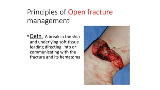
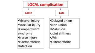
Fracture is break in the structural continuity of the bone.
Based on
etiology
• Traumatic direct
indirect
• Pathologic
• Stress
Based on
communication
• Open
• Closed
Based on
shape
• Simple
• Wedge
• complex
• Proximal and distal segment fractures
Based on communication Closed and open
Based on shape
simple
Wedge
Complex
• Fracture usually occurs after a trauma
• Estimate the energy applied
E=1/2mv2
• Take a short history AMPLE
At the scene of an accident
Establish the airway
protect the cervical spine
 Ensure ventilation
 Arrest hemorrhage and combat shock
 Give analgesia (pain killer)
 Splint fracture with what ever available
 Transport
Hospital care
Golden hour
• Follow the ATLS protocol.
Primary survey; with simultaneous resuscitation
A- Airway maintenance with cervical spine control
B- breathing and oxygenation
C- circulation and control bleeding
D- disability
E-exposure and avoid hypothermia
Secondary survey;
complete history and thorough physical examination
History
 Patients age and mechanism of injury
Occupation
 Pain is the most presenting symptom
 Local bruising and swelling
 Numbness and loss of mov’t
 Associated injury
Blood in urine ,abdominal pain, difficulty with breathing or transient loss of consciousness.
 Previous injuries or any musculoskeletal abnormality – to avoid x-ray confusion
If the trauma is trivial , suspect of pathological lesion
Physical examination
• General signs
Assessed with primary survey
• Local signs
Includes
Injured area
Adjacent joint
Peripheral neurovaculature
 Fracture cannot always be on the site of trauma
 Includes 3 steps
Look
 Swelling and bruising
 Skin break ,if it is open fracture
 Deformity - Posture of distal extremity
 Shortening
 Color of the skin
 Atrophy
Feel
 Swelling
 Tenderness
 Temperature
 Peripheral pulse
 Capillary filling
 Peripheral sensation
Move
• Range of motion if the patient is comfortable
N.B. No attempt should be made to elicit crypts
Sure signs of fracture
visible fracture
Deformity
Length discrepancy
Movement disorder
Crypitus
oRoutine investigations
 CBC
 ESR
 Blood group and RH
 RBS
oIndicated investigations
 Urine analysis
 Chest x ray
 Abdominal u/s
 Organ function test
X-ray
Rule of two
Two views; AP and lateral
Fracture or dislocation may not be seen in single x-ray
Two joints;
To visualize angulations of a bone with associated joint dislocation
Two occasions
Some fractures are notoriously
absent soon after the injury
.e.g. scaphoid fracture.
Two limbs
In children to avoid confusion
between fracture and
immature epiphysis
Special investigations
• Computerised tomography
 Magnetic resonance imaging
Soft-tissue problems and fractures
 Bone scan
Stress fractures and tumors
• Diagnosis alone is not enough, describing the fracture is also needed
1- Site of fracture
2- Extent of fracture
3- Fracture line
4- Displacement
5- Stability
6- Skin damage
AIM
• the aims of treatment are to restore function safely
with minimal complications.
• The first principle is to consider whether any
intervention is necessary; many fractures require no
or just symptomatic treatment.
Treatment of fracture
A high velocity injury should always be
treated according to the Advanced Trauma Life
Support (ATLS) guidelines with attention to
Airway, Breathing and Circulation on
presentation
The patient should be optimally resuscitated
before any fracture treatment is considered
“ treat the patient, not only the
fracture “
5 Rs
• Resuscitation
• Recognition
• Reduction
• Retention
• Rehabilitation
Tx. Of closed fracture
• The interventional management of
the fracture has three components:
• Appropriate reduction
• Stabilisation(Immobilization)
• Rehabilitation
Reduction, ‘if necessary
is manipulation of the #ed bone to improve the position of the fragments &
restore to acceptable position and function
Aim
I. adequate apposition and
II. normal alignment of the bone fragments
In many fractures reduction is unnecessary, either
• because there is little or no displacement or
• because the displacement is immaterial to the final result
There are two methods of
reduction:
closed and
open.
closed reduction
• Under appropriate anaesthesia and muscle relaxation
• most suitable for fractures with intact soft tissues
Used for
minimally displaced #
most # in children and
 # that are not unstable after reduction
OPEN REDUCTION
• indicated:
(1) when closed reduction fails, either
• difficulty in controlling the fragments or
• ST are interposed between them;
(2) large articular fragment that needs accurate positioning or
(3) avulsion fractures.
(4) first step to internal fixation.
METHODS OF IMMOBILISATION
1. Cast or external splint
2. traction
3. Functional bracing
4. external fixation
5. internal fixation.
splint
• For some fractures a splint made from metal, wood or plastic is more
appropriate
ex. Ready made splint, or a plastic collar for certain injuries of the
cervical spine.
casts
• plaster of Paris cast standard for most #s.
esp. distal limb fractures and
for most children’s fractures
• speed of union is neither greater nor less than with traction
• plaster slab – when there is significant ST swelling
 decrease risk of neurovascular damage,
 skin damage and
 compartment syndrome
FUNCTIONAL BRACING
• Use plaster of Paris
• prevente joint stiffness while still permitting fracture splintage and loading
• Segments of a cast are applied only over the shafts of the bones, leaving the
joints free;
• the cast segments are connected by metal or plastic hinges that allow movement
in one plane.
• most widely for fractures of the femur or tibia
• applied only when the fracture is beginning to unite
i.e. after 3–6 weeks of traction or conventional cast.
TRACTION
• Traction is pulling to change or hold the position of fracture fragments to balance
pull of the muscles
• Used When it is difficult or impossible to hold the fragments in proper position
by a plaster or splint.
• The problem is speed
2 types of traction
I. skin traction
II. skeletal traction
Skin traction
indications
• when small force requires to maintain traction
• temporal traction only for few days
• for children
Contraindication
- force required > 5kg
- skin damage
-infection at the area
- allergy
Skeletal traction
indications
force required greater than 5kg
skin damage requiring cleaning
 the force applied is 20% of total body weight
Internal fixation
1. # that cannot be reduced except by operation.
2. # that are inherently unstable and prone to
re-displace after reduction
3. # that unite poorly and slowly ,ex. femoral neck #
4. Pathological #
5. Multiple #s
6. #s in patients who present nursing
difficulties paraplegics
very elderly
multiple injuries
Types of internal fixation
Interfragmentary screws
Wires
• Transfixing wires
• often passed percutaneously
• can hold major fracture fragments together.
• used in situations where fracture healing is predictably quick
• Usually Cast is applied as supplementary support.
• tension-band wiring
• loops of wire passed around two bone fragments and then tightened to
compress the fragments together
• for olecranon or # of the patella
Plates and screws
• This form of fixation is useful for treating
• metaphyseal fractures of long bones and
• diaphyseal fractures of the radius and ulna
Intramedullary nails
• suitable for long bones
• rotational forces are resisted by introducing transverse interlocking
screws
Complications of internal fixation
• Most of the complications of internal fixation are due to
poor technique,
poor equipment or
poor operating
• conditions:
Iatrogenic infection
Non-union
Implant failure
Refracture
EXTERNAL FIXATION
• Indications
1. # associated with severe STI or contaminated
2. # around joints with swollen ST
3. Patients with severe multiple injuries, esp. if there are
• bilateral femoral fractures,
• Pelvic fractures with severe bleeding, and
• those with limb and associated chest or head injuries.
4. Ununited #
5. Infected #
REHABILITATION
“restore function”
• Improved results in the treatment of fracture
not only to the injured parts but also to the
patient as a whole
• The objectives are
• reduce oedema,
• by combination of elevation and exercise
• preserve joint movement,
• restore muscle power and
• guide the patient back to normal activity:
Principles of Open fracture
management
•Defn. A break in the skin
and underlying soft tissue
leading directing into or
communicating with the
fracture and its hematoma
• Infection is the most feared complication of compound fractures and
may cause delayed healing, non union, sepsis or even death.
• It is a surgical emergency
Treatment…
• The object is
 to clean the wound and, whenever necessary,
to remove all dead and devitalised tissue and all extraneous
material
• Treatment is determined by the
type of fracture,
The nature of the STI
Wound size and
the degree of contamination.

Principles….
Urgent wound and fracture debridement.
Early definitive wound cover.
Antibiotic prophylaxis
Prophylaxis against tetanus
Stabilization of the fracture
treatment of the fracture itself should
follow the general principles already suggested for closed fractures
Complication of Fracture
A. General / systemic
i) Early
•Shock
•Hemorrhage
•fat embolism ARDS
•Infection
•Crush syndrome
Late complication
• Complication associated with prolonged
recumbancy
•DVT
•Bed sore
•Osteoporosis
•Nephrolithiasis
EARLY
•Visceral injury
•Vascular injury
•Compartment
syndrome
•Nerve injury
•Haemarthrosis
•Infection
•Delayed union
•Non-union
•Malunion
•Joint stiffness
•AVN
•Osteoarthritis
LATE
LOCAL complication
Dislocation
• Def: : complete loss of contact between joint
surfaces.
• Subluxation: partial lose of joint surfaces
contact with each other.
• This is also described as ‘incomplete’ or ‘partial’
dislocation.
Diagnosis
Signs and Symptoms
• Pain and deformity of the joint
• Inability to bear weight on the affected limb
• Neurologic compromise
History
• Traumatic injury, as from a fall or car accident
Physical Exam
• A complete neurologic examination is important,
and the strength and sensation distally should
be noted.
• Pulses should be palpated and, if not found,
Doppler ultrasound should be performed.
• The remainder of the extremity should be
evaluated for swelling or deformity.
Tests
Imaging
• Radiography:
• Plain radiographs of the joint should be taken in 2
planes to show the direction of dislocation.
• Radiographs are repeated after reduction.
• CT is used to evaluate intra-articular fracture
patterns.
• MRI is used to evaluate ligament and soft-tissue
damage around the joint.
Treatment
Initial Stabilization
• All joint dislocations should be reduced as soon
as possible.
• Radiographs should be taken 1st to confirm the
dislocation and to show fractures.
General Measures
• Depending on the nature of the injury, reduction
may require no anesthesia, intra-articular joint
injection, conscious sedation, or full anesthesia.
• The hip joint often requires sedation or a full anesthetic with
muscle relaxant because the muscle forces around the joint can
be great.
• After reduction, joint stability should be
confirmed by taking the joint through its ROM.
• Unstable joints should be braced or placed in
traction after reduction and usually require
surgical stabilization.
• Plain radiographs should be taken in 2 views to
confirm reduction.
• CT is used in dislocations of the hip and shoulder
to assess for fractures or intra-articular
fragments
Activity
• Dislocations that are stable after reduction and
do not have fractures usually should be
mobilized quickly.
• Immobilization for a week may help with soft-
tissue pain and swelling.
Special Therapy
Physical Therapy
• Patients with stable dislocations should begin an
assisted program in Range Of Movment and joint
strengthening.
Surgery
• Indications for surgery include:
• Unstable joint dislocations
• Periarticular fractures:
• .
• In some cases, ruptured or torn periarticular soft tissue may be treated with
surgery.
• Acute shoulder dislocations in young adults have a high risk of recurrent instability.
• Irreducible joint dislocations:
• Soft tissues such as tendons, nerves, or arteries may be caught in the joint.
• Open reduction is required.
• Intra-articular osteochondral fragments
Follow-up
• Confirmation should be made after reduction, taking care to assure
joint congruity.
• Patients should be reassessed in 1 to 2 weeks.
• If the joint is stable, early ROM should be started.
• Dislocation may put the patients at risk for instability, osteonecrosis,
or posttraumatic arthritis.
AMPUTATIONS
64
• An amputation is removal or excision of part or whole of a limb.
Indications
1- Dead limb (Gangrene)
• - Due to: - Atherosclerosis
• - Embolism
• - Major arterial injury
• - Diabetic gangrene…
2- Deadly limb
• - Life threatening infection (e.g. Gas gangrene) or malignancies which can’t be controlled by other
local measures
3- Dead loss
• - Severe soft tissue injury especially associated with major nerve injury, which may occur in
compound fractures.
Level of amputation
65
The choice for the level of amputation depends on:
• - Age
• - The nature and extent of the pathology e.g. Neoplasm, trauma
• - The vascularity of tissues
• - Presence of infection
• - Status of the joints
• - Access to the various types of prostheses
Generally, the most distal level that will heal and still provide a functional stump is selected.
• In the upper limb, attempt should be made to conserve every possible inch.
• In the lower limb, the most important factor is to try and conserve the knee joint whenever
possible.
• Amputations performed in the face of infection should be left open for a later closure.
Complications of amputation
66
• Edema
• Hematoma
• Secondary and reactionary hemorrhage
• Infection
• Ischemic necrosis
• Flexion contracture
• Chronic pain-psychogenic, neuromas, etc.

Fracture

  • 1.
    Fracture is breakin the structural continuity of the bone.
  • 2.
    Based on etiology • Traumaticdirect indirect • Pathologic • Stress Based on communication • Open • Closed Based on shape • Simple • Wedge • complex
  • 3.
    • Proximal anddistal segment fractures
  • 4.
    Based on communicationClosed and open
  • 5.
  • 6.
  • 7.
  • 8.
  • 9.
    • Fracture usuallyoccurs after a trauma • Estimate the energy applied E=1/2mv2 • Take a short history AMPLE
  • 10.
    At the sceneof an accident Establish the airway protect the cervical spine  Ensure ventilation  Arrest hemorrhage and combat shock  Give analgesia (pain killer)  Splint fracture with what ever available  Transport
  • 11.
    Hospital care Golden hour •Follow the ATLS protocol. Primary survey; with simultaneous resuscitation A- Airway maintenance with cervical spine control B- breathing and oxygenation C- circulation and control bleeding D- disability E-exposure and avoid hypothermia Secondary survey; complete history and thorough physical examination
  • 12.
    History  Patients ageand mechanism of injury Occupation  Pain is the most presenting symptom  Local bruising and swelling  Numbness and loss of mov’t  Associated injury Blood in urine ,abdominal pain, difficulty with breathing or transient loss of consciousness.  Previous injuries or any musculoskeletal abnormality – to avoid x-ray confusion If the trauma is trivial , suspect of pathological lesion
  • 13.
    Physical examination • Generalsigns Assessed with primary survey • Local signs Includes Injured area Adjacent joint Peripheral neurovaculature  Fracture cannot always be on the site of trauma  Includes 3 steps
  • 14.
    Look  Swelling andbruising  Skin break ,if it is open fracture  Deformity - Posture of distal extremity  Shortening  Color of the skin  Atrophy
  • 15.
    Feel  Swelling  Tenderness Temperature  Peripheral pulse  Capillary filling  Peripheral sensation
  • 16.
    Move • Range ofmotion if the patient is comfortable N.B. No attempt should be made to elicit crypts
  • 17.
    Sure signs offracture visible fracture Deformity Length discrepancy Movement disorder Crypitus
  • 18.
    oRoutine investigations  CBC ESR  Blood group and RH  RBS oIndicated investigations  Urine analysis  Chest x ray  Abdominal u/s  Organ function test
  • 19.
    X-ray Rule of two Twoviews; AP and lateral Fracture or dislocation may not be seen in single x-ray Two joints; To visualize angulations of a bone with associated joint dislocation
  • 20.
    Two occasions Some fracturesare notoriously absent soon after the injury .e.g. scaphoid fracture. Two limbs In children to avoid confusion between fracture and immature epiphysis
  • 21.
    Special investigations • Computerisedtomography  Magnetic resonance imaging Soft-tissue problems and fractures  Bone scan Stress fractures and tumors
  • 22.
    • Diagnosis aloneis not enough, describing the fracture is also needed 1- Site of fracture 2- Extent of fracture 3- Fracture line 4- Displacement 5- Stability 6- Skin damage
  • 23.
    AIM • the aimsof treatment are to restore function safely with minimal complications. • The first principle is to consider whether any intervention is necessary; many fractures require no or just symptomatic treatment. Treatment of fracture
  • 24.
    A high velocityinjury should always be treated according to the Advanced Trauma Life Support (ATLS) guidelines with attention to Airway, Breathing and Circulation on presentation The patient should be optimally resuscitated before any fracture treatment is considered “ treat the patient, not only the fracture “
  • 25.
    5 Rs • Resuscitation •Recognition • Reduction • Retention • Rehabilitation
  • 26.
    Tx. Of closedfracture • The interventional management of the fracture has three components: • Appropriate reduction • Stabilisation(Immobilization) • Rehabilitation
  • 27.
    Reduction, ‘if necessary ismanipulation of the #ed bone to improve the position of the fragments & restore to acceptable position and function Aim I. adequate apposition and II. normal alignment of the bone fragments In many fractures reduction is unnecessary, either • because there is little or no displacement or • because the displacement is immaterial to the final result
  • 28.
    There are twomethods of reduction: closed and open.
  • 29.
    closed reduction • Underappropriate anaesthesia and muscle relaxation • most suitable for fractures with intact soft tissues Used for minimally displaced # most # in children and  # that are not unstable after reduction
  • 30.
    OPEN REDUCTION • indicated: (1)when closed reduction fails, either • difficulty in controlling the fragments or • ST are interposed between them; (2) large articular fragment that needs accurate positioning or (3) avulsion fractures. (4) first step to internal fixation.
  • 31.
    METHODS OF IMMOBILISATION 1.Cast or external splint 2. traction 3. Functional bracing 4. external fixation 5. internal fixation.
  • 32.
    splint • For somefractures a splint made from metal, wood or plastic is more appropriate ex. Ready made splint, or a plastic collar for certain injuries of the cervical spine.
  • 33.
    casts • plaster ofParis cast standard for most #s. esp. distal limb fractures and for most children’s fractures • speed of union is neither greater nor less than with traction • plaster slab – when there is significant ST swelling  decrease risk of neurovascular damage,  skin damage and  compartment syndrome
  • 34.
    FUNCTIONAL BRACING • Useplaster of Paris • prevente joint stiffness while still permitting fracture splintage and loading • Segments of a cast are applied only over the shafts of the bones, leaving the joints free; • the cast segments are connected by metal or plastic hinges that allow movement in one plane. • most widely for fractures of the femur or tibia • applied only when the fracture is beginning to unite i.e. after 3–6 weeks of traction or conventional cast.
  • 35.
    TRACTION • Traction ispulling to change or hold the position of fracture fragments to balance pull of the muscles • Used When it is difficult or impossible to hold the fragments in proper position by a plaster or splint. • The problem is speed 2 types of traction I. skin traction II. skeletal traction
  • 36.
    Skin traction indications • whensmall force requires to maintain traction • temporal traction only for few days • for children Contraindication - force required > 5kg - skin damage -infection at the area - allergy
  • 37.
    Skeletal traction indications force requiredgreater than 5kg skin damage requiring cleaning  the force applied is 20% of total body weight
  • 38.
    Internal fixation 1. #that cannot be reduced except by operation. 2. # that are inherently unstable and prone to re-displace after reduction 3. # that unite poorly and slowly ,ex. femoral neck # 4. Pathological # 5. Multiple #s 6. #s in patients who present nursing difficulties paraplegics very elderly multiple injuries
  • 39.
    Types of internalfixation Interfragmentary screws
  • 40.
    Wires • Transfixing wires •often passed percutaneously • can hold major fracture fragments together. • used in situations where fracture healing is predictably quick • Usually Cast is applied as supplementary support.
  • 41.
    • tension-band wiring •loops of wire passed around two bone fragments and then tightened to compress the fragments together • for olecranon or # of the patella
  • 42.
    Plates and screws •This form of fixation is useful for treating • metaphyseal fractures of long bones and • diaphyseal fractures of the radius and ulna
  • 43.
    Intramedullary nails • suitablefor long bones • rotational forces are resisted by introducing transverse interlocking screws
  • 44.
    Complications of internalfixation • Most of the complications of internal fixation are due to poor technique, poor equipment or poor operating • conditions: Iatrogenic infection Non-union Implant failure Refracture
  • 45.
    EXTERNAL FIXATION • Indications 1.# associated with severe STI or contaminated 2. # around joints with swollen ST 3. Patients with severe multiple injuries, esp. if there are • bilateral femoral fractures, • Pelvic fractures with severe bleeding, and • those with limb and associated chest or head injuries. 4. Ununited # 5. Infected #
  • 46.
    REHABILITATION “restore function” • Improvedresults in the treatment of fracture not only to the injured parts but also to the patient as a whole • The objectives are • reduce oedema, • by combination of elevation and exercise • preserve joint movement, • restore muscle power and • guide the patient back to normal activity:
  • 47.
    Principles of Openfracture management •Defn. A break in the skin and underlying soft tissue leading directing into or communicating with the fracture and its hematoma
  • 48.
    • Infection isthe most feared complication of compound fractures and may cause delayed healing, non union, sepsis or even death. • It is a surgical emergency
  • 49.
    Treatment… • The objectis  to clean the wound and, whenever necessary, to remove all dead and devitalised tissue and all extraneous material • Treatment is determined by the type of fracture, The nature of the STI Wound size and the degree of contamination. 
  • 50.
    Principles…. Urgent wound andfracture debridement. Early definitive wound cover. Antibiotic prophylaxis Prophylaxis against tetanus Stabilization of the fracture treatment of the fracture itself should follow the general principles already suggested for closed fractures
  • 51.
    Complication of Fracture A.General / systemic i) Early •Shock •Hemorrhage •fat embolism ARDS •Infection •Crush syndrome
  • 52.
    Late complication • Complicationassociated with prolonged recumbancy •DVT •Bed sore •Osteoporosis •Nephrolithiasis
  • 53.
    EARLY •Visceral injury •Vascular injury •Compartment syndrome •Nerveinjury •Haemarthrosis •Infection •Delayed union •Non-union •Malunion •Joint stiffness •AVN •Osteoarthritis LATE LOCAL complication
  • 54.
    Dislocation • Def: :complete loss of contact between joint surfaces. • Subluxation: partial lose of joint surfaces contact with each other. • This is also described as ‘incomplete’ or ‘partial’ dislocation.
  • 55.
    Diagnosis Signs and Symptoms •Pain and deformity of the joint • Inability to bear weight on the affected limb • Neurologic compromise
  • 56.
    History • Traumatic injury,as from a fall or car accident Physical Exam • A complete neurologic examination is important, and the strength and sensation distally should be noted. • Pulses should be palpated and, if not found, Doppler ultrasound should be performed. • The remainder of the extremity should be evaluated for swelling or deformity.
  • 57.
    Tests Imaging • Radiography: • Plainradiographs of the joint should be taken in 2 planes to show the direction of dislocation. • Radiographs are repeated after reduction. • CT is used to evaluate intra-articular fracture patterns. • MRI is used to evaluate ligament and soft-tissue damage around the joint.
  • 58.
    Treatment Initial Stabilization • Alljoint dislocations should be reduced as soon as possible. • Radiographs should be taken 1st to confirm the dislocation and to show fractures.
  • 59.
    General Measures • Dependingon the nature of the injury, reduction may require no anesthesia, intra-articular joint injection, conscious sedation, or full anesthesia. • The hip joint often requires sedation or a full anesthetic with muscle relaxant because the muscle forces around the joint can be great. • After reduction, joint stability should be confirmed by taking the joint through its ROM. • Unstable joints should be braced or placed in traction after reduction and usually require surgical stabilization.
  • 60.
    • Plain radiographsshould be taken in 2 views to confirm reduction. • CT is used in dislocations of the hip and shoulder to assess for fractures or intra-articular fragments Activity • Dislocations that are stable after reduction and do not have fractures usually should be mobilized quickly. • Immobilization for a week may help with soft- tissue pain and swelling.
  • 61.
    Special Therapy Physical Therapy •Patients with stable dislocations should begin an assisted program in Range Of Movment and joint strengthening. Surgery • Indications for surgery include: • Unstable joint dislocations • Periarticular fractures: • .
  • 62.
    • In somecases, ruptured or torn periarticular soft tissue may be treated with surgery. • Acute shoulder dislocations in young adults have a high risk of recurrent instability. • Irreducible joint dislocations: • Soft tissues such as tendons, nerves, or arteries may be caught in the joint. • Open reduction is required. • Intra-articular osteochondral fragments
  • 63.
    Follow-up • Confirmation shouldbe made after reduction, taking care to assure joint congruity. • Patients should be reassessed in 1 to 2 weeks. • If the joint is stable, early ROM should be started. • Dislocation may put the patients at risk for instability, osteonecrosis, or posttraumatic arthritis.
  • 64.
    AMPUTATIONS 64 • An amputationis removal or excision of part or whole of a limb. Indications 1- Dead limb (Gangrene) • - Due to: - Atherosclerosis • - Embolism • - Major arterial injury • - Diabetic gangrene… 2- Deadly limb • - Life threatening infection (e.g. Gas gangrene) or malignancies which can’t be controlled by other local measures 3- Dead loss • - Severe soft tissue injury especially associated with major nerve injury, which may occur in compound fractures.
  • 65.
    Level of amputation 65 Thechoice for the level of amputation depends on: • - Age • - The nature and extent of the pathology e.g. Neoplasm, trauma • - The vascularity of tissues • - Presence of infection • - Status of the joints • - Access to the various types of prostheses Generally, the most distal level that will heal and still provide a functional stump is selected. • In the upper limb, attempt should be made to conserve every possible inch. • In the lower limb, the most important factor is to try and conserve the knee joint whenever possible. • Amputations performed in the face of infection should be left open for a later closure.
  • 66.
    Complications of amputation 66 •Edema • Hematoma • Secondary and reactionary hemorrhage • Infection • Ischemic necrosis • Flexion contracture • Chronic pain-psychogenic, neuromas, etc.

Editor's Notes

  • #24 NB. In any sustained trauma or high velocity injury initially we should assess ABC’s of life
  • #26 Phases Emergency care Definitive care What do u do as emergency care to the fracture Pain management Manage blood loss Splinting Open fracture
  • #27 If the overlying skin remains intact it is a closed (or simple)fracture; if the skin or one of the body cavities is breached it is an open (or compound) fracture, liable to contamination and infection
  • #48 -One-third of patients with open fractures are multiply injured. So the nedd ATLS and wound care
  • #50 Management is based on Type, so, type 1 and type 3 are not managed equally type 3 is kind of an ACUTE. More susceptible to infection Type 3 may need grafting, amputaton
  • #52 Fat globules from the fracture site embolize to different vasculature's like to pulmonary, or may ascend to vertebral arteries- circule of wills to any of the cerebral vessels, and causing blood occlusion CNS- agitation, irritable, confusion… Skin-petechia. +A minute red or purple spot on the surface of the skin as the result of tiny hemorrhages of blood vessels in the skin.
  • #53 Read the mechanism of nephrolithiasis and constipation
  • #54 EAARLY Affect mainly the soft tissues LATE Months after injury , affect mainly bones and joints