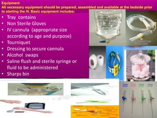
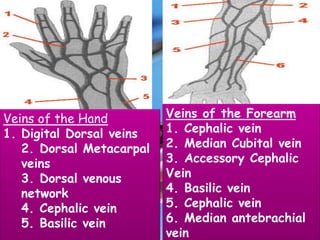
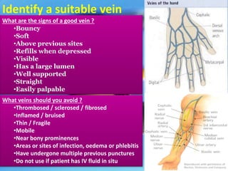
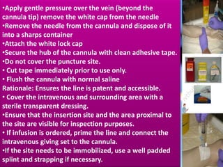
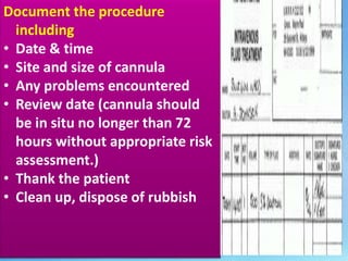
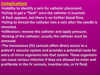
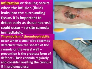
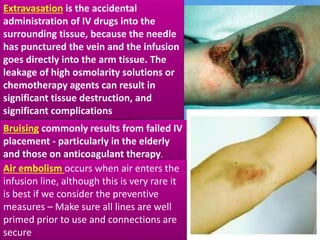
2. Insertion of Peripheral IV Line
âThe aim of intravenous management
is safe, effective delivery of
treatment without discomfort or
tissue damage and without
compromising venous access,
especially if long term therapy is
proposedâ
3. Indications:
â˘Fluid and electrolyte replacement
â˘Administration of medicines
â˘Administration of blood/blood
products
â˘Administration of Total Parenteral
Nutrition
â˘Hemodynamic monitoring,
Blood sampling
4. Advantages
â˘Immediate effect
â˘Control over the rate of administration
â˘Patient cannot tolerate drugs / fluids
orally
â˘Some drugs cannot be absorbed by any
other route
â˘Pain and irritation is avoided compared to
some substances when given SC/IM
5. Equipment
All necessary equipment should be prepared, assembled and available at the bedside prior
to starting the IV. Basic equipment includes:
⢠Tray contains
⢠Non Sterile Gloves
⢠IV cannula (appropriate size
according to age and purpose)
⢠Tourniquet
⢠Dressing to secure cannula
⢠Alcohol swaps
⢠Saline flush and sterile syringe or
fluid to be administered
⢠Sharps bin
6. Veins of the Hand
1. Digital Dorsal veins
2. Dorsal Metacarpal
veins
3. Dorsal venous
network
4. Cephalic vein
5. Basilic vein
Veins of the Forearm
1. Cephalic vein
2. Median Cubital vein
3. Accessory Cephalic
Vein
4. Basilic vein
5. Cephalic vein
6. Median antebrachial
vein
7. Preparation of the patient and environment:
⢠Check and verify the physicianâs orders
⢠Gather equipment
⢠Explain the procedure to the patient
⢠Provide privacy
⢠Restraint the site, in case of children
⢠Check the vital signs and record
⢠Adjust the height of the bed for
comfortable working of the Nurse
⢠Place the patient in comfortable position
⢠Select a site on a Non âDominant arm
⢠Provide a good source of light
⢠Call for assistance if needed
8. Identify a suitable vein
What are the signs of a good vein ?
â˘Bouncy
â˘Soft
â˘Above previous sites
â˘Refills when depressed
â˘Visible
â˘Has a large lumen
â˘Well supported
â˘Straight
â˘Easily palpable
What veins should you avoid ?
â˘Thrombosed / sclerosed / fibrosed
â˘Inflamed / bruised
â˘Thin / Fragile
â˘Mobile
â˘Near bony prominences
â˘Areas or sites of infection, oedema or phlebitis
â˘Have undergone multiple previous punctures
â˘Do not use if patient has IV fluid in situ
9. Choose a suitable vein In adults, use long straight
veins in an upper extremity away from the joints for
catheter insertion â in preference to sites on the
lower extremities. If possible avoid veins in the
dominant hand and use distal veins first.
Do not insert cannula on the side of MASTECTOMYor
AV shunts/Gortex. Transfer catheter inserted in a
lower extremity site to an upper extremity site as
soon as the latter is available.
In pediatric patients, it is recommended that the
cannula be inserted into the scalp, hand, or foot site
in preference to a leg, arm, or ante cubital fossa site
(Category II).
10. Encourage venous filling by:
Correctly applying a tourniquet (A tourniquet should
.
be applied to the patientâs upper arm. The tourniquet
should be applied at a pressure which is high enough
to impede venous distension but not to restrict
arterial flow)
Opening & closing the fist
Lowering the limb below the heart
11. PROCEDURE
â˘Check and verify the physician order
⢠Identify and Explain procedure to
.
patient/parent.
â˘Wash hands with antiseptic soap.
â˘Don gloves
â˘Strict adherence to hand washing and
aseptic technique remains the cornerstone
of prevention of cannula related infections
â˘Apply the tourniquet above insertion site
â˘For pediatric patient, an assistantâs hand
used both as a tourniquet and restraint, is
often more acceptable to a child than a
tourniquet.
â˘Disinfect the selected site with skin prep
and allow to dry.
â˘Do not touch the skin with the fingers after
preparation solution has been applied.
12. â˘Inspect the cannula before insertion to ensure that
the needle is fully inserted into the plastic cannula
and that the cannula tip is not damaged.
.
â˘Do not touch the shaft or tip of the cannula.
⢠Ensure that the bevel of the cannula is facing
upwards.
Rationale: Facilitates the piercing of the skin by the
bevel.
â˘Hold the cannula in your dominant hand, stretch
the skin over the vein to anchor the vein with your
non-dominant hand (Do not re palpate the
vein)
â˘Insert the needle (bevel side up) at an angle of 15-
30o to the skin (this will depend on vein depth.)
Observe for blood in the flashback chamber
â˘Partially withdraw the needle and advance the
cannula.
⢠Release the tourniquet
13. â˘Apply gentle pressure over the vein (beyond the
cannula tip) remove the white cap from the needle
â˘Remove the needle from the cannula and dispose of it
.
into a sharps container
â˘Attach the white lock cap
â˘Secure the hub of the cannula with clean adhesive tape.
â˘Do not cover the puncture site.
⢠Cut tape immediately prior to use only.
⢠Flush the cannula with normal saline
Rationale: Ensures the line is patent and accessible.
⢠Cover the intravenous and surrounding area with a
sterile transparent dressing.
â˘Ensure that the insertion site and the area proximal to
the site are visible for inspection purposes.
⢠If infusion is ordered, prime the line and connect the
intravenous giving set to the cannula.
â˘If the site needs to be immobilized, use a well padded
splint and strapping if necessary.
14. Document the procedure
.
including
⢠Date & time
⢠Site and size of cannula
⢠Any problems encountered
⢠Review date (cannula should
be in situ no longer than 72
hours without appropriate risk
assessment.)
⢠Thank the patient
⢠Clean up, dispose of rubbish
15. Complications
.â˘Inability to identify a vein for catheter placement.
â˘Failing to get a âflashâ once the catheter is inserted.
â˘A flash appears, but there is no further blood flow.
â˘Failing to thread the catheter into a vein after the needle is
retracted.
â˘Infiltration; remove the catheter and apply pressure.
â˘Kinking of the catheter; usually the catheter must be
removed.
â˘The intravenous (IV) cannula offers direct access to a
patient's vascular system and provides a potential route for
entry of micro organisms into that system. These organisms
can cause serious infection if they are allowed to enter and
proliferate in the IV cannula, insertion site, or IV fluid.
16. IV-Site Infection: Does not
.
produce much (if any) pus
or inflammation at the IV
site. This is the most
common cannula-related
infection, may be the most
difficult to identify
Cellulites: Warm, red and
often tender skin
surrounding the site of
cannula insertion; pus is
rarely detectable.
17. Infiltration or tissuing occurs
.
when the infusion (fluid)
leaks into the surrounding
tissue. It is important to
detect early as tissue necrosis
could occur â re-site cannula
immediately.
Thrombolism / thrombophlebitis
occur when a small clot becomes
detached from the sheath of the
cannula or the vessel wall â
prevention is the greatest form of
defence. Flush cannula regularly
and consider re-siting the cannula
if in prolonged use.
18. Extravasation is the accidental
administration of IV drugs into the
.
surrounding tissue, because the needle
has punctured the vein and the infusion
goes directly into the arm tissue. The
leakage of high osmolarity solutions or
chemotherapy agents can result in
significant tissue destruction, and
significant complications
Bruising commonly results from failed IV
placement - particularly in the elderly
and those on anticoagulant therapy.
Air embolism occurs when air enters the
infusion line, although this is very rare it
is best if we consider the preventive
measures â Make sure all lines are well
primed prior to use and connections are
secure
19. Haematoma occurs when blood leaks out
of the infusion site. The common cause
.
of this is using cannula that are not
tapered at the distal end. It will also
occur if on insertion the cannula has
penetrated through the other side of the
vessel wall â apply pressure to the site
for approximately 4 minutes and elevate
the limb.
Phlebitis is common in IV therapy and can be cause in many
ways. It is inflammation of a vein (redness and pain at the
infusion site) â prevention can be using aseptic insertion
techniques, choosing the smallest gauge cannula possible
for the prescribed treatment, secure the cannula properly to
prevent movement and carry out regular checks of the
infusion site.