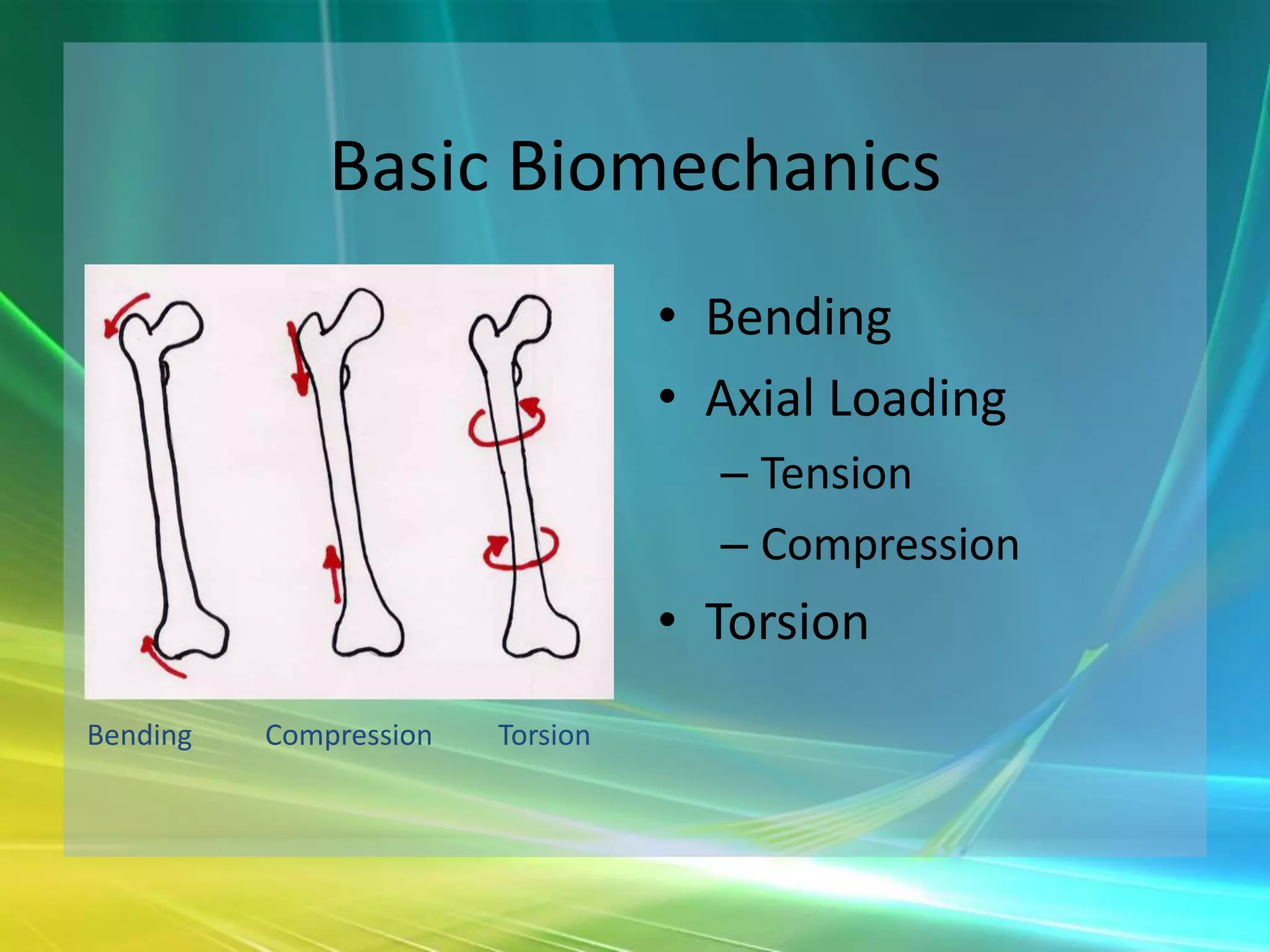
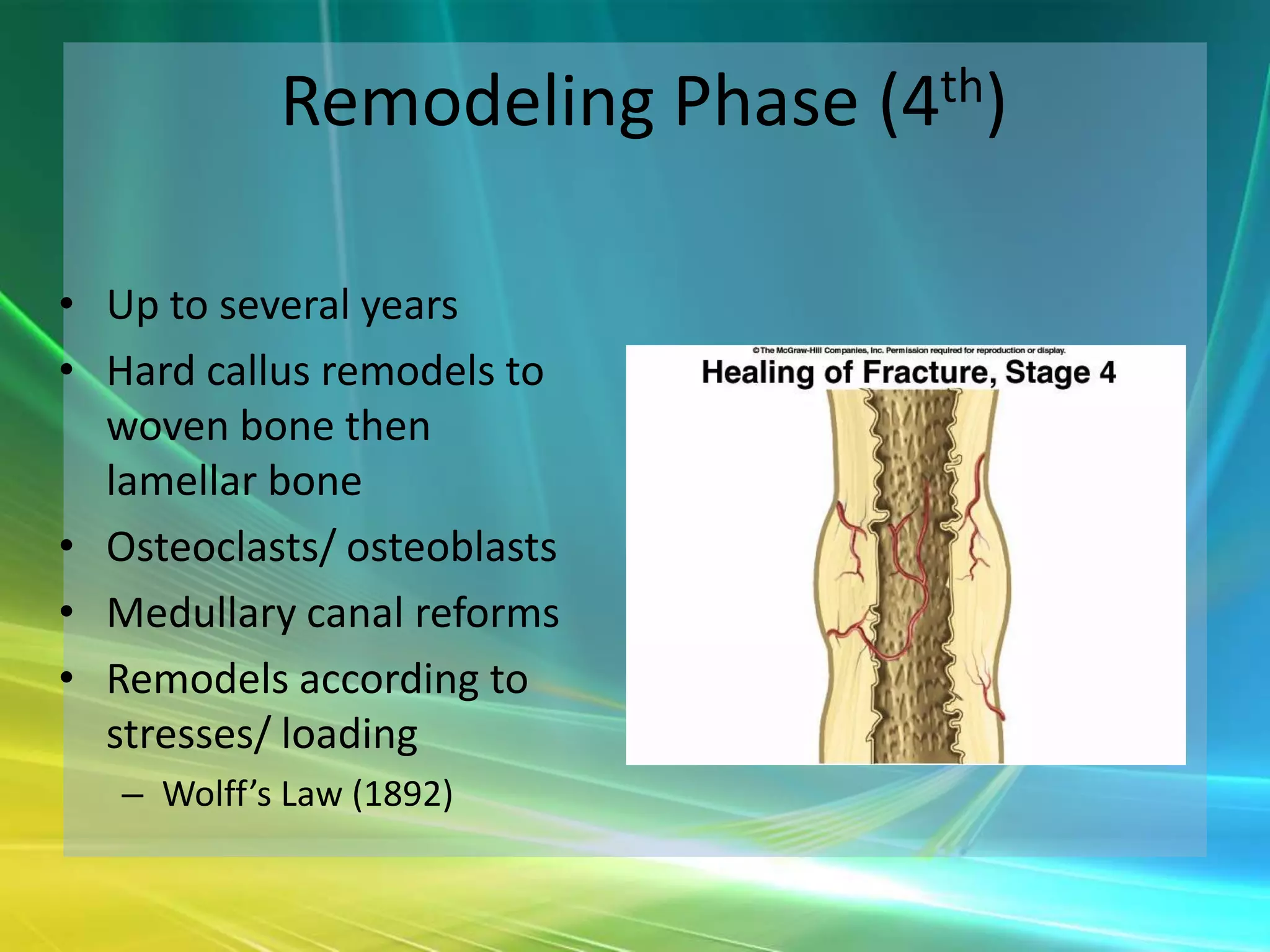
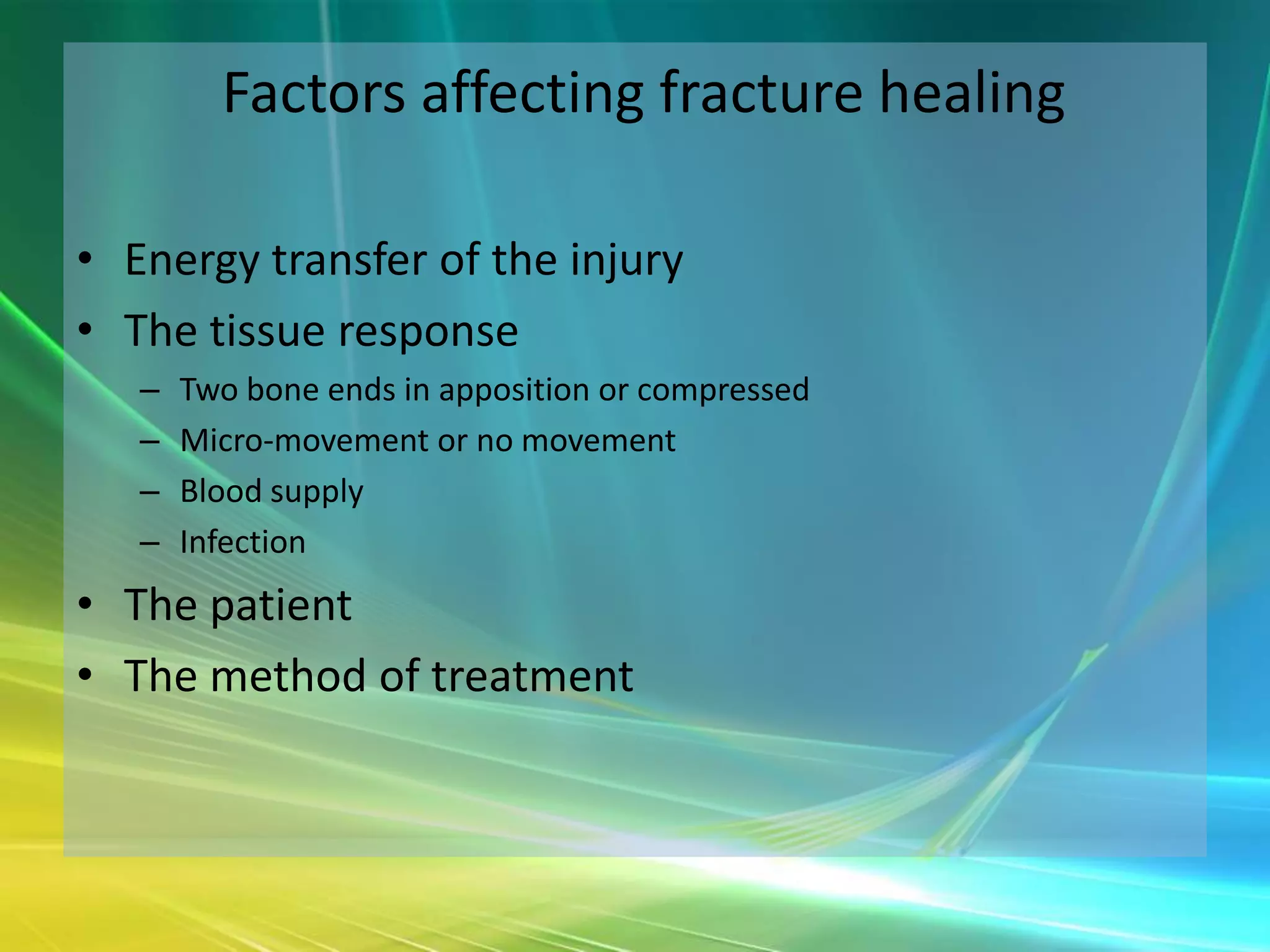
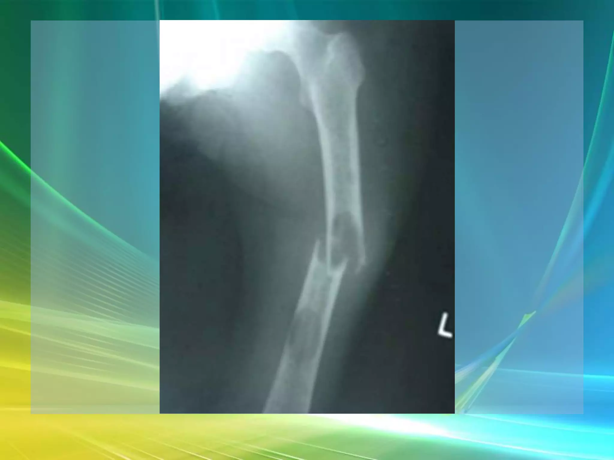
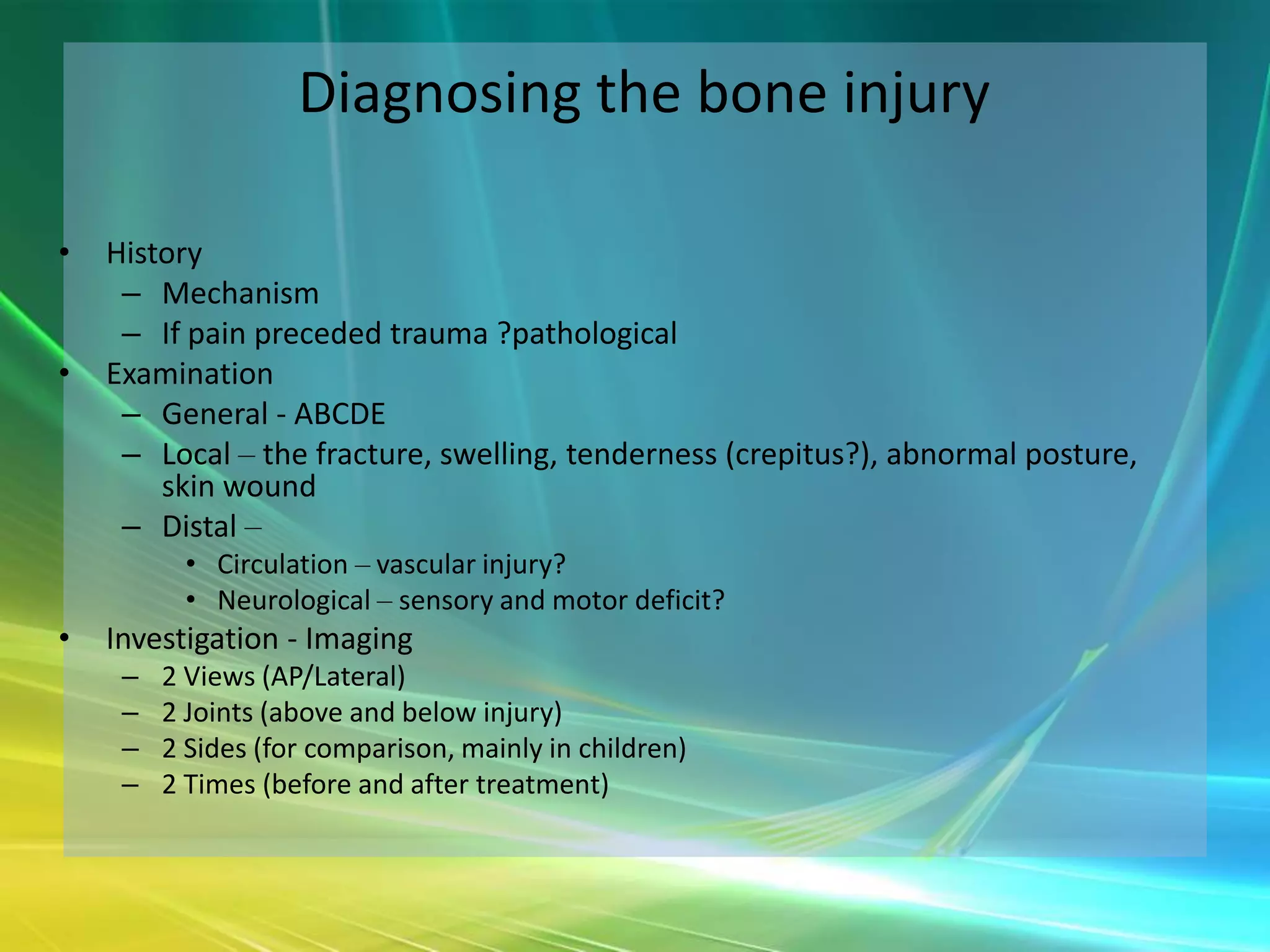
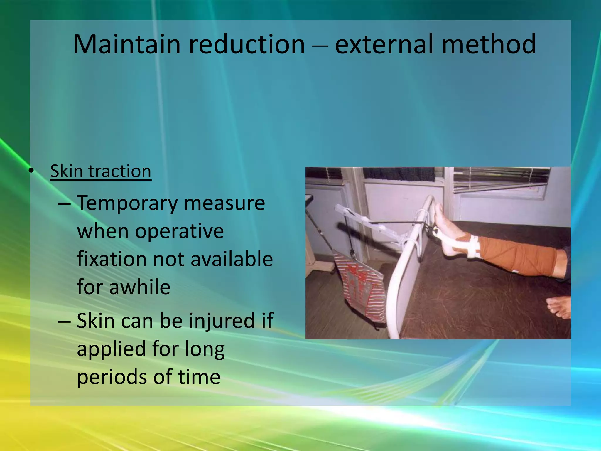
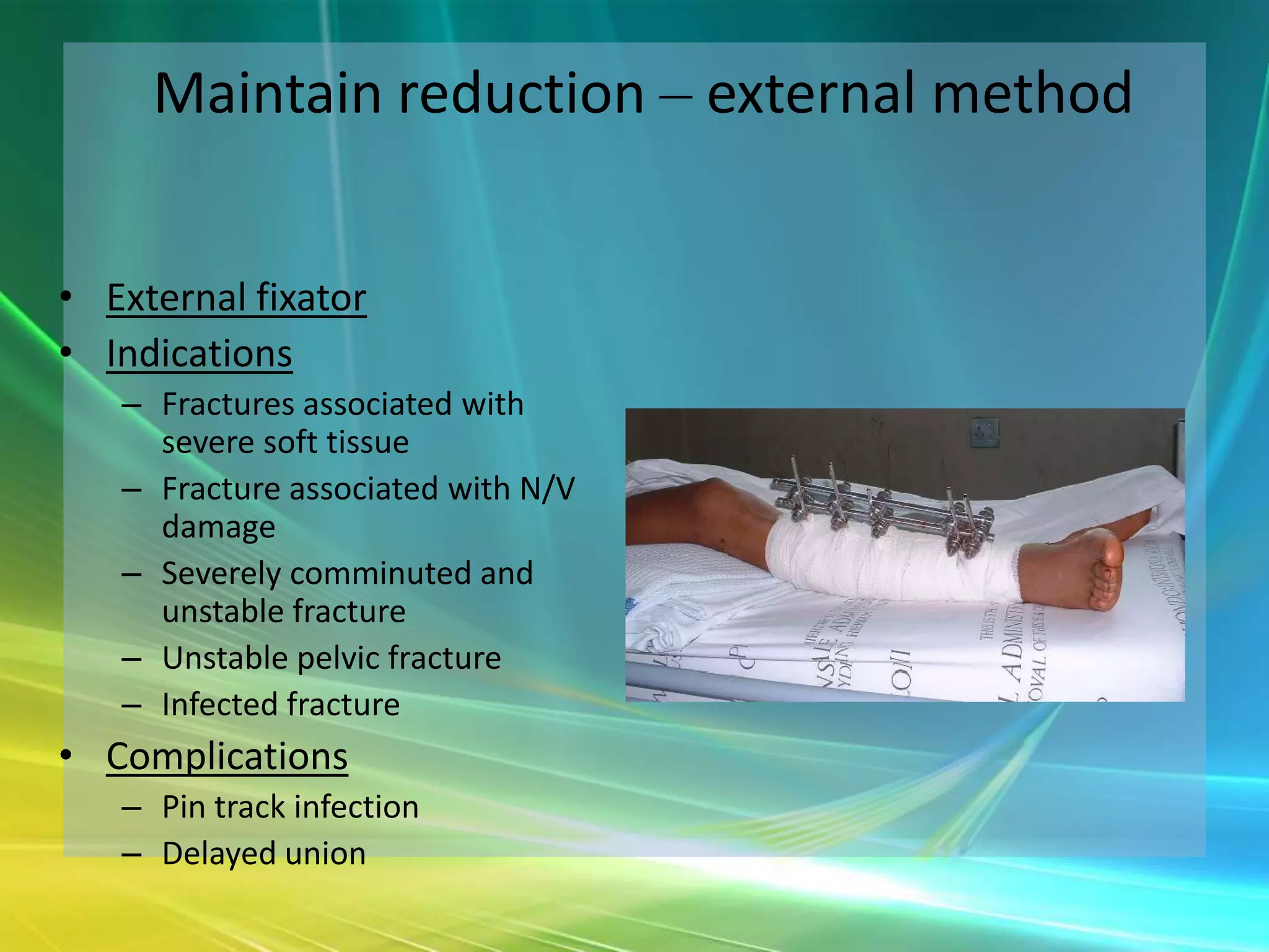
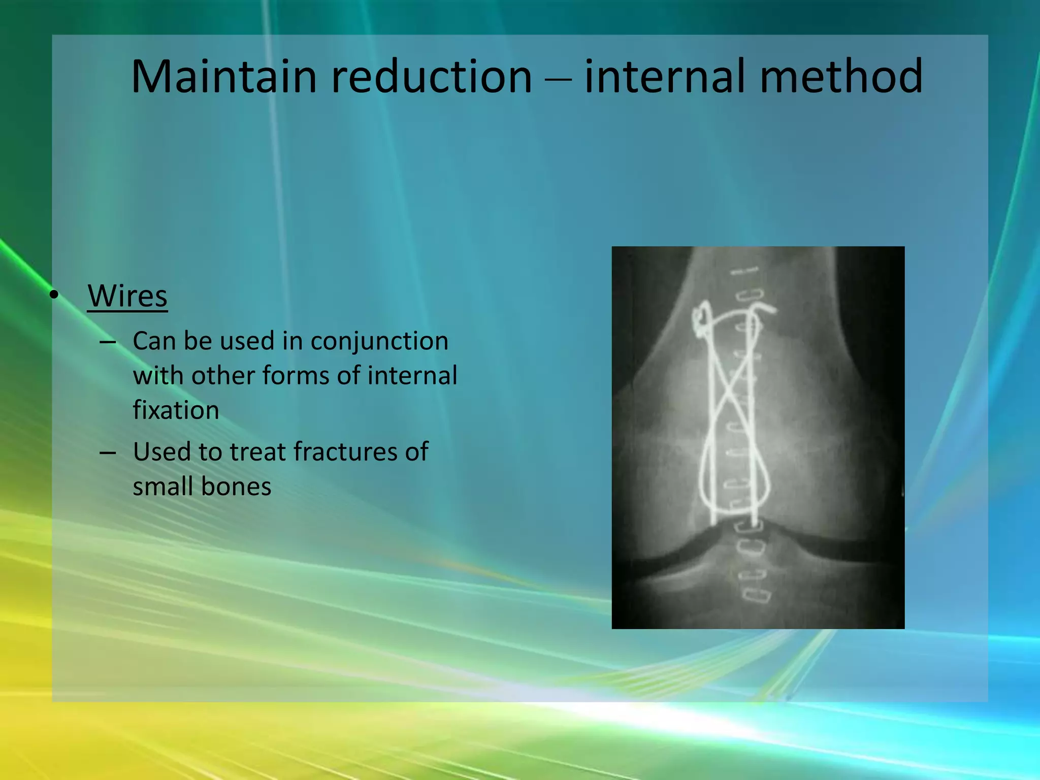
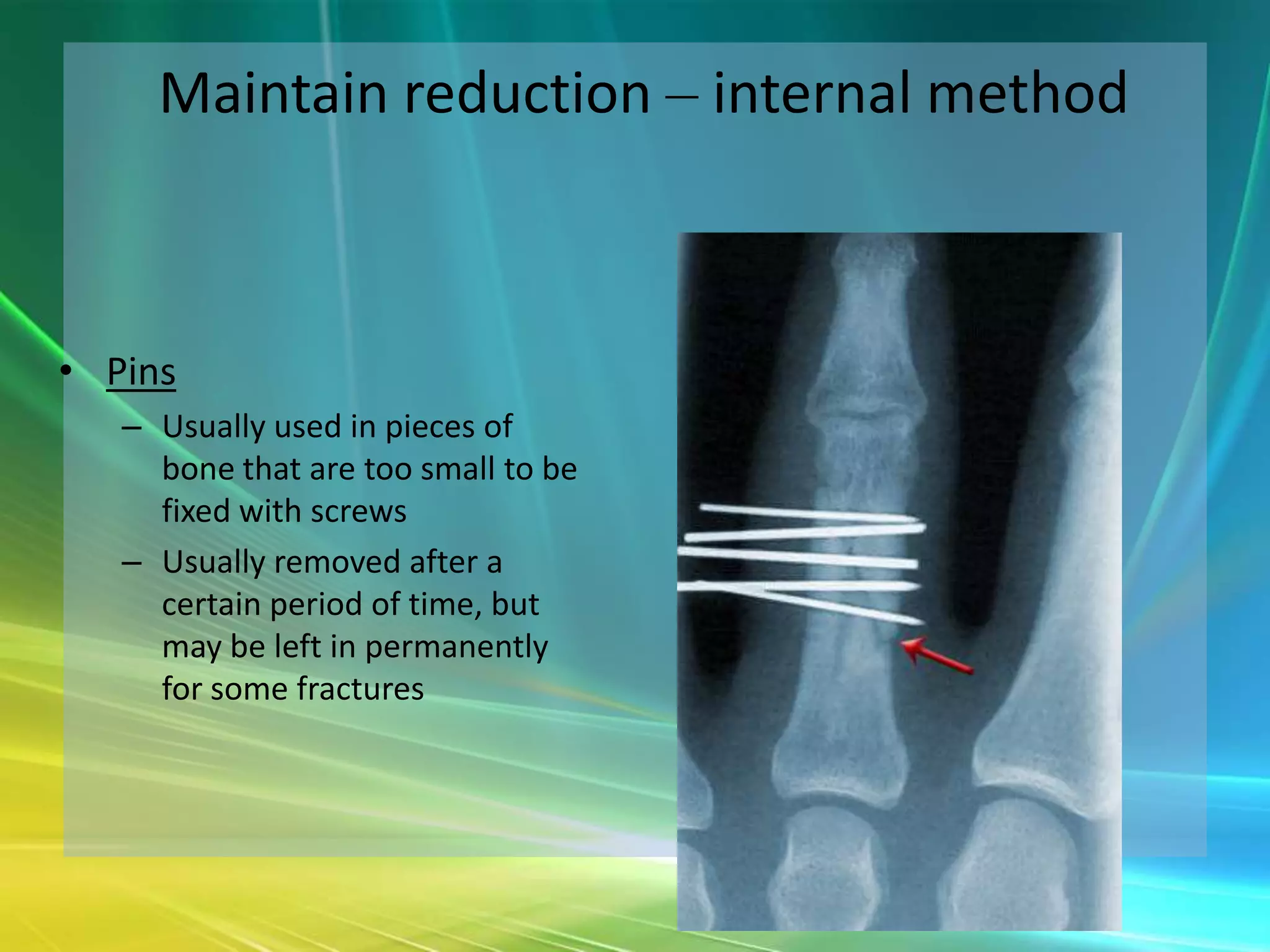
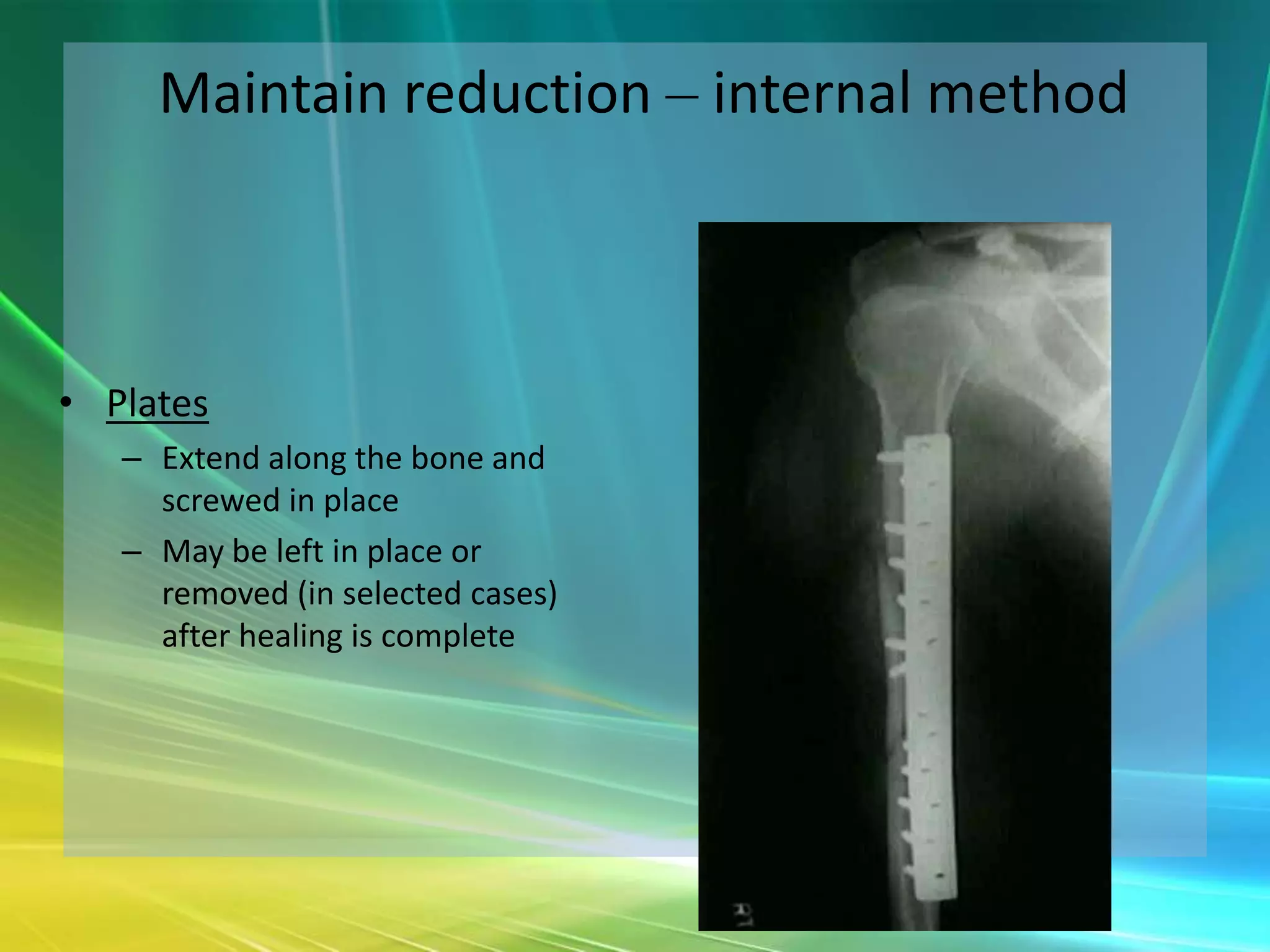
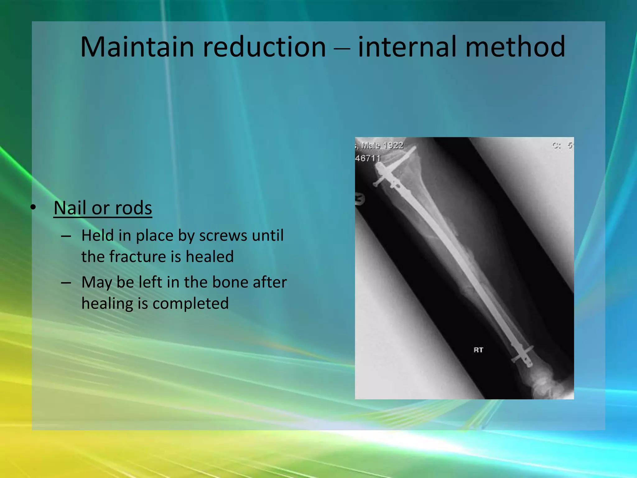
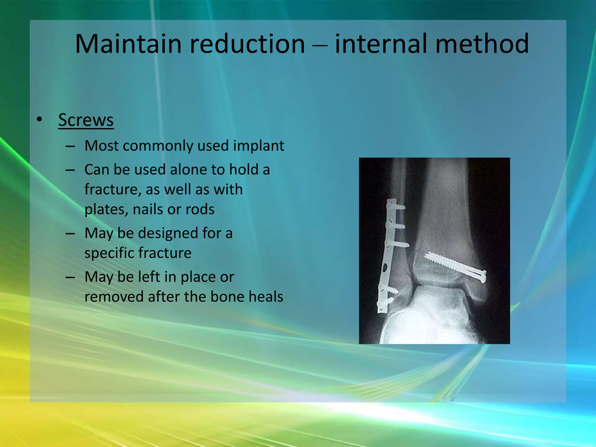
Orthopaedic Trauma

         Mr Hiren Divecha
                 ST3 T&O
               19/1/2011
Definitions
Trauma & Basic Management
Trauma
• Golden Hour of Trauma
  – Rapid transport of a severely injured patient to a
    trauma center for definitive care
  – Initial treatment has a significantly higher chance
    for survival during this period
Trauma Evaluation
• ATLS
  – Advanced Trauma and Life Support

  – A standardized protocol for the evaluation and
    treatment of victims of trauma
  – Developed by a Nebraska orthopaedic surgeon
    who was involved in a trauma and was not
    satisfied with the lack of a protocol for such
    patients
ATLS
• Airway (+ cervical spine
  immobilisation)

• Breathing (+ high flow O2)

• Circulation

• Disability

• Exposure
Primary Survey
• Rapid assessment of ABC’s and addressing life
  threatening problems
   – establishing airway and ventilation, placing chest tubes,
     control active hemorrhage
• Place large bore IV’s and begin fluid replacement for
  patients in shock
• Trauma x-rays
   – chest, pelvis and lateral C-Spine
Secondary Survey
• Assessing entire patient for
  other non-life threatening
  injuries.
• Orthopaedic assessment of
  skeleton
   – splint fractures
   – reduce dislocations
   – evaluate distal pulses and
     peripheral nerve function
• Obtain Xray or CT of
  affected areas when pt is
  stable
Trauma Assessment
• History  Mechanism of Injury
• Palpation
• Note swelling, Lacerations
• Painful ROM
• Crepitus- that grating feeling
  when two bone ends rub against
  each other
• Abnormal Motion- ie the tibia
  bends in the middle
• Check pulses, sensory exam, and
  motor testing if possible
• Assess for lacerations that communicate with
  the fracture
  – Closed Fracture= intact skin over fracture
  – Open Fracture= laceration communicating with
    fracture (often referred to as a compound fracture
    by lay persons)
Emergency Skeletal Issues
• Hemorrhage control from Pelvis Fractures in pt with
  labile blood pressure (shock)
   – Close pelvic volume
• Hemorrhage control from open fractures
   – Direct pressure
• Restore pulses by realigning fractures and
  dislocations
Urgent Skeletal Issues
•   Irrigation and Debridement of open fractures
•   Reduction of dislocations
•   Splinting of fractures
•   Compartment syndromes
Fracture Basics
Basic Biomechanics
                                  • Bending
                                  • Axial Loading
                                    – Tension
                                    – Compression
                                  • Torsion
Bending   Compression   Torsion
Describing The Fracture
•   Mechanism of injury
     – traumatic, pathological, stress
•   Anatomical site
     – bone and location in bone
•   Fracture geometry/ type
• Displacement
     – three planes of angulation
     – translation
     – shortening
     – rotation
•   Articular involvement
     – Involving joint
     – Fracture – dislocation
•   Soft tissue injury
     – Closed vs open
     – nerves, vessels, tendons, tissue loss
Fracture Mechanics
Reading X-rays
• Say what it is
  – what anatomic structure are you looking at and
    how many different views are there
• Regional Location
  –   Epiphysis, metaphysis
  –   Diaphysis (rule of 1/3rds)
  –   Intra/extra-articular
• Fracture geometry/ type
  – Transverse, Oblique, Spiral
Reading X-rays
• Condition of the bone
  – Comminution (3 or more parts)
  – Segmental (middle fragment)
  – Butterfly segment
• Deformity
  – Angulation (varus/valgus, anterior/posterior)
  – Translation
  – Rotation
  – Shortening/ distraction
Fracture Pattern
• Transverse
• Produced by a
  distracting or tensile
  force
Fracture Pattern
• Spiral
• Produced by a twisting/
  torsional force
Fracture Pattern
• Butterfly
• Produced by pure
  bending force
Fracture Pattern
• Comminuted
• Multifragmentary
• High energy transfer!!
Location-Diaphysis
• Shaft portion of bone
Location-Metaphysis
• The ends of the bone (if
  the fracture goes into a
  joint it is described as
  intra- articular)
What do you see?
What do you see?
What do you see?
What do you see?
Fracture Management
Goals of fracture treatment

• Restore patient to optimal functional state
• Prevent fracture and soft tissue complications
• Get fracture to heal and in satisfactory position for optimal
  functional recovery
• Rehabilitate as early as possible
Inflammatory/ Hematoma Phase (1st)

• Up to 1 week
• Acute inflammation
• Hematoma formation
  (48-72 hours)
• Inflammatory cytokines
• Fibroblasts –
  granulation tissue
• Angiogenesis
Soft Callus Phase(2nd)

• 1 week – 1 month
• Chondroblasts +
  fibroblasts
• Fibrous tissue +
  cartilage + woven bone
Hard Callus Phase (3rd)

• 1 – 4 months
• Soft callus resorbed and
  replaced by osteoid
  from osteoblasts
• Osteoid mineralised
  (hydroxyapatite)
• United, solid, pain free
Remodeling Phase (4th)

• Up to several years
• Hard callus remodels to
  woven bone then
  lamellar bone
• Osteoclasts/ osteoblasts
• Medullary canal reforms
• Remodels according to
  stresses/ loading
   – Wolff’s Law (1892)
Factors affecting fracture healing

• Energy transfer of the injury
• The tissue response
   –   Two bone ends in apposition or compressed
   –   Micro-movement or no movement
   –   Blood supply
   –   Infection
• The patient
• The method of treatment
Diagnosing the bone injury

•   History
     – Mechanism
     – If pain preceded trauma ?pathological
•   Examination
     – General - ABCDE
     – Local – the fracture, swelling, tenderness (crepitus?), abnormal posture,
        skin wound
     – Distal –
          • Circulation – vascular injury?
          • Neurological – sensory and motor deficit?
•   Investigation - Imaging
     –   2 Views (AP/Lateral)
     –   2 Joints (above and below injury)
     –   2 Sides (for comparison, mainly in children)
     –   2 Times (before and after treatment)
Treatment of fractures

•   Reduce
•   Maintain reduction (+ hold until union)
•   Rehabilitate – restore function
•   Prevent or treat complications
Maintain reduction

• How?
  – External method
    • POP (+ equivalents), traction, external fixator
  – Internal method
    • Wires, pins, plates, nails, screws
Maintain reduction – external method


• POP
• Mould with palms
   – Adv – cheap,easy to use,
     convenient, can be
     moulded
   – Disadv – susceptibility to
     damage (disintegrates
     when wet), up to 48hrs
     to dry
Maintain reduction – external method


• Resin cast
• Adv
   – lighter and stronger
   – sets in 5-10mins
   – max strength in 30mins
• Disadv
   – Cost
   – more difficult to apply/remove
   – more rigid with greater risk of
     complications eg. swelling and
     pressure necrosis
Maintain reduction – external method


• Skin traction
   – Temporary measure
     when operative
     fixation not available
     for awhile
   – Skin can be injured if
     applied for long
     periods of time
Maintain reduction – external method


• Skeletal traction
   – Requires invasive
     procedure for longer
     term traction requiring
     heavier weights
   – Complications
     associated with pin
     insertion eg. infection
Maintain reduction – external method


• External fixator
• Indications
   – Fractures associated with
     severe soft tissue
   – Fracture associated with N/V
     damage
   – Severely comminuted and
     unstable fracture
   – Unstable pelvic fracture
   – Infected fracture
• Complications
   – Pin track infection
   – Delayed union
Maintain reduction – internal method


• Advantages
   – Shorter hospital stay
   – Enables individuals to return to fxn earlier
   – Reduces incidence of non and mal-union
• Indications
   – Fractures that need operative fixation
   – Inherently unstable fractures prone to re-displacement after reduction
     (eg. mid-shaft femoral fractures)
   – Pathological fracture
   – Polytrauma (minimise ARDS)
   – Patients with nursing difficulties (paraplegics, v. elderly, multiple
     trauma)
Maintain reduction – internal method
• Stainless steel, titanium, cobalt

• Complications
   – Infection
   – Non-union
   – Implant failure
   – Re-fracture
Maintain reduction – internal method


• Wires
   – Can be used in conjunction
     with other forms of internal
     fixation
   – Used to treat fractures of
     small bones
Maintain reduction – internal method


• Pins
   – Usually used in pieces of
     bone that are too small to be
     fixed with screws
   – Usually removed after a
     certain period of time, but
     may be left in permanently
     for some fractures
Maintain reduction – internal method


• Plates
   – Extend along the bone and
     screwed in place
   – May be left in place or
     removed (in selected cases)
     after healing is complete
Maintain reduction – internal method


• Nail or rods
   – Held in place by screws until
     the fracture is healed
   – May be left in the bone after
     healing is completed
Maintain reduction – internal method

• Screws
   – Most commonly used implant
   – Can be used alone to hold a
     fracture, as well as with
     plates, nails or rods
   – May be designed for a
     specific fracture
   – May be left in place or
     removed after the bone heals
Maintain reduction – how long?


• Judge each case on its own merits
• X-ray in POP for position; out of POP to clinically assess state
  of healing
• Sticky – “Deformable but not displaceable”
• Union (weeks)
   – Incomplete repair; Part moves as one; Local tenderness; Local pain on
     stress; See fracture line on-x-ray
• Consolidation (months)
   – Complete repair; No external protection needed; Upper limb 6/52;
     Lower limb 12/52; Half for child; Double for transverse fractures
Complications of fractures
• Early                                   • Late
   –   Visceral/ vascular/ nerve injury      –   Mal-union
   –   Haemarthrosis                         –   Delayed union
   –   Infection                             –   Non-union
   –   Fat embolism                          –   Tendon rupture
   –   Compartment syndrome                  –   Myositis ossificans
                                             –   Osteonecrosis
                                             –   Complex regional pain syndrome
                                             –   Osteoarthritis and joint stiffness
Thanks for listening !

     Any questions

Orthopaedic Trauma - The Basics

  • 1.
    Orthopaedic Trauma Mr Hiren Divecha ST3 T&O 19/1/2011
  • 3.
  • 4.
    Trauma & BasicManagement
  • 5.
    Trauma • Golden Hourof Trauma – Rapid transport of a severely injured patient to a trauma center for definitive care – Initial treatment has a significantly higher chance for survival during this period
  • 7.
    Trauma Evaluation • ATLS – Advanced Trauma and Life Support – A standardized protocol for the evaluation and treatment of victims of trauma – Developed by a Nebraska orthopaedic surgeon who was involved in a trauma and was not satisfied with the lack of a protocol for such patients
  • 8.
    ATLS • Airway (+cervical spine immobilisation) • Breathing (+ high flow O2) • Circulation • Disability • Exposure
  • 9.
    Primary Survey • Rapidassessment of ABC’s and addressing life threatening problems – establishing airway and ventilation, placing chest tubes, control active hemorrhage • Place large bore IV’s and begin fluid replacement for patients in shock • Trauma x-rays – chest, pelvis and lateral C-Spine
  • 10.
    Secondary Survey • Assessingentire patient for other non-life threatening injuries. • Orthopaedic assessment of skeleton – splint fractures – reduce dislocations – evaluate distal pulses and peripheral nerve function • Obtain Xray or CT of affected areas when pt is stable
  • 11.
    Trauma Assessment • History Mechanism of Injury • Palpation • Note swelling, Lacerations • Painful ROM • Crepitus- that grating feeling when two bone ends rub against each other • Abnormal Motion- ie the tibia bends in the middle • Check pulses, sensory exam, and motor testing if possible
  • 12.
    • Assess forlacerations that communicate with the fracture – Closed Fracture= intact skin over fracture – Open Fracture= laceration communicating with fracture (often referred to as a compound fracture by lay persons)
  • 13.
    Emergency Skeletal Issues •Hemorrhage control from Pelvis Fractures in pt with labile blood pressure (shock) – Close pelvic volume • Hemorrhage control from open fractures – Direct pressure • Restore pulses by realigning fractures and dislocations
  • 14.
    Urgent Skeletal Issues • Irrigation and Debridement of open fractures • Reduction of dislocations • Splinting of fractures • Compartment syndromes
  • 15.
  • 16.
    Basic Biomechanics • Bending • Axial Loading – Tension – Compression • Torsion Bending Compression Torsion
  • 17.
    Describing The Fracture • Mechanism of injury – traumatic, pathological, stress • Anatomical site – bone and location in bone • Fracture geometry/ type • Displacement – three planes of angulation – translation – shortening – rotation • Articular involvement – Involving joint – Fracture – dislocation • Soft tissue injury – Closed vs open – nerves, vessels, tendons, tissue loss
  • 18.
  • 20.
    Reading X-rays • Saywhat it is – what anatomic structure are you looking at and how many different views are there • Regional Location – Epiphysis, metaphysis – Diaphysis (rule of 1/3rds) – Intra/extra-articular • Fracture geometry/ type – Transverse, Oblique, Spiral
  • 21.
    Reading X-rays • Conditionof the bone – Comminution (3 or more parts) – Segmental (middle fragment) – Butterfly segment • Deformity – Angulation (varus/valgus, anterior/posterior) – Translation – Rotation – Shortening/ distraction
  • 22.
    Fracture Pattern • Transverse •Produced by a distracting or tensile force
  • 23.
    Fracture Pattern • Spiral •Produced by a twisting/ torsional force
  • 24.
    Fracture Pattern • Butterfly •Produced by pure bending force
  • 25.
    Fracture Pattern • Comminuted •Multifragmentary • High energy transfer!!
  • 26.
  • 27.
    Location-Metaphysis • The endsof the bone (if the fracture goes into a joint it is described as intra- articular)
  • 28.
  • 29.
  • 30.
  • 31.
  • 32.
  • 33.
    Goals of fracturetreatment • Restore patient to optimal functional state • Prevent fracture and soft tissue complications • Get fracture to heal and in satisfactory position for optimal functional recovery • Rehabilitate as early as possible
  • 34.
    Inflammatory/ Hematoma Phase(1st) • Up to 1 week • Acute inflammation • Hematoma formation (48-72 hours) • Inflammatory cytokines • Fibroblasts – granulation tissue • Angiogenesis
  • 35.
    Soft Callus Phase(2nd) •1 week – 1 month • Chondroblasts + fibroblasts • Fibrous tissue + cartilage + woven bone
  • 36.
    Hard Callus Phase(3rd) • 1 – 4 months • Soft callus resorbed and replaced by osteoid from osteoblasts • Osteoid mineralised (hydroxyapatite) • United, solid, pain free
  • 37.
    Remodeling Phase (4th) •Up to several years • Hard callus remodels to woven bone then lamellar bone • Osteoclasts/ osteoblasts • Medullary canal reforms • Remodels according to stresses/ loading – Wolff’s Law (1892)
  • 38.
    Factors affecting fracturehealing • Energy transfer of the injury • The tissue response – Two bone ends in apposition or compressed – Micro-movement or no movement – Blood supply – Infection • The patient • The method of treatment
  • 40.
    Diagnosing the boneinjury • History – Mechanism – If pain preceded trauma ?pathological • Examination – General - ABCDE – Local – the fracture, swelling, tenderness (crepitus?), abnormal posture, skin wound – Distal – • Circulation – vascular injury? • Neurological – sensory and motor deficit? • Investigation - Imaging – 2 Views (AP/Lateral) – 2 Joints (above and below injury) – 2 Sides (for comparison, mainly in children) – 2 Times (before and after treatment)
  • 41.
    Treatment of fractures • Reduce • Maintain reduction (+ hold until union) • Rehabilitate – restore function • Prevent or treat complications
  • 42.
    Maintain reduction • How? – External method • POP (+ equivalents), traction, external fixator – Internal method • Wires, pins, plates, nails, screws
  • 43.
    Maintain reduction –external method • POP • Mould with palms – Adv – cheap,easy to use, convenient, can be moulded – Disadv – susceptibility to damage (disintegrates when wet), up to 48hrs to dry
  • 44.
    Maintain reduction –external method • Resin cast • Adv – lighter and stronger – sets in 5-10mins – max strength in 30mins • Disadv – Cost – more difficult to apply/remove – more rigid with greater risk of complications eg. swelling and pressure necrosis
  • 45.
    Maintain reduction –external method • Skin traction – Temporary measure when operative fixation not available for awhile – Skin can be injured if applied for long periods of time
  • 46.
    Maintain reduction –external method • Skeletal traction – Requires invasive procedure for longer term traction requiring heavier weights – Complications associated with pin insertion eg. infection
  • 47.
    Maintain reduction –external method • External fixator • Indications – Fractures associated with severe soft tissue – Fracture associated with N/V damage – Severely comminuted and unstable fracture – Unstable pelvic fracture – Infected fracture • Complications – Pin track infection – Delayed union
  • 48.
    Maintain reduction –internal method • Advantages – Shorter hospital stay – Enables individuals to return to fxn earlier – Reduces incidence of non and mal-union • Indications – Fractures that need operative fixation – Inherently unstable fractures prone to re-displacement after reduction (eg. mid-shaft femoral fractures) – Pathological fracture – Polytrauma (minimise ARDS) – Patients with nursing difficulties (paraplegics, v. elderly, multiple trauma)
  • 49.
    Maintain reduction –internal method • Stainless steel, titanium, cobalt • Complications – Infection – Non-union – Implant failure – Re-fracture
  • 50.
    Maintain reduction –internal method • Wires – Can be used in conjunction with other forms of internal fixation – Used to treat fractures of small bones
  • 51.
    Maintain reduction –internal method • Pins – Usually used in pieces of bone that are too small to be fixed with screws – Usually removed after a certain period of time, but may be left in permanently for some fractures
  • 52.
    Maintain reduction –internal method • Plates – Extend along the bone and screwed in place – May be left in place or removed (in selected cases) after healing is complete
  • 53.
    Maintain reduction –internal method • Nail or rods – Held in place by screws until the fracture is healed – May be left in the bone after healing is completed
  • 54.
    Maintain reduction –internal method • Screws – Most commonly used implant – Can be used alone to hold a fracture, as well as with plates, nails or rods – May be designed for a specific fracture – May be left in place or removed after the bone heals
  • 55.
    Maintain reduction –how long? • Judge each case on its own merits • X-ray in POP for position; out of POP to clinically assess state of healing • Sticky – “Deformable but not displaceable” • Union (weeks) – Incomplete repair; Part moves as one; Local tenderness; Local pain on stress; See fracture line on-x-ray • Consolidation (months) – Complete repair; No external protection needed; Upper limb 6/52; Lower limb 12/52; Half for child; Double for transverse fractures
  • 56.
    Complications of fractures •Early • Late – Visceral/ vascular/ nerve injury – Mal-union – Haemarthrosis – Delayed union – Infection – Non-union – Fat embolism – Tendon rupture – Compartment syndrome – Myositis ossificans – Osteonecrosis – Complex regional pain syndrome – Osteoarthritis and joint stiffness
  • 57.
    Thanks for listening! Any questions