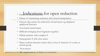
The document discusses fractures, dislocations, and their treatment. It defines fractures and describes different types including closed/open, pathological, and stress fractures. Signs and symptoms of fractures and dislocations are outlined. The principles of diagnosing and treating fractures are described, including reduction, splinting, and casting. Factors that influence fracture healing are also mentioned.
Call Now (412) 486-5100 - At Dr. Nigro Ankle and Foot Care we have been committed to providing you with the most advanced podiatric care in a compassionate and caring environment since 1990. Dr. Nigro Foot and Ankle Care physicians take a whole-person approach to your feet and ankles, because your overall wellness can influence the health of your feet. Visit us at http://PittsburghFootandAnkle.com
This slide is a brief overview of Femoral shaft fractures for undergraduate medical students (MBBS) . Video lecture of the content is available on
https://www.youtube.com/watch?v=4rHXKtG36HA
Feel free to drop in any comments or questions
Arthroscopic ACL Reconstruction By Dr Shekhar ShrivastavDelhiArthroscopy
Arthroscopic Acl Reconstruction By Dr Shekhar Shrivastav.
HOW NORMAL KNEE WORKS ?
The knee is the largest joint in the body, and one of the most easily injured. It is made up of the lower end of the thigh bone(femur), the upper end of the shin bone (tibia), and the knee cap (patella), which slides in a groove on the end of the femur. Four bands of tissue, the anterior and posterior cruciate ligaments, and the medial and lateral collateral ligaments connect the femur and the tibia and provide joint stability. The surfaces where the femur, tibia and patella touch are covered with articular cartilage, a smooth substance that cushions the bones and enables them to glide freely. Semicircular rings of tough fibrous-cartilage tissue called the lateral and medial menisci act as shock absorbers and stabilizers.
WHAT IS THE ROLE OF ACL ?
ACL along with other ligaments of the knee joint and meniscus provides stability to the knee joint.
WHAT IS LIGAMENT RECONSTRUCTION ( ACL ) ?
Ligament reconstruction involves replacing the torn ligament with a tendon (graft) from your knee and fixing the graft in place with screws. This procedure is performed with the use of the arthroscope. The anterior cruciate ligament (ACL) is the most common ligament requiring reconstruction procedures. The torn ligament is excised arthroscopically and new ligament is prepared by ligament grafts taken from your own body. Bony tunnels are prepared in femur and tibia using specialized instruments through which the new ligament is passed and fixed with special screws. This procedure requires relative rest or leave from your work or studies for about 2-3 weeks after which you will be allowed normal day to day activities.
WHEN CAN THE PATIENT BE AMBULATED AFTER SURGERY ?
The patient can walk from the same evening of the surgery. Initially the patient is advised to walk with a brace and a walking cane. Strengthening and range of motion exercises for the knee are started from the next day. The patient is discharged from the hospital 2nd or 3rd day after surgery. The patient can walk without support by 10-14 days depending on muscle strengthening. Slow Jogging and other strenuous activities are permitted after 3 months and the patient can return to active sports only 8-9 months after surgery.
Torn ACL Reconstructed ACL
For Further Queries contact your Orthopedic Surgeon at
+ 91 9971192233
Vertebral Fracture Management Professor presented by Opinder Sahota, Orthogeriatric Medicine & Consultant Physician QMC, Nottingham University Hospitals NHS Trust.
Presented at the FLS Champions' Summit, February 2016.
Total shoulder arthroplasty and reverse TSA - Hussain AlgawahmedHussainAlgawahmedMBB
Discussion of the clinical presentation of shoulder arthritis followed by treatment options and the mechanical basis of total and reverse total shoulder arthroplasty
A tibial shaft fracture occurs along the length of the bone, below the knee and above the ankle. It typically takes a major force to cause this type of broken leg. Motor vehicle collisions, for example, are a common cause of tibial shaft fractures.
this presentation explain about the fracture, don't miss to take a look on it, it will help you, you will find a useful knowledge through this a brief presentation.
This slide includes general principles of fracture management. This is just a basic idea. I have tried to include figures as well as videos. But unfortunately videos wont play here.
Prix Galien International 2024 Forum ProgramLevi Shapiro
June 20, 2024, Prix Galien International and Jerusalem Ethics Forum in ROME. Detailed agenda including panels:
- ADVANCES IN CARDIOLOGY: A NEW PARADIGM IS COMING
- WOMEN’S HEALTH: FERTILITY PRESERVATION
- WHAT’S NEW IN THE TREATMENT OF INFECTIOUS,
ONCOLOGICAL AND INFLAMMATORY SKIN DISEASES?
- ARTIFICIAL INTELLIGENCE AND ETHICS
- GENE THERAPY
- BEYOND BORDERS: GLOBAL INITIATIVES FOR DEMOCRATIZING LIFE SCIENCE TECHNOLOGIES AND PROMOTING ACCESS TO HEALTHCARE
- ETHICAL CHALLENGES IN LIFE SCIENCES
- Prix Galien International Awards Ceremony
Report Back from SGO 2024: What’s the Latest in Cervical Cancer?bkling
Are you curious about what’s new in cervical cancer research or unsure what the findings mean? Join Dr. Emily Ko, a gynecologic oncologist at Penn Medicine, to learn about the latest updates from the Society of Gynecologic Oncology (SGO) 2024 Annual Meeting on Women’s Cancer. Dr. Ko will discuss what the research presented at the conference means for you and answer your questions about the new developments.
Lung Cancer: Artificial Intelligence, Synergetics, Complex System Analysis, S...Oleg Kshivets
RESULTS: Overall life span (LS) was 2252.1±1742.5 days and cumulative 5-year survival (5YS) reached 73.2%, 10 years – 64.8%, 20 years – 42.5%. 513 LCP lived more than 5 years (LS=3124.6±1525.6 days), 148 LCP – more than 10 years (LS=5054.4±1504.1 days).199 LCP died because of LC (LS=562.7±374.5 days). 5YS of LCP after bi/lobectomies was significantly superior in comparison with LCP after pneumonectomies (78.1% vs.63.7%, P=0.00001 by log-rank test). AT significantly improved 5YS (66.3% vs. 34.8%) (P=0.00000 by log-rank test) only for LCP with N1-2. Cox modeling displayed that 5YS of LCP significantly depended on: phase transition (PT) early-invasive LC in terms of synergetics, PT N0—N12, cell ratio factors (ratio between cancer cells- CC and blood cells subpopulations), G1-3, histology, glucose, AT, blood cell circuit, prothrombin index, heparin tolerance, recalcification time (P=0.000-0.038). Neural networks, genetic algorithm selection and bootstrap simulation revealed relationships between 5YS and PT early-invasive LC (rank=1), PT N0—N12 (rank=2), thrombocytes/CC (3), erythrocytes/CC (4), eosinophils/CC (5), healthy cells/CC (6), lymphocytes/CC (7), segmented neutrophils/CC (8), stick neutrophils/CC (9), monocytes/CC (10); leucocytes/CC (11). Correct prediction of 5YS was 100% by neural networks computing (area under ROC curve=1.0; error=0.0).
CONCLUSIONS: 5YS of LCP after radical procedures significantly depended on: 1) PT early-invasive cancer; 2) PT N0--N12; 3) cell ratio factors; 4) blood cell circuit; 5) biochemical factors; 6) hemostasis system; 7) AT; 8) LC characteristics; 9) LC cell dynamics; 10) surgery type: lobectomy/pneumonectomy; 11) anthropometric data. Optimal diagnosis and treatment strategies for LC are: 1) screening and early detection of LC; 2) availability of experienced thoracic surgeons because of complexity of radical procedures; 3) aggressive en block surgery and adequate lymph node dissection for completeness; 4) precise prediction; 5) adjuvant chemoimmunoradiotherapy for LCP with unfavorable prognosis.
Couples presenting to the infertility clinic- Do they really have infertility...Sujoy Dasgupta
Dr Sujoy Dasgupta presented the study on "Couples presenting to the infertility clinic- Do they really have infertility? – The unexplored stories of non-consummation" in the 13th Congress of the Asia Pacific Initiative on Reproduction (ASPIRE 2024) at Manila on 24 May, 2024.
TEST BANK for Operations Management, 14th Edition by William J. Stevenson, Ve...kevinkariuki227
TEST BANK for Operations Management, 14th Edition by William J. Stevenson, Verified Chapters 1 - 19, Complete Newest Version.pdf
These lecture slides, by Dr Sidra Arshad, offer a quick overview of physiological basis of a normal electrocardiogram.
Learning objectives:
1. Define an electrocardiogram (ECG) and electrocardiography
2. Describe how dipoles generated by the heart produce the waveforms of the ECG
3. Describe the components of a normal electrocardiogram of a typical bipolar leads (limb II)
4. Differentiate between intervals and segments
5. Enlist some common indications for obtaining an ECG
Study Resources:
1. Chapter 11, Guyton and Hall Textbook of Medical Physiology, 14th edition
2. Chapter 9, Human Physiology - From Cells to Systems, Lauralee Sherwood, 9th edition
3. Chapter 29, Ganong’s Review of Medical Physiology, 26th edition
4. Electrocardiogram, StatPearls - https://www.ncbi.nlm.nih.gov/books/NBK549803/
5. ECG in Medical Practice by ABM Abdullah, 4th edition
6. ECG Basics, http://www.nataliescasebook.com/tag/e-c-g-basics
NVBDCP.pptx Nation vector borne disease control programSapna Thakur
NVBDCP was launched in 2003-2004 . Vector-Borne Disease: Disease that results from an infection transmitted to humans and other animals by blood-feeding arthropods, such as mosquitoes, ticks, and fleas. Examples of vector-borne diseases include Dengue fever, West Nile Virus, Lyme disease, and malaria.
Flu Vaccine Alert in Bangalore Karnatakaaddon Scans
As flu season approaches, health officials in Bangalore, Karnataka, are urging residents to get their flu vaccinations. The seasonal flu, while common, can lead to severe health complications, particularly for vulnerable populations such as young children, the elderly, and those with underlying health conditions.
Dr. Vidisha Kumari, a leading epidemiologist in Bangalore, emphasizes the importance of getting vaccinated. "The flu vaccine is our best defense against the influenza virus. It not only protects individuals but also helps prevent the spread of the virus in our communities," he says.
This year, the flu season is expected to coincide with a potential increase in other respiratory illnesses. The Karnataka Health Department has launched an awareness campaign highlighting the significance of flu vaccinations. They have set up multiple vaccination centers across Bangalore, making it convenient for residents to receive their shots.
To encourage widespread vaccination, the government is also collaborating with local schools, workplaces, and community centers to facilitate vaccination drives. Special attention is being given to ensuring that the vaccine is accessible to all, including marginalized communities who may have limited access to healthcare.
Residents are reminded that the flu vaccine is safe and effective. Common side effects are mild and may include soreness at the injection site, mild fever, or muscle aches. These side effects are generally short-lived and far less severe than the flu itself.
Healthcare providers are also stressing the importance of continuing COVID-19 precautions. Wearing masks, practicing good hand hygiene, and maintaining social distancing are still crucial, especially in crowded places.
Protect yourself and your loved ones by getting vaccinated. Together, we can help keep Bangalore healthy and safe this flu season. For more information on vaccination centers and schedules, residents can visit the Karnataka Health Department’s official website or follow their social media pages.
Stay informed, stay safe, and get your flu shot today!
263778731218 Abortion Clinic /Pills In Harare ,sisternakatoto
263778731218 Abortion Clinic /Pills In Harare ,ABORTION WOMEN’S CLINIC +27730423979 IN women clinic we believe that every woman should be able to make choices in her pregnancy. Our job is to provide compassionate care, safety,affordable and confidential services. That’s why we have won the trust from all generations of women all over the world. we use non surgical method(Abortion pills) to terminate…Dr.LISA +27730423979women Clinic is committed to providing the highest quality of obstetrical and gynecological care to women of all ages. Our dedicated staff aim to treat each patient and her health concerns with compassion and respect.Our dedicated group ABORTION WOMEN’S CLINIC +27730423979 IN women clinic we believe that every woman should be able to make choices in her pregnancy. Our job is to provide compassionate care, safety,affordable and confidential services. That’s why we have won the trust from all generations of women all over the world. we use non surgical method(Abortion pills) to terminate…Dr.LISA +27730423979women Clinic is committed to providing the highest quality of obstetrical and gynecological care to women of all ages. Our dedicated staff aim to treat each patient and her health concerns with compassion and respect.Our dedicated group of receptionists, nurses, and physicians have worked together as a teamof receptionists, nurses, and physicians have worked together as a team wwww.lisywomensclinic.co.za/
micro teaching on communication m.sc nursing.pdfAnurag Sharma
Microteaching is a unique model of practice teaching. It is a viable instrument for the. desired change in the teaching behavior or the behavior potential which, in specified types of real. classroom situations, tends to facilitate the achievement of specified types of objectives.
3. FRACTURES
• Definition: A break in the continuity of a bone with associated damage to the
surrounding soft tissues.
• The majority of fractures are managed non surgically.
• Optimal treatment depends on multiple factors, including the location and type of
fracture.
• In some instances (such as a minimally displaced fracture of the middle phalanx of one
of the lesser toes) no treatment at all other than those for symptoms of pain generally
are necessary
4. • In most other fractures, some form of immobilization is the treatment of choice, this might involve a
simple sling (as for a midshaft clavicle fracture) or a more robust splint or brace.
• When more complete immobilization is desired, immobilization in a circumferential plaster or fiberglass
cast is often ideal.
5.
6.
7. Types of fracture
• 1. Closed (simple) fracture: there is no laceration of the skin overlying the
fracture.
• 2. Open (compound) fracture: An open fracture is a fracture hematoma that
communicates with an epithelial surface, e.g. skin or a pelvic fracture may
communicate with a ruptured rectum or a rib fracture may penetrate a lung.
Open fractures are potentially infected and need urgent treatment.
• 3. Pathological fracture: fracture occurs in a diseased or abnormal bone.
• Stress fracture: due to repeated shearing force in a healthy bone. E.g. athletes.
9. Transverse fracture: This is usually caused by a force applied directly to the site at
which the fracture occurs
Spiral or oblique fracture: This is produced by a twisting force applied distant from
the site of the fracture, usually at each end of a long bone such as the tibia.
Greenstick fracture: This occurs in children whose bones are soft and yielding. The
bone bends without fracturing across completely, the cortex on the concave side usually
remaining intact.
Crush fracture: This occurs in cancellous bone as a result of a compression force
Burst fracture: This usually occurs in a short bone, such as a vertebra from strong
direct pressure; in the vertebrae this usually occurs as a result of impaction of the disc.
11. • Avulsion fracture: This is caused by traction, a bony fragment usually being torn
off by a tendon or ligament.
• Fracture dislocation or subluxation: This is a fracture which involves a joint and
results in malalignment of the joint surfaces
• Complicated fracture: when there is important soft - tissue damage to nerves,
vessels or internal organs.
• Impacted fracture: when the fragments are driven into one another.
• Stable fracture: fracture held in anatomical position firmly by soft tissue
attachment.
• Unstable fracture: Is one which is displaced or has the potential to displace.
• Intra-articular fracture: is one in which the fracture involves the joint surface.
Usually these fractures require operative anatomical reduction if there is
displacement.
15. Signs & Symptoms
• Tenderness is almost invariable with a recent fracture, assuming the patient is
conscious.
• Deformity may or may not be evident. The limb may be bent or shortened, or
there may be a step in the alignment of the bone or joint.
• Swelling is usual when the fracture is fairly superfi cial; gross swelling usually
implies a vascular rupture.
• Local temperature increase is essentially part of the inflammatory response which
rapidly follows the injury and may be evident even if the damage is confined to the
soft tissues.
16. • Abnormal mobility or crepitus , i.e. grating of the fracture ends, may be
noticed. Vigorous attempts to elicit it should be avoided.
• Loss of function is almost always found to some extent. The patient usually
has difficulty in moving the adjacent joints.
• Having diagnosed a fracture or joint injury, the presence and extent of any
wound should be , and the area examined for evidence of ischaemia and nerve
or other important soft – tissue damage.
17. Dislocations
• Sometimes a dislocated joint will spontaneously reduce before your
assessment.
• Confirm the dislocation by taking a patient history.
• A dislocation that does not reduce is a serious problem.
18. Signs and symptoms
• Marked deformity.
• Swelling.
• Pain that is aggravated by any attempt at
movement.
• Tenderness on palpation.
• Virtually complete loss of normal joint motion.
• Numbness or impaired circulation to the limb or
digit.
19. Sprains
• A sprain occurs when a joint is twisted or stretched beyond its normal range
of motion.
• Alignment generally returns to a fairly normal position, although there may
be some displacement.
• Severe deformity does not typically occur.
20. Sprains
• Signs and symptoms
• Point tenderness
• Swelling and ecchymosis
• Pain
• Instability of the joint
21. Strain
• A strain is an injury to a muscle and/or tendon that results from a violent
muscle contraction or from excessive stretching.
• Often no deformity is present and only minor swelling is noted at the site
of the injury.
22. Compartment Syndrome
• Most often occurs with a fractured tibia or forearm of children
• Typically develops within 6 to 12 hours after injury, as a result of:
• Excessive bleeding
• A severely crushed extremity
• The rapid return of blood to an ischemic limb
23. Compartment Syndrome
• This syndrome is characterized by:
• Pain that is out of proportion to the injury
• Pain on passive stretching of muscles within the compartment
• Pallor
• Decreased sensation
• Decreased power
24. Diagnosing fracture
• History: A brief history is essential in order to assess the mechanism of injury and
to raise suspicion of other, less apparent, injuries.
• Pain. This is the commonest symptom, but varies with the site and instability of the
fracture.
• Loss of function. There is almost always some impairment of function in the
injured area, so that patient may be unable to move the limb at all, or may use it with
difficulty.
• Loss of sensation or motor power. This is a particularly important symptom,
suggesting nerve or vascular complications.
25. History Taking
• Investigate the chief complaint.
• Obtain a medical history and be alert for injury-specific signs and symptoms and any
pertinent negatives.
• Obtain a SAMPLE history for all trauma patients.
• OPQRST is too lengthy when matters of ABCs require immediate attention.
26. Secondary Assessment
• More detailed examination of the patient to reveal hidden injuries
• Physical examinations
• If significant trauma has occurred, start with a full-body scan
• Begin with the head and work systematically toward the feet.
• Assess the musculoskeletal system.
27. Secondary Assessment
• Physical examinations (cont’d)
• When lacerations are present in an extremity, consider an open fracture.
• Any injury or deformity of the bone may be associated with vessel or nerve injury.
28. Investigation
• Full Blood Count
• Plain x-ray A-P and lateral views
• Computed tomography (CT) scanning has become a useful aid in diagnosing
the more difficult injuries, particularly fractures of the pelvis, spine and
complex intra - articular fractures. Unfortunately this is not usually done in
our part of the world.
29.
30. Factors that influence the healing of fractures
Unfavorable factors.
• Impairment of blood supply.
• Infection.
• Excessive movement.
• Presence of tumor.
• Interposition of soft tissue.
• Any form of Nicotine.
32. Open fractures
• The treatment of these fractures is an orthopaedic emergency.
• The most important consideration when dealing with an open fracture is to reduce
the risks of infection.
• In order to achieve this, the wound often needs to be extended. Loose fragments of
bone are devitalized and so should be removed, as should bone deep to areas where
the periosteum has been stripped. This process of cleaning and removing
devitalized tissue is termed debridement.
• All communicating wounds should be left open and covered with a sterile dressing,
with a view to later closure when infection has been avoided or overcome.
33. • Primary closure is desirable if it can be achieved safely, if the degree of
contamination and soft - tissue damage is minimal and if the time from the accident
is not too great, usually less than 6 hours.
• To this must be added the that the patient should be kept under observation,
preferably in hospital.
• Antibiotics should always be given after culture swabs have been taken.
• Anti tetanus injection should be given
• Correct shock if any.
35. Indications for open reduction
• Failure of closed reduction
• Failure of maintaining reduction after closed manipulation
• Fracture that cannot be reduced by closed means eg. displaced
epiphyseal fractures
• Associated arterial injury
• Difficulty bringing short fragments together
• Elderly patients with complex #
• Intracapsular # with slow union
• Major avulsion fractures where there is loss of function of a joint or
muscle group
• Non‐unions
• Re‐ implantations of limbs or extremities
36. Relative Indications for OR of fractures
• Delayed unions
• Multiple fractures to assist in care and general management
• Unable to maintain a reduction
• Pathological fractures
• To assist in nursing care
• To reduce morbidity due to prolonged immobilisation
• For fractures in which closed methods are known to be
ineffective
37. Adv and disadv of open reduction
• Adv
• Allows early movement
• Disadv
• Sepsis
• Delay bone repair
38. Closed reduction and fixation
• The fracture segments are manipulated with a force opposite in direction to
the one that produced the fracture to restore nomal anatomy.
• Indicated in simple fractures.
39. Maintaining fracture reduction
• Stability is achieved by one of the following
techniques:
1 Intrinsic stability. Some fractures require no
additional stabilization
2 External splintage.
3 Internal fixation.
40. External splintage
• Many fractures can be adequately immobilized with a simple device, such as a splint made of
wire, metal or polythene, bandaged in place, and a sling or crutches may be used to avoid
load - bearing.
• These devices are often used to relieve pain rather than to secure immobilization.
• Casting: Plaster of Paris is still widely used for making open or closed casts, jointed casts,
splints.
• Cast bracing: Is a hinged or jointed cast. It has been used particularly for fractures of the
femur and tibia.
• Traction: Is pulling bones directly or indirectly in order to reduce and hold fractures.
41. Complications of fractures
• Early ‐ visceral injury
• ‐ vascular injury
• ‐ nerve injury
• ‐ compartment syndrome
• ‐ haemarthrosis
• ‐ infection
• ‐ gas gangrene
• ‐ fracture blisters
• ‐ plaster and pressure sores
• Late ‐ delayed union