Downloaded 2,439 times

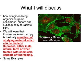
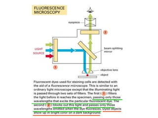
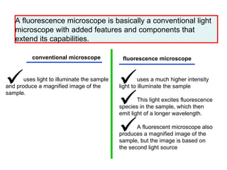
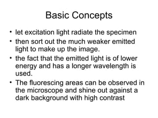

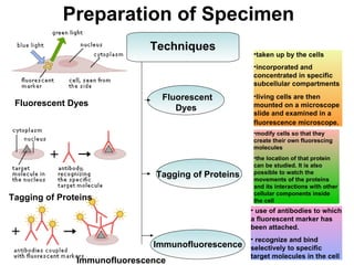
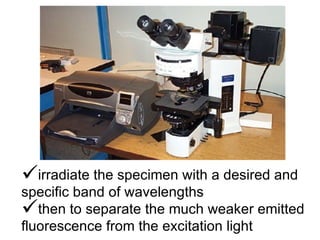
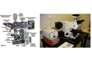

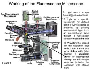
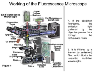
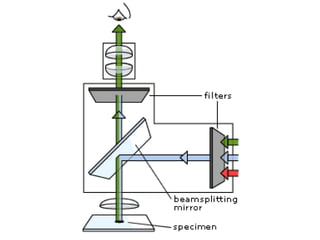
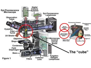
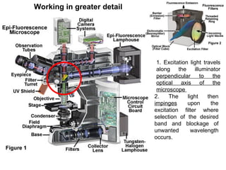
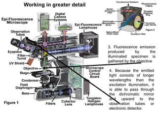
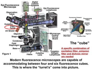
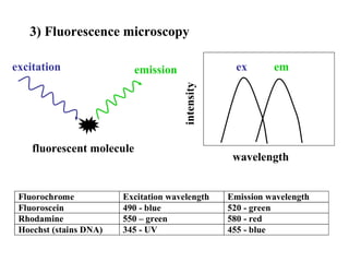
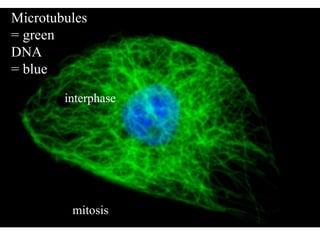
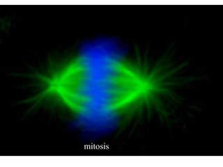


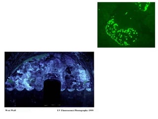
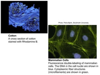
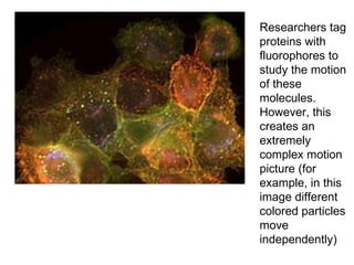


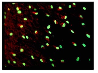
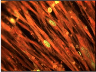

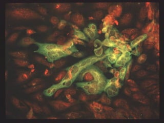
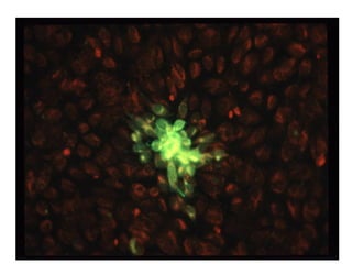
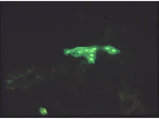
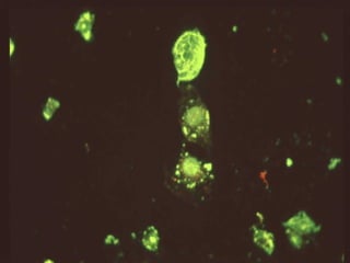

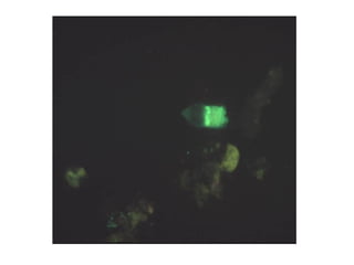

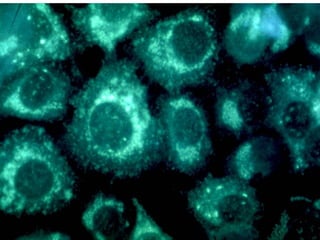


Fluorescence microscopy uses fluorescence to visualize specimens. It works by exciting fluorescent molecules in the sample with high intensity light, causing them to emit light of a longer wavelength. This emitted light is then filtered and used to produce a magnified image of the sample. Modern fluorescence microscopes allow multiple fluorescence filters to be used, and fluorescent markers like dyes, proteins, and antibodies can be introduced to tag specific structures in cells or proteins of interest. This technique is widely used in medical and biological research to study structures and track molecules within living cells.


