E-Study material
For 5thsemester Botany Honours (CBCS)
Course Code: BD501T
Core Course VII: Genetics
DSE Course – I: Analytical Techniques in Plant Sciences
Unit 1: Imaging and related techniques
Debajit Saikia
Assistant Professor
Department of Botany
CNB College, Bokakhat
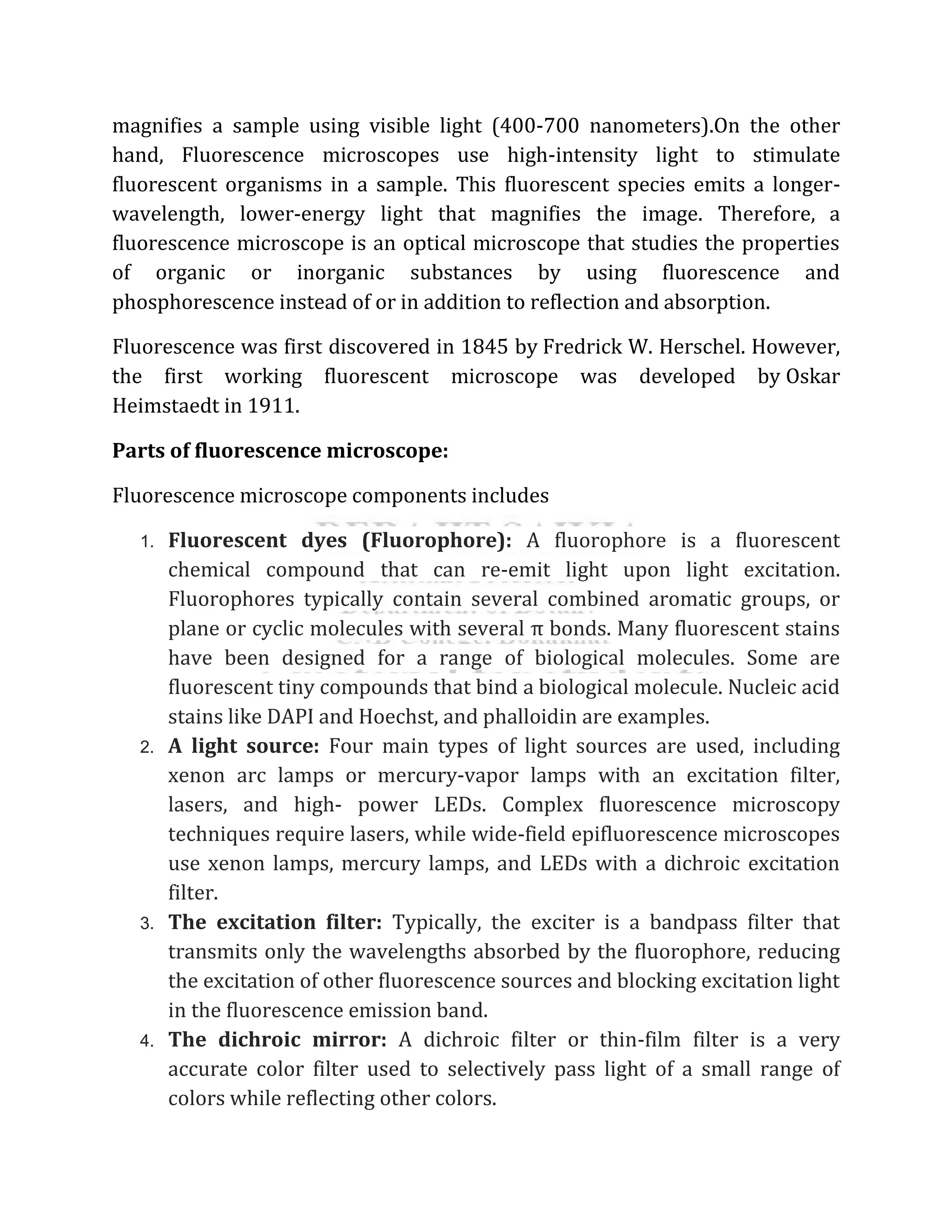
Fluorescence microscopy:
Question: What do you mean by fluorescence microscope? Who discovered
it? Describe briefly its parts, types and working principle with diagram.
State briefly the application of fluorescence microscope.
A fluorescence microscope is similar to a regular light microscope, but it has
several extra qualities that enhance its usefulness. The typical microscope
2.
magnifies a sampleusing visible light (400-700 nanometers).On the other
hand, Fluorescence microscopes use high-intensity light to stimulate
fluorescent organisms in a sample. This fluorescent species emits a longer-
wavelength, lower-energy light that magnifies the image. Therefore, a
fluorescence microscope is an optical microscope that studies the properties
of organic or inorganic substances by using fluorescence and
phosphorescence instead of or in addition to reflection and absorption.
Fluorescence was first discovered in 1845 by Fredrick W. Herschel. However,
the first working fluorescent microscope was developed by Oskar
Heimstaedt in 1911.
Parts of fluorescence microscope:
Fluorescence microscope components includes
1. Fluorescent dyes (Fluorophore): A fluorophore is a fluorescent
chemical compound that can re-emit light upon light excitation.
Fluorophores typically contain several combined aromatic groups, or
plane or cyclic molecules with several π bonds. Many fluorescent stains
have been designed for a range of biological molecules. Some are
fluorescent tiny compounds that bind a biological molecule. Nucleic acid
stains like DAPI and Hoechst, and phalloidin are examples.
2. A light source: Four main types of light sources are used, including
xenon arc lamps or mercury-vapor lamps with an excitation filter,
lasers, and high- power LEDs. Complex fluorescence microscopy
techniques require lasers, while wide-field epifluorescence microscopes
use xenon lamps, mercury lamps, and LEDs with a dichroic excitation
filter.
3. The excitation filter: Typically, the exciter is a bandpass filter that
transmits only the wavelengths absorbed by the fluorophore, reducing
the excitation of other fluorescence sources and blocking excitation light
in the fluorescence emission band.
4. The dichroic mirror: A dichroic filter or thin-film filter is a very
accurate color filter used to selectively pass light of a small range of
colors while reflecting other colors.
3.
5. The emissionfilter: The emitter is typically a bandpass filter that
passes only the wavelengths emitted by the fluorophore and blocks all
undesired light outside this band – especially the excitation light. By
blocking unwanted excitation energy (including UV and IR) or sample
and system autofluorescence, optical filters ensure the darkest
background.
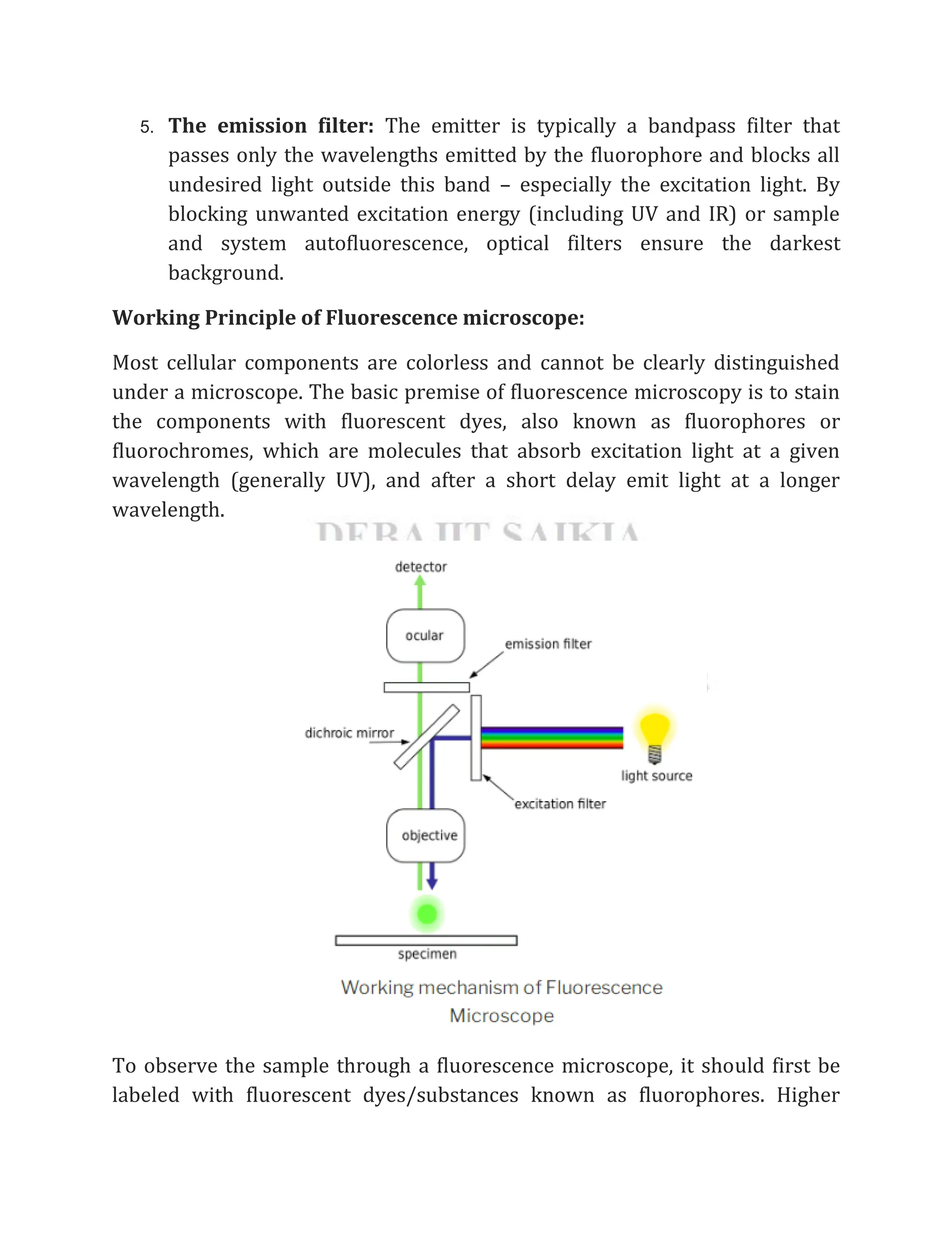
Working Principle of Fluorescence microscope:
Most cellular components are colorless and cannot be clearly distinguished
under a microscope. The basic premise of fluorescence microscopy is to stain
the components with fluorescent dyes, also known as fluorophores or
fluorochromes, which are molecules that absorb excitation light at a given
wavelength (generally UV), and after a short delay emit light at a longer
wavelength.
To observe the sample through a fluorescence microscope, it should first be
labeled with fluorescent dyes/substances known as fluorophores. Higher
4.
energy shorter wavelengthlights (UV rays or blue light) generated from
xenon arc lamp or mercury vapor arc lamp passes through the excitation
filter. The excitation filter allows only the short wavelength of light to pass
through and removes all other non-specific wavelengths of light. The filtered
light is reflected by the dichroic filter and falls on the fluorophore-labeled
sample. The fluorochrome absorbs shorter wavelength rays and emits rays of
longer wavelength (lower energy) that pass through the emission filter. The
emission filter blocks (suppresses) any residual excitation light and passes the
desired longer emission wavelengths to the detector. Thus the microscope
forms glowing images of the fluorochrome-labeled microorganisms against a
dark background.
To the observer, the background is dark, as there is no visible light and only
the labeled specimen (cells, microorganisms, etc.) appear bright (fluoresce).
Types of Fluorescence microscope:
Fluorescence microscopy is one of the most used imaging modalities in
molecular biology and living specimens. To increase image contrast and
spatial resolution, different type of fluorescence microscopy has been
developed. However there are 4 main types of fluorescence microscopy:
a) Epifluorescence microscopes: It is the most common type of
fluorescence microscope. In this microscope, excitation of the
fluorophore and detection of the fluorescence are done through the
same light path (i.e., through the objective).
b) Confocal microscope: In this type of fluorescence microscope,
high‐resolution imaging of thick specimens (without physical
sectioning) can be analyzed using fluorescent-labeled dye.
c) Multiphoton microscope: In this type of microscope, multiphoton
fluorescence excitation captures high-resolution three-dimensional
images of specimens tagged with highly specific fluorophores.
d) Total internal reflection fluorescence (TIRF) microscope :Total
internal reflection fluorescence microscopy (TIRFM) exploits the unique
properties of an induced evanescent wave or field in a limited specimen
5.
region immediately adjacentto the interface between two media having
different refractive indices.
Applications of Fluorescence Microscope:
1. Fluorescence microscopy is widely used in diagnostic microbiology and
microbial ecology for enumerating bacteria in natural environments.
Some organisms, such as Pseudomonas, fluoresce naturally when
irradiated with ultraviolet light. Other organisms, such as
Mycobacterium tuberculosis and Treponema pallidum, are treated with
fluorochrome. Acid-fast bacilli (AFB) in sputum or CSF are detected
when stained with auramine fluorescent dye. Detection of Trichomonas
vaginalis, intracellular gonococci, and other parasites when stained by
acridine orange. In immunodiagnosis of infectious diseases, using both
direct and indirect antibody techniques. Immunofluorescence is
especially useful in diagnosing syphilis and rabies.
2. It is used to identify structures in fixed and live biological samples.
3. Fluorescence microscopy is a common tool for today’s life science
research because it allows the use of multicolor staining, labeling of
structures within cells, and the measurement of the physiological state
of a cell.
4. It is used to imaging structural components of small specimens, such as
cells
5. It is used to conducting viability studies on cell populations (are they
alive or dead?)
6. It is used to imaging the genetic material within a cell (DNA and RNA)
7. It is used to viewing specific cells within a larger population with
techniques such as FISH
Advantages of Fluorescence Microscope:
1. Fluorescence microscopy is the most popular method for studying the
dynamic behavior exhibited in live-cell imaging.
2. This stems from its ability to isolate individual proteins with a high
degree of specificity amidst non-fluorescing material.
6.
3. The sensitivityis high enough to detect as few as 50 molecules per cubic
micrometer.
4. Different molecules can now be stained with different colors, allowing
multiple types of the molecule to be tracked simultaneously.
5. These factors combine to give fluorescence microscopy a clear
advantage over other optical imaging techniques, for both in vitro and in
vivo imaging.
Limitations of Fluorescence Microscope:
Fluorophores gradually lose their ability to fluoresce as they are illuminated
in photobleaching. Photobleaching can severely limit the time a sample can be
observed by fluorescence microscopy. However, several techniques exist to
reduce photobleaching, such as using more robust fluorophores, minimizing
illumination, or using photoreactive scavenger chemicals.
Fluorescence microscopy has enabled the analysis of live cells, but fluorescent
molecules generate reactive chemical species under illumination that
enhances the phototoxic effect, to which live cells are susceptible.
Fluorescence microscopy only allows observation of the specific structures
labeled for fluorescence. For example, observing a tissue sample prepared
with a fluorescent DNA stain by fluorescence microscopy only reveals the
organization of the DNA within the cells and reveals nothing else about the
cell morphologies.


![[DSC Europe 25] Hans Kleinsman - The Compliance Gearbox: How Tax Tech Mediate...](https://cdn.slidesharecdn.com/ss_thumbnails/dxdytie1toel0hr90bjs-2-251212103250-174fdbe7-thumbnail.jpg?width=640&height=640&fit=bounds)


![[DSC Europe 25] Branko Urosevic -Rethinking Financial Talent: Integrating Cod...](https://cdn.slidesharecdn.com/ss_thumbnails/8jjrus8ttko6qj64f58f-3-251212103250-642c6374-thumbnail.jpg?width=640&height=640&fit=bounds)
![[DSC Europe 25] Milan Sekuloski - Data, Defence, and Development: Cybersecuri...](https://cdn.slidesharecdn.com/ss_thumbnails/dfrkwwx4qly6atqpbl4z-4-251209104645-c3d4b0ca-thumbnail.jpg?width=640&height=640&fit=bounds)
![[DSC Europe 25] Dunja Adzic Jovanovic - AI and Cybersecurity: Defending Data ...](https://cdn.slidesharecdn.com/ss_thumbnails/o1zylpbhrtwnixxq2xj8-7-251211083048-185086f6-thumbnail.jpg?width=640&height=640&fit=bounds)
![[DSC Europe 25] Behzad Hosseini - AI Agents in the Wild: Deploying Models tha...](https://cdn.slidesharecdn.com/ss_thumbnails/3qtejajvsjqrzwfept2c-10-251212103250-7f2b1068-thumbnail.jpg?width=640&height=640&fit=bounds)

![[DSC Europe 25] Marko Krstic - Understanding the AI Threat Landscape - Risks,...](https://cdn.slidesharecdn.com/ss_thumbnails/tiyim1ins5jvbrvzpzla-2-251209104645-c69d3553-thumbnail.jpg?width=640&height=640&fit=bounds)

![[DSC Europe 25] Bassam Maharmeh - Artificial Intelligence: Opportunities and ...](https://cdn.slidesharecdn.com/ss_thumbnails/thhfmr2fqpawzj7hsjpg-5-251211083048-2c23204f-thumbnail.jpg?width=640&height=640&fit=bounds)
![[DSC Europe 25] Kaja Kandare - LLM as a judge.pptx](https://cdn.slidesharecdn.com/ss_thumbnails/arxyccaxsdsd1ba99wjw-7-251212104007-2b4e3f64-thumbnail.jpg?width=640&height=640&fit=bounds)