Fluorescence Microscopy , principles, components, fluorescent dyes and probes, working mechanism, types of fluorescence microscopy and applications in research and medicine
Introduction
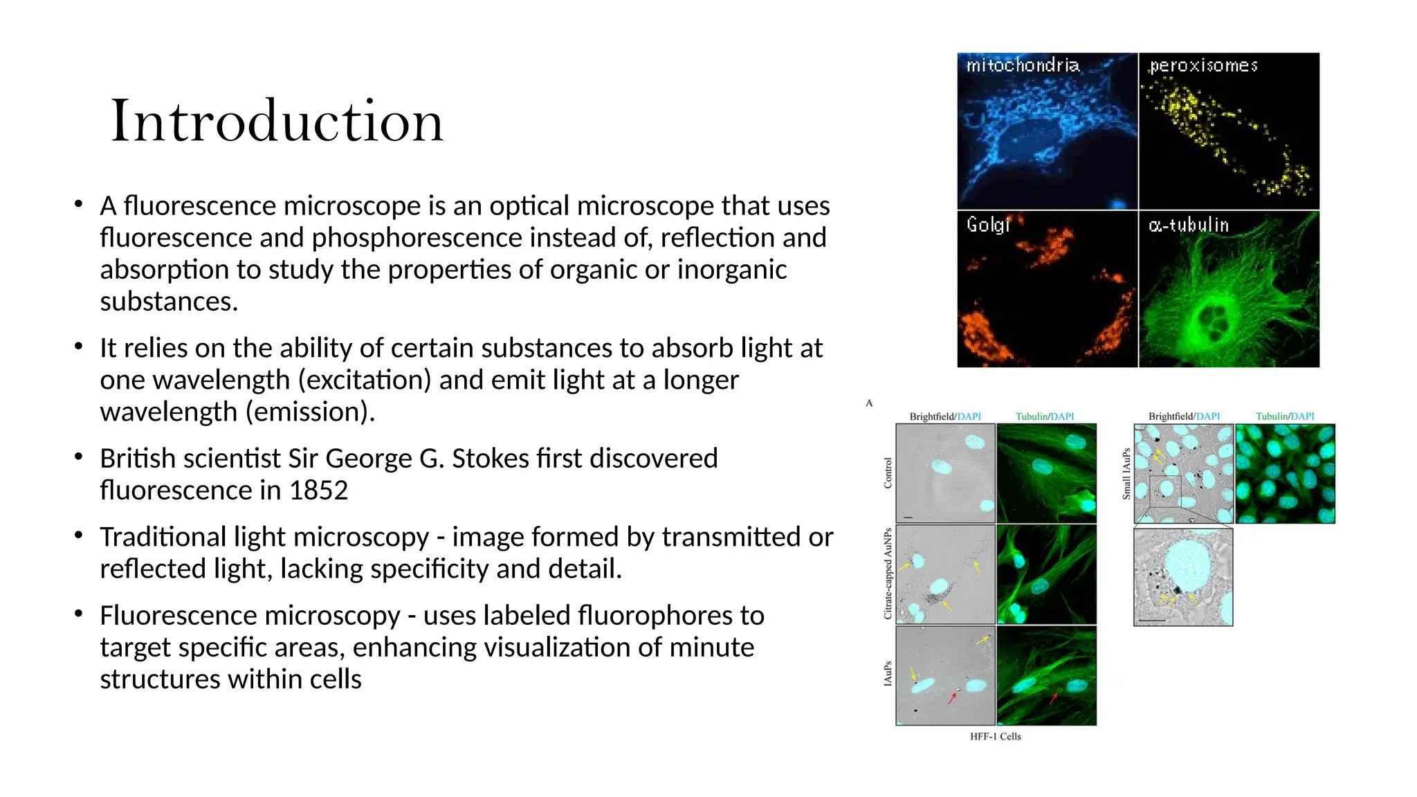
• A fluorescencemicroscope is an optical microscope that uses
fluorescence and phosphorescence instead of, reflection and
absorption to study the properties of organic or inorganic
substances.
• It relies on the ability of certain substances to absorb light at
one wavelength (excitation) and emit light at a longer
wavelength (emission).
• British scientist Sir George G. Stokes first discovered
fluorescence in 1852
• Traditional light microscopy - image formed by transmitted or
reflected light, lacking specificity and detail.
• Fluorescence microscopy - uses labeled fluorophores to
target specific areas, enhancing visualization of minute
structures within cells
3.
Principles of Fluorescence
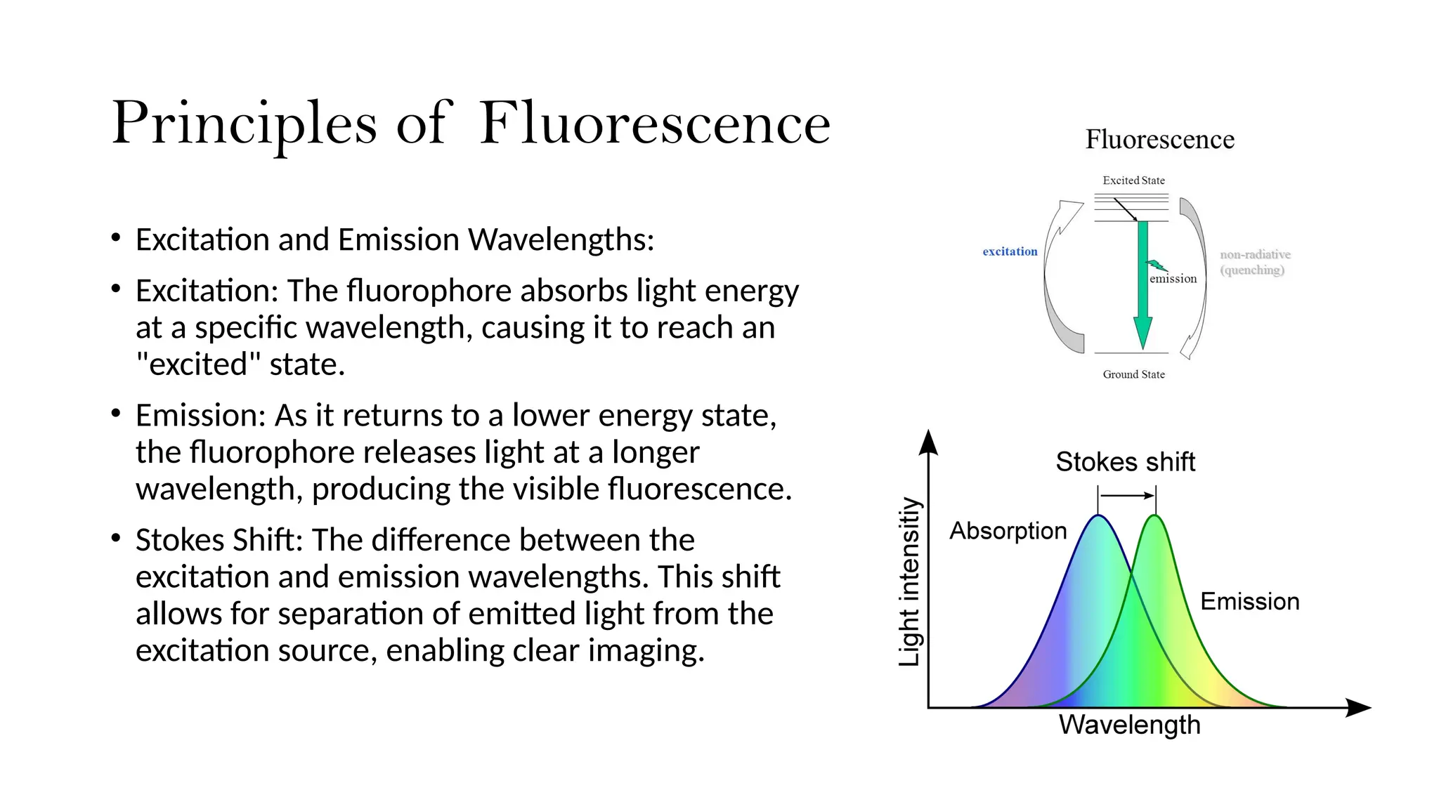
•Excitation and Emission Wavelengths:
• Excitation: The fluorophore absorbs light energy
at a specific wavelength, causing it to reach an
"excited" state.
• Emission: As it returns to a lower energy state,
the fluorophore releases light at a longer
wavelength, producing the visible fluorescence.
• Stokes Shift: The difference between the
excitation and emission wavelengths. This shift
allows for separation of emitted light from the
excitation source, enabling clear imaging.
4.
Components of aFluorescence Microscope
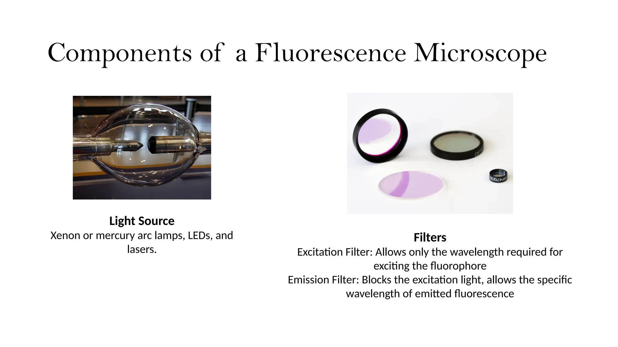
Light Source
Xenon or mercury arc lamps, LEDs, and
lasers.
Filters
Excitation Filter: Allows only the wavelength required for
exciting the fluorophore
Emission Filter: Blocks the excitation light, allows the specific
wavelength of emitted fluorescence
5.
Components of aFluorescence Microscope
Dichroic Mirror (or Beam Splitter)
Splits the excitation and emission paths,
so only emitted light reaches the
detector.
Objective Lens
Collects the emitted light and
magnifies the image.
High numerical aperture (NA)
objectives are preferred as they
increase resolution and
brightness.
Detector
CCD (Charge-Coupled Device) cameras,
CMOS cameras, or photomultiplier tubes
(PMTs) in confocal microscopy.
Captures the emitted fluorescence and
converts it into an image.
7.
Fluorescent Dyes andProbes
• Fluorescent dyes are organic
compounds that emit light
when exposed to certain
wavelengths of light.
• Fluorescent probes are
compounds designed to bind
specifically to certain target
molecules (e.g., proteins,
nucleic acids, ions) or cellular
structures in biological
samples.
Commonly used-
• DAPI: Binds to DNA, used to
stain cell nuclei in blue.
• FITC (Fluorescein
isothiocyanate): Labels proteins,
emits green fluorescence.
• GFP (Green Fluorescent Protein)
and RFP (Red Fluorescent
Protein) : Genetically fused to
proteins, allowing visualization
of protein dynamics in live cells.
8.
Working Mechanism ofFluorescence
Microscopy
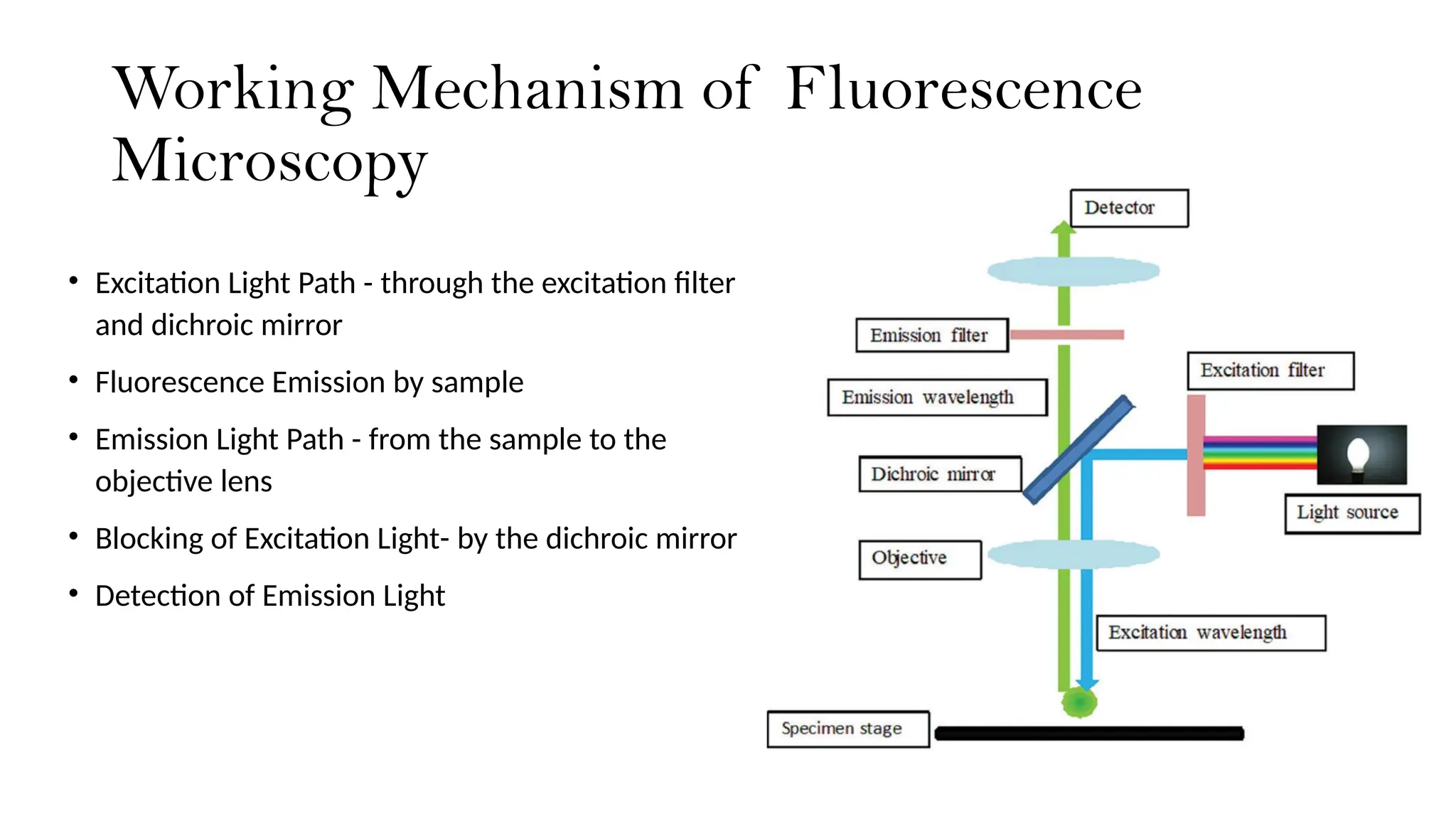
• Excitation Light Path - through the excitation filter
and dichroic mirror
• Fluorescence Emission by sample
• Emission Light Path - from the sample to the
objective lens
• Blocking of Excitation Light- by the dichroic mirror
• Detection of Emission Light
9.
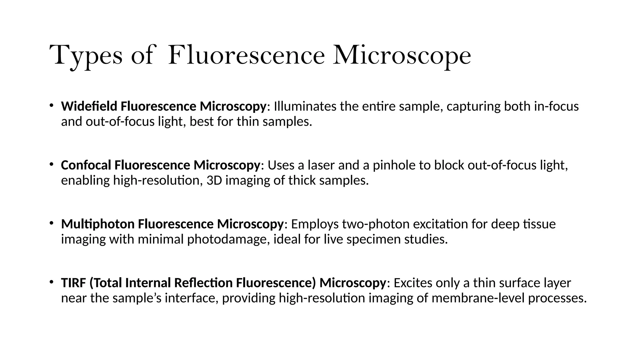
Types of FluorescenceMicroscope
• Widefield Fluorescence Microscopy: Illuminates the entire sample, capturing both in-focus
and out-of-focus light, best for thin samples.
• Confocal Fluorescence Microscopy: Uses a laser and a pinhole to block out-of-focus light,
enabling high-resolution, 3D imaging of thick samples.
• Multiphoton Fluorescence Microscopy: Employs two-photon excitation for deep tissue
imaging with minimal photodamage, ideal for live specimen studies.
• TIRF (Total Internal Reflection Fluorescence) Microscopy: Excites only a thin surface layer
near the sample’s interface, providing high-resolution imaging of membrane-level processes.
10.
Applications in Researchand
Medicine
• Cell and Tissue Imaging : visualize specific cell structures,
such as nuclei, mitochondria, and cytoskeleton using
dyes like DAPI for staining DNA enables visualization of
cell nuclei.
• Immunofluorescence : uses antibodies tagged with
fluorescent dyes to bind to specific proteins or antigens
within cells, detecting biomarkers associated with
diseases like cancer
• Live-Cell Imaging : proteins like GFP and RFP enable
visualization of living cells in real-time, allowing
observation of dynamic cellular processes like cell
division, protein trafficking, and cellular responses to
stimuli.
Immunofluorescence
11.
Applications in Researchand
Medicine
• Diagnosis and Pathogen Detection : used in medical
diagnostics to identify pathogens and abnormal cells in
patient samples (bacteria, viruses, and cancer cells)
• Neuroscience Applications : used to map neural
circuits and visualize neurotransmitter activity in brain
tissue. eg- Calcium imaging to observe neural activity
or using fluorescent tags to trace nerve connections.
• Genetic and Molecular Research : Techniques like
Fluorescence In Situ Hybridization (FISH) allow for the
localization of specific DNA or RNA sequences within
cells studying gene expression patterns and identifying
genetic abnormalities.
Calcium imaging in neurons
FISH technique
Editor's Notes
#2 Fluorescence is the emission of light by a substance that has absorbed radiation while phosphorescence is a specific type of photoluminescence related to fluorescence in which the emission of light is a little delayed as compared to fluorescence.
Fluorochromes are photoreactive chemicals that can absorb energy via the interaction of an orbital electron in the molecule's atomic structure with a photon of light.
#6 The filters and the dichroic mirror are often plugged in together in a filter cube.
#8 The excitation light passes through the excitation filter and is directed to the dichroic mirror. This reflects the light through the objective towards the specimen.
Fluorochromes in the specimen are excited and emit photons. This emission light passes back through the objective to the dichroic mirror.
The emitted light has an appropriate wavelength and is able to pass. Excitation light that is reflected by the specimen is not able to pass through the dichroic mirror and will be blocked.
If excitation light is able to pass through the dichroic mirror it will be blocked when it reaches the emission filter. Light passing through the emission filter can be measured with a detector.