Fluorescence microscopy is a powerful technique used in many fields of science, especially medical sciences. It works by exciting fluorescent molecules in a specimen with one wavelength of light, which causes them to emit a different wavelength of light. A fluorescence microscope uses filters to isolate the emitted light and form an image with high contrast against a dark background. This allows very sensitive observations and measurements by detecting the natural or dye-induced fluorescence of cells and molecules.
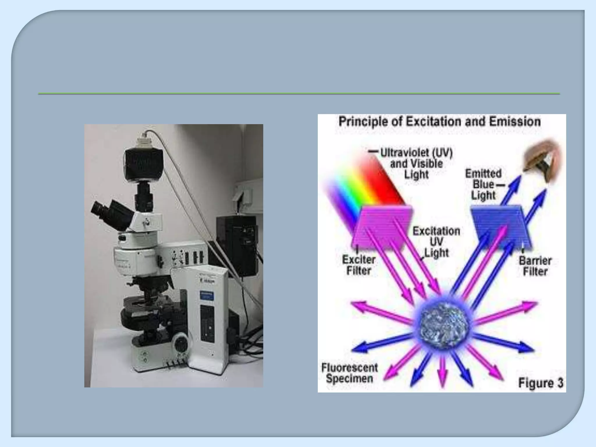
![Principle of Fluorescence
The specimen is illuminated with light of a specific wavelength (or
wavelengths) which is absorbed by the fluorophores, causing them
to emit light of longer wavelengths (i.e., of a different color than the
absorbed light). The illumination light is separated from the much
weaker emitted fluorescence through the use of a spectral emission
filter. Typical components of a fluorescence microscope are a light
source (xenon arc lamp or mercury-vapor lamp), the excitation
filter, the dichroic mirror (or dichroic beamsplitter), and theemission
filter (see figure below). The filters and the dichroic are chosen to
match the spectral excitation and emission characteristics of the
fluorophore used to label the specimen.[1] In this manner, the
distribution of a single fluorophore (color) is imaged at a time.
Multi-color images of several types of fluorophores must be
composed by combining several single-color images.[1]
Most fluorescence microscopes in use are epifluorescence
microscopes (i.e., excitation and observation of the fluorescence are
from above (epi–) the specimen). These microscopes have become
an important part in the field of biology, opening the doors for more
advanced microscope designs, such as the confocal microscope and
the total internal reflection fluorescence microscope (TIRF).](https://image.slidesharecdn.com/123456-130331053809-phpapp02/75/123456-5-2048.jpg)